Recognise Organelles and Other Cell Structures in Eukaryotic Cells
🌿 Cell Surface Membrane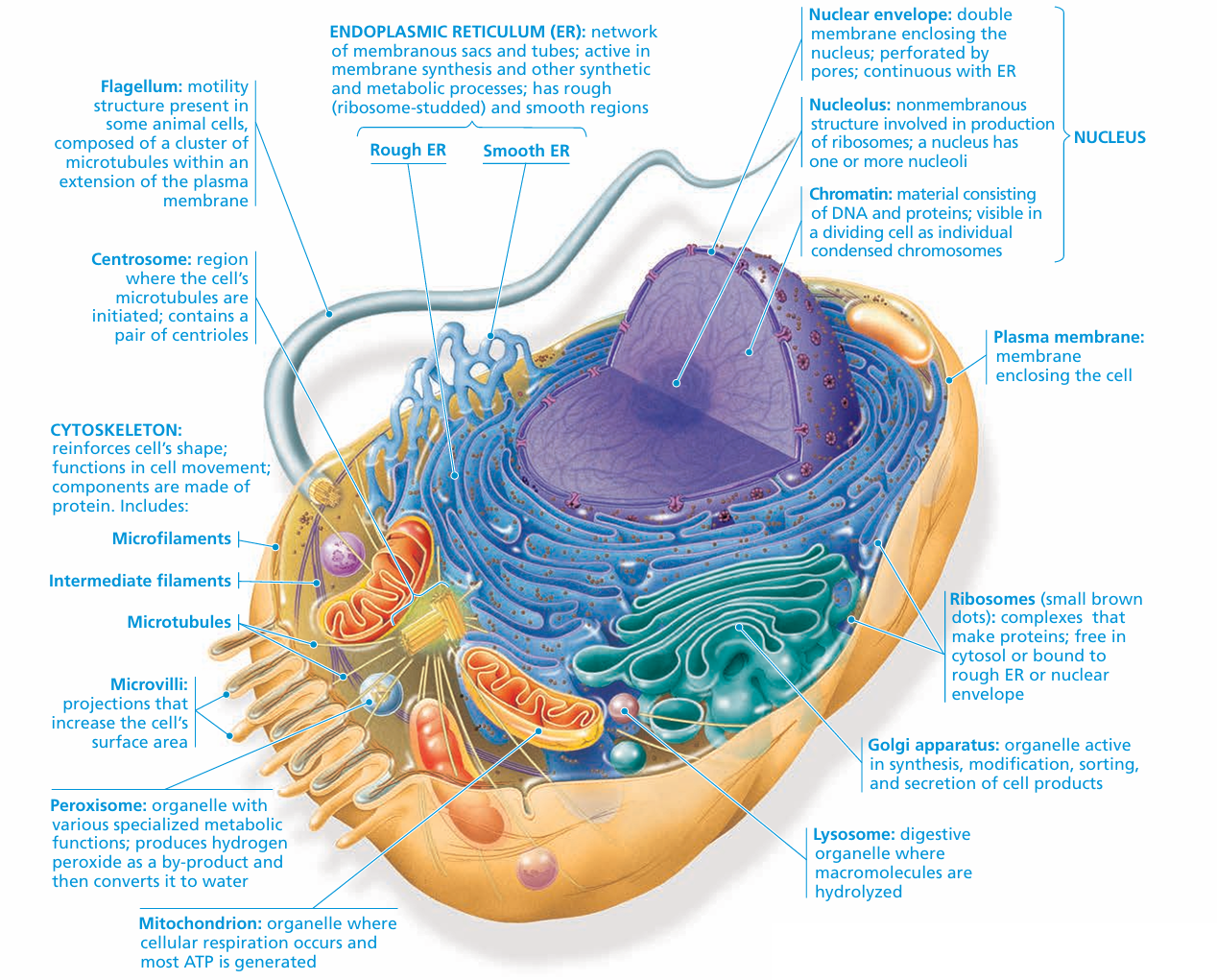
Structure: Thin phospholipid bilayer with embedded proteins.
Function: Controls entry and exit of substances (selectively permeable barrier).
🧠 Nucleus, Nuclear Envelope & Nucleolus
Nucleus: Spherical organelle containing DNA, controls cellular activities.
Nuclear Envelope: Double membrane with pores; allows material exchange with cytoplasm.
Nucleolus: Dense region inside nucleus; synthesises ribosomal RNA and assembles ribosomes.
🔬 Rough Endoplasmic Reticulum (RER)
Structure: Flattened sacs with ribosomes attached.
Function: Synthesises and transports proteins.
🔬 Smooth Endoplasmic Reticulum (SER)
Structure: Tubular network without ribosomes.
Function: Synthesises lipids and steroids; detoxifies substances.
📦 Golgi Body (Golgi Apparatus or Golgi Complex)
Structure: Stack of flattened membranous sacs.
Function: Modifies, sorts, packages, and transports proteins and lipids.
⚡ Mitochondria
Structure: Double membrane; inner folds called cristae; contains small circular DNA.
Function: Site of aerobic respiration; produces ATP (energy).
🧪 Ribosomes
Structure: Composed of RNA and protein; 80S in cytoplasm, 70S in chloroplasts and mitochondria.
Function: Synthesise proteins.
🗑️ Lysosomes
Structure: Membrane-bound sacs with digestive enzymes.
Function: Break down waste materials, old organelles, and pathogens.
⚙️ Centrioles and Microtubules
Centrioles: Cylindrical structures made of microtubules, involved in cell division (mainly animal cells).
Microtubules: Tubular proteins maintaining cell shape and aiding intracellular transport.
🌬️ Cilia
Structure: Hair-like projections with microtubule arrangement.
Function: Move fluids or the cell itself.
🔍 Microvilli
Structure: Finger-like extensions of the cell membrane.
Function: Increase surface area for absorption.
🌞 Chloroplasts
Structure: Double membrane with internal thylakoid stacks; contains chlorophyll and small circular DNA.
Function: Conduct photosynthesis.
🧱 Cell Wall
Structure: Rigid outer layer composed mainly of cellulose in plants.
Function: Provides structural support and protection.
🔗 Plasmodesmata
Structure: Channels through cell walls connecting adjacent plant cells.
Function: Facilitate communication and transport between cells.
💧 Large Permanent Vacuole and Tonoplast (Plant Cells)
Large Permanent Vacuole: Fluid-filled sac storing water, nutrients, and waste.
Tonoplast: Membrane surrounding the vacuole.
Function: Maintains turgor pressure; stores substances; isolates harmful materials.
📊 Summary Table
| Organelle/Structure | Structure Highlights | Function |
|---|---|---|
| Cell surface membrane | Phospholipid bilayer with proteins | Regulates substance movement |
| Nucleus + nuclear envelope | Double membrane with pores | Controls cell activities |
| Nucleolus | Dense body inside nucleus | Produces ribosomal RNA |
| Rough ER | Flattened sacs with ribosomes | Protein synthesis and transport |
| Smooth ER | Tubular without ribosomes | Lipid synthesis, detoxification |
| Golgi Body | Stacked membranous sacs | Modifies and packages proteins |
| Mitochondria | Double membrane, cristae, circular DNA | ATP production (respiration) |
| Ribosomes | RNA and protein; 80S & 70S types | Protein synthesis |
| Lysosomes | Membrane sacs with enzymes | Digestion and waste removal |
| Centrioles & microtubules | Cylindrical & tubular structures | Cell division, structural support |
| Cilia | Microtubule-based hair-like projections | Movement of cell or fluids |
| Microvilli | Membrane projections | Increase absorption surface area |
| Chloroplasts | Double membrane, thylakoids, DNA | Photosynthesis |
| Cell wall | Rigid cellulose layer | Support and protection |
| Plasmodesmata | Channels through cell walls | Cell-to-cell communication |
| Large vacuole + tonoplast | Membrane sac | Storage and turgor maintenance |
Organelles have specific structures tailored for their functions.
Some organelles like mitochondria and chloroplasts contain their own DNA.
Plant cells have unique features like cell wall, chloroplasts, plasmodesmata, and large vacuole.
Describe and Interpret Photomicrographs, Electron Micrographs, and Drawings of Typical Plant and Animal Cells
🌱 Types of Images
- Photomicrographs: Photographs taken through a light microscope; magnifications up to ×1500; show overall cell shapes and large organelles but limited detail.
- Electron Micrographs: Images from electron microscopes (TEM or SEM); magnifications up to ×1,000,000; reveal ultrastructure like membranes, ribosomes, and detailed organelles.
- Drawings: Hand-drawn or digital illustrations based on observations; highlight key features clearly with labels and simplify complex details for clarity.
🔍 Describing Typical Plant Cells
- Photomicrographs & Drawings:

- Cell wall (rigid outline)
- Large central vacuole (clear area)
- Chloroplasts (green structures in live samples)
- Nucleus, sometimes visible near the edge
- Cytoplasm surrounding organelles
- Electron Micrographs:
- Detailed double membranes of nucleus and chloroplasts
- Thylakoid membranes inside chloroplasts
- Mitochondria with cristae
- Ribosomes as small dots
- Tonoplast membrane around vacuole
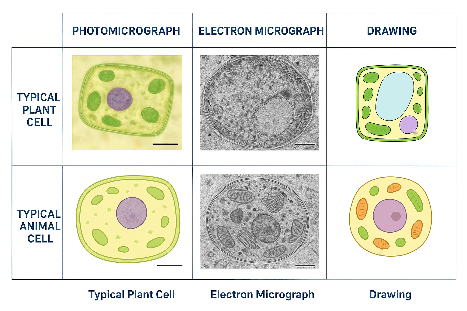
🔍 Describing Typical Animal Cells
- Photomicrographs & Drawings:
- Irregular shape without a cell wall
- Nucleus often centrally located
- Cytoplasm filling cell interior
- Visible organelles like mitochondria (if stained)
- May show cilia or microvilli on surface
- Electron Micrographs:
- Clear nuclear envelope with pores
- Detailed mitochondria structure
- Centrioles near the nucleus
- Lysosomes as membrane-bound vesicles
- Microtubules forming cytoskeleton
🧩 Interpreting Images: Key Points
- Identify cell type (plant vs animal) by presence or absence of cell wall, chloroplasts, and vacuole.
- Look for organelles like nucleus, mitochondria, ER, Golgi, lysosomes.
- Note shape and size differences: plant cells are more regular and rectangular; animal cells are irregular.
- Use scale bars and magnification info to estimate sizes.
- Recognise staining patterns in photomicrographs (e.g., darker nucleus).
- Understand grayscale images in electron micrographs showing fine details, membranes, and internal structures.
📊 Summary Table: Features in Different Image Types
| Feature/Organelle | Photomicrograph | Electron Micrograph | Drawing |
|---|---|---|---|
| Cell wall | Visible in plant cells as rigid boundary | Clear double-layered structure | Clear outline |
| Nucleus | Visible, sometimes stained dark | Detailed envelope and nucleolus | Clearly labelled spherical |
| Chloroplasts | Green spots in plant cells | Internal thylakoid membranes | Oval with internal stacks |
| Mitochondria | Occasionally visible | Detailed cristae and membranes | Small oval with folds |
| Vacuole | Large clear area in plant cells | Tonoplast membrane visible | Large central sac |
| Cilia/Microvilli | Visible on cell surface (animal) | Microtubule arrangement visible | Short projections |
🧠 Key Tips for Describing and Interpreting
- Start with overall cell shape and size.
- Identify unique plant or animal features.
- Mention visible organelles and their appearance.
- Use scientific terms and correct labels.
- Refer to the scale bar to estimate size where possible.
- Describe staining or contrast effects in photomicrographs.
Cells Use ATP from Respiration for Energy-Requiring Processes
🌱 What is ATP?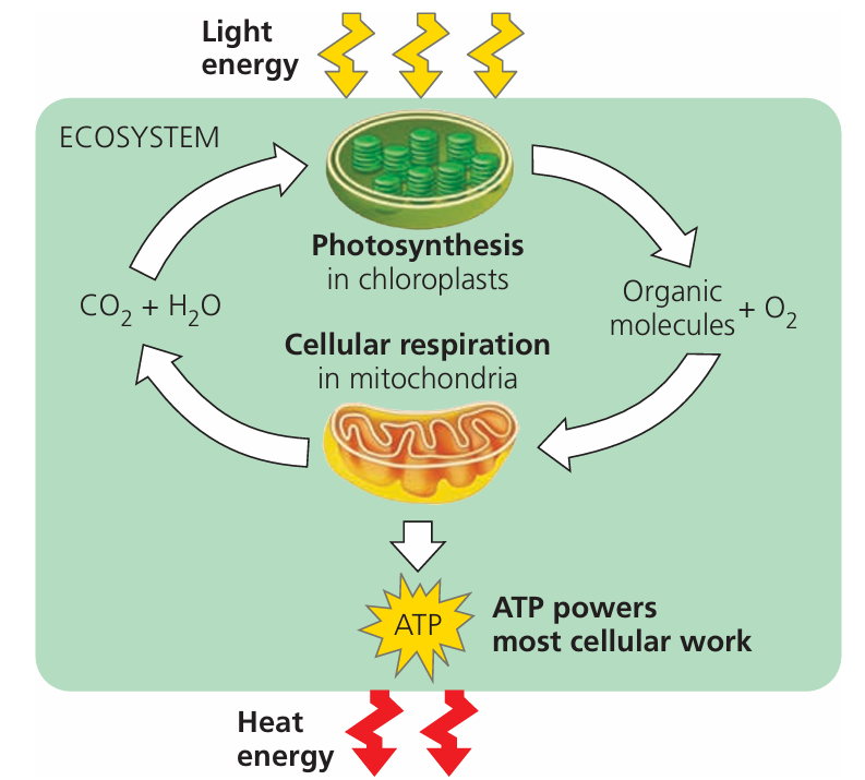
- ATP (Adenosine Triphosphate) is the energy currency of the cell.
- It stores and transports energy within cells.
🔥 How is ATP Produced?
- ATP is produced during cellular respiration in mitochondria.
- Respiration breaks down glucose (or other molecules) to release energy, which is stored in ATP.
⚙️ Role of ATP in Cells
- Provides energy needed for energy-requiring processes such as:
- Active transport: Moving substances against concentration gradients across membranes.
- Protein synthesis: Building proteins from amino acids.
- Cell division: Processes like mitosis and cytokinesis.
- Muscle contraction: Allowing movement in muscle cells.
- Metabolic reactions: Various chemical reactions that require energy input.
- Movement of organelles: Transporting materials inside the cell.
ATP generated by respiration is essential for powering all cellular activities that require energy.
Without ATP, cells cannot perform vital functions necessary for life.
Key Structural Features of a Prokaryotic Cell (Typical Bacterium)
🌱 Basic Characteristics
- Unicellular: Prokaryotes exist as single, independent cells.
- Size: Generally small, about 1–5 µm in diameter.
🧱 Cell Wall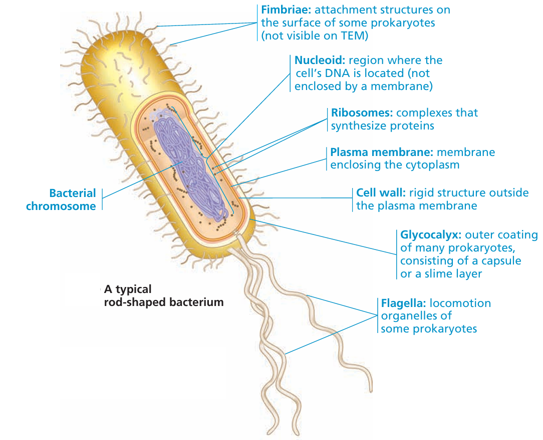
- Made of peptidoglycan, a strong, mesh-like polymer that provides shape and protection.
- Unlike plant cells, no cellulose; distinct bacterial structure.
🧬 Genetic Material
- Contains a single, circular DNA molecule located in the nucleoid region (no true nucleus).
- DNA is not enclosed in a membrane-bound nucleus.
🧪 Ribosomes
- Have 70S ribosomes, smaller than eukaryotic 80S ribosomes.
- Ribosomes are the site of protein synthesis.
🚫 Absence of Membrane-Bound Organelles
- No mitochondria, chloroplasts, endoplasmic reticulum, or Golgi apparatus.
- Cellular processes occur in the cytoplasm or at the cell membrane.
| Feature | Description |
|---|---|
| Unicellular | Single-celled organism |
| Size | 1–5 µm diameter |
| Cell Wall | Peptidoglycan layer |
| DNA | Circular, free in cytoplasm |
| Ribosomes | 70S type |
| Membrane-bound organelles | Absent |
Prokaryotic cells are simpler than eukaryotic cells, lacking nucleus and membrane-bound organelles, but have unique features like peptidoglycan walls and 70S ribosomes essential for their survival.