Fluid Mosaic Model of Membrane Structure
🌱 Overview
- Proposed by Singer and Nicolson (1972).
- Describes the plasma membrane as a flexible, dynamic structure made up of a phospholipid bilayer with proteins embedded in it.
🧩 Phospholipid Bilayer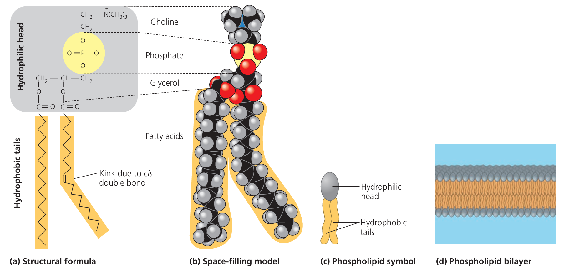
- Phospholipids have:
- Hydrophilic (polar) phosphate heads → attract water.
- Hydrophobic (non-polar) fatty acid tails → repel water.
- In water, phospholipids arrange themselves into a bilayer:
- Heads face outward towards aqueous environments (inside cytoplasm & outside cell).
- Tails face inward, shielded from water.
🌊 Hydrophobic & Hydrophilic Interactions
- Hydrophobic tails avoid contact with water → cause bilayer formation.
- Hydrophilic heads interact with water on both membrane surfaces.
- This arrangement creates a stable barrier that is selectively permeable.
🧬 Proteins in the Membrane
- Integral (intrinsic) proteins:
- Span across the bilayer or are embedded within it.
- Often function as transport channels, carriers, or receptors.
- Peripheral (extrinsic) proteins:
- Loosely attached to surface (inside or outside).
- Often involved in cell signalling or structural support.
🎯 “Fluid” Aspect
- Phospholipids and proteins can move laterally within the layer.
- This fluidity allows:
- Membrane repair
- Flexibility
- Dynamic changes in protein arrangement
🧩 “Mosaic” Aspect
- The membrane is a patchwork of:
- Proteins
- Phospholipids
- Cholesterol
- Glycolipids & Glycoproteins
- Cholesterol helps regulate fluidity and stability.
📌 Key Functions from the Structure
- Selective permeability
- Cell signalling
- Transport of molecules
- Cell recognition
✅ Summary:
The fluid mosaic model explains how hydrophobic and hydrophilic interactions drive phospholipids into a bilayer, with proteins embedded in a dynamic, flexible structure. This arrangement supports selective transport, cell communication, and structural integrity.
The fluid mosaic model explains how hydrophobic and hydrophilic interactions drive phospholipids into a bilayer, with proteins embedded in a dynamic, flexible structure. This arrangement supports selective transport, cell communication, and structural integrity.
Arrangement of Cholesterol, Glycolipids & Glycoproteins in Cell Surface Membranes
🌱 Cholesterol
- Location: Found between phospholipid molecules in both layers of the bilayer.
- Structure: Small, rigid, hydrophobic molecule with a small polar hydroxyl group.
- Role in arrangement:
- Hydroxyl group aligns near phosphate heads (hydrophilic region).
- Rigid steroid ring structure inserts into hydrophobic tails.
- Function:
- Reduces phospholipid movement → membrane stability.
- Prevents packing of tails at low temperatures → maintains fluidity.
🌿 Glycolipids

- Location: Lipids with a carbohydrate chain attached; always in outer layer of the bilayer.
- Arrangement:
- Lipid portion embedded in the hydrophobic region of bilayer.
- Carbohydrate chain extends into the extracellular fluid.
- Function:
- Cell recognition & signalling.
- Acts as a receptor for certain molecules.
- Contributes to glycocalyx (carbohydrate-rich cell surface).
🧩 Glycoproteins
- Location: Proteins with carbohydrate chains attached; carbohydrate part always projects into extracellular space.
- Arrangement:
- Protein portion can be integral (spanning membrane) or peripheral.
- Carbohydrate chain faces outward, forming part of glycocalyx.
- Function:
- Cell-cell recognition (e.g., immune responses).
- Acts as receptors for hormones & neurotransmitters.
- Helps with cell adhesion.
📌 Integrated Arrangement in the Fluid Mosaic Model
| Component | Location in Membrane | Orientation | Main Role |
|---|---|---|---|
| Cholesterol | Between phospholipid tails in both layers | Hydroxyl group near heads, rings in tails | Stability & fluidity |
| Glycolipids | Outer layer only | Carbohydrate chain outward | Cell recognition, signalling |
| Glycoproteins | Outer layer (protein in bilayer) | Carbohydrate chain outward | Receptors, recognition, adhesion |
✅ Summary:
In the cell surface membrane, cholesterol sits between phospholipids to regulate fluidity, glycolipids anchor into the outer layer with carbohydrates extending outward, and glycoproteins project carbohydrate chains from membrane proteins into the extracellular space for recognition, signalling, and adhesion.
In the cell surface membrane, cholesterol sits between phospholipids to regulate fluidity, glycolipids anchor into the outer layer with carbohydrates extending outward, and glycoproteins project carbohydrate chains from membrane proteins into the extracellular space for recognition, signalling, and adhesion.
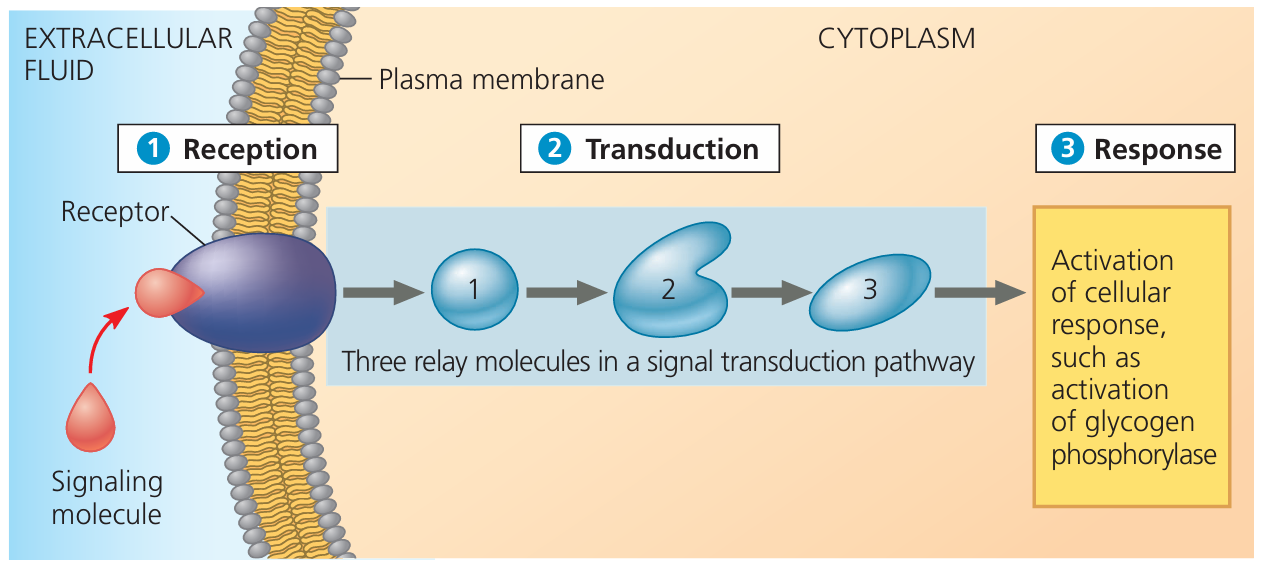
Stages in the Process of Cell Signalling
🧪 1. Secretion of Ligands
- Ligand = a chemical messenger (e.g., hormone, neurotransmitter).
- Produced and secreted by specific cells in response to a stimulus.
- Examples: Hormones (insulin, adrenaline), Neurotransmitters (acetylcholine).
🚚 2. Transport of Ligands to Target Cells
- Ligands travel from secreting cell to target cell via:
- Bloodstream (endocrine signalling – hormones).
- Diffusion across synaptic cleft (paracrine signalling – neurotransmitters).
- Ligand remains intact and active until reaching its target.
🎯 3. Binding to Cell Surface Receptors
- Target cells have specific receptor proteins on their surface membranes.
- Receptors have complementary shapes to their ligand → ensures specificity.
- Binding causes a conformational change in the receptor, triggering a response inside the cell.
🌀 4. Triggering the Response
- Receptor activation leads to:
- Activation of enzymes inside cell.
- Opening/closing ion channels.
- Activation of second messengers (e.g., cAMP) → amplifies signal.
- Changes in gene expression.
📊 Overview Table
| Stage | Key Event | Example |
|---|---|---|
| 1. Secretion | Ligand released from signalling cell | Insulin secreted by pancreas |
| 2. Transport | Ligand moves to target | Blood carries insulin to muscles |
| 3. Binding | Ligand binds receptor | Insulin binds receptor on muscle cell membrane |
| 4. Response | Cellular change occurs | Glucose uptake increases |
✅ Summary Box
Cell signalling is a specific communication system where ligands secreted by one cell travel to target cells, bind to complementary receptors, and trigger specific cellular responses. This ensures coordination between cells in multicellular organisms.
Cell signalling is a specific communication system where ligands secreted by one cell travel to target cells, bind to complementary receptors, and trigger specific cellular responses. This ensures coordination between cells in multicellular organisms.