Home / CIE AS & A Level / CIE AS & A Level biology 9700 / CIE AS/A Level Biology -5.1 Replication and division of nuclei and cells- Study Notes

CIE AS/A Level Biology -5.1 Replication and division of nuclei and cells- Study Notes

CIE AS/A Level Biology -5.1 Replication and division of nuclei and cells- Study Notes- New Syllabus

CIE AS/A Level Biology -5.1 Replication and division of nuclei and cells- Study Notes- New Syllabus

Ace A level Biology Exam with CIE AS/A Level Biology -5.1 Replication and division of nuclei and cells- Study Notes- New Syllabus 

Key Concepts:

  •  describe the structure of a chromosome, limited to:
    • DNA
    • histone proteins
    • sister chromatids
    • centromere
    • telomeres
  •  explain the importance of mitosis in the production of genetically identical daughter cells during:
    • growth of multicellular organisms
    • replacement of damaged or dead cells
    • repair of tissues by cell replacement
    • asexual reproduction
  • outline the mitotic cell cycle, including:
    • interphase (growth in G1 and G2 phases and DNA replication in S phase)
    • mitosis
    • cytokinesis
  • outline the role of telomeres in preventing the loss of genes from the ends of chromosomes during DNA replication
  •  outline the role of stem cells in cell replacement and tissue repair by mitosis
  • explain how uncontrolled cell division can result in the formation of a tumour

CIE AS/A Level Biology 9700-Study Notes- All Topics

Structure of a Chromosome

📜 1. DNA

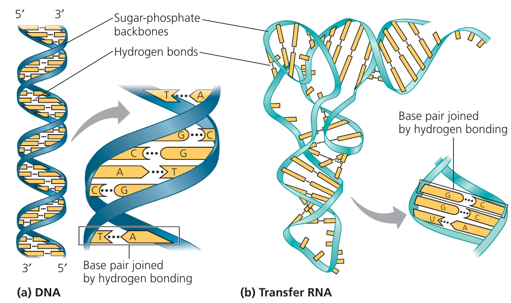
  • Definition: Long, double-helical molecule made of nucleotides.
  • Contains genetic instructions for making proteins.
  • In eukaryotic chromosomes, DNA is tightly coiled to fit inside the nucleus.

🧵 2. Histone Proteins

  • Definition: Special proteins that DNA wraps around to form nucleosomes.
  • Help in compacting DNA and regulating gene activity.
  • Act like “spools” to organise and stabilise the DNA structure

🧬 3. Sister Chromatids

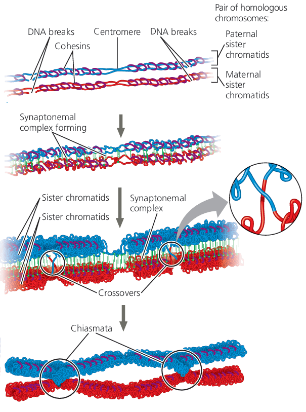
  • Definition: After DNA replication, each chromosome has two identical copies called sister chromatids.
  • They are joined together at a central point called the centromere.
  • Separated during cell division (mitosis/meiosis) to ensure each new cell gets a copy.

🎯 4. Centromere

  • Definition: Region that holds sister chromatids together.
  • Attachment point for spindle fibres during cell division.
  • Can be located centrally, near one end, or at the end, influencing chromosome shape.

🔒 5. Telomeres

  • Definition: Repetitive DNA sequences at the ends of chromosomes.
  • Protect chromosome ends from damage and prevent them from fusing with other chromosomes.
  • Shorten with each cell division – linked to cell ageing.
StructureDescriptionFunction
DNADouble helix molecule of genetic codeStores genetic information
HistonesProteins DNA wraps aroundPackaging & gene regulation
Sister ChromatidsIdentical copies of a chromosomeEnsure accurate DNA distribution
CentromereRegion joining sister chromatidsAttachment for spindle fibres
TelomeresRepetitive DNA endsProtects chromosome ends
🧠 Key Takeaways
Chromosomes are DNA + proteins (mainly histones).
Sister chromatids are identical – joined at a centromere.
Telomeres protect and maintain chromosome stability.

Importance of Mitosis in Producing Genetically Identical Daughter Cells

📌 Definition Recap

  • Definition: Mitosis is a type of cell division in which a parent cell produces two genetically identical daughter cells.
  • The chromosome number remains the same as in the original cell (diploid → diploid).

🌱 1. Growth of Multicellular Organisms

  • Multicellular organisms grow by increasing the number of cells through mitosis.
  • Each new cell is genetically identical, ensuring all body cells have the same DNA for correct functioning.
  • Example: Growth of a human from a zygote to an adult.

🩹 2. Replacement of Damaged or Dead Cells

  • Cells that are worn out or lost (e.g., skin cells, blood cells) are replaced via mitosis.
  • Maintains tissue structure and function by replacing them with identical copies.
  • Example: Replacement of red blood cells in bone marrow.

🛠 3. Repair of Tissues by Cell Replacement

  • Mitosis helps in healing wounds by producing new cells to replace damaged ones.
  • The identical genetic material ensures the repaired area functions normally.
  • Example: Regrowth of epidermis after a cut.

🌿 4. Asexual Reproduction

  • In some organisms, mitosis is the basis for reproduction without gametes.
  • Offspring are genetically identical to the parent (clones).
  • Example: Binary fission in protozoa, budding in hydra, vegetative propagation in plants.
Role of MitosisExampleImportance
GrowthIncrease in human heightMaintains genetic consistency
ReplacementSkin cell renewalSustains normal function
RepairWound healingRestores damaged tissues
Asexual reproductionPotato tuber sproutingProduces identical offspring
🧠 Key Takeaways
Mitosis ensures genetic stability in all new cells.
Critical for growth, maintenance, repair, and reproduction.
Prevents genetic variation – important for preserving organism traits.

 Mitotic Cell Cycle

📌 Overview

  • The mitotic cell cycle is the process by which eukaryotic cells grow, duplicate their DNA, and divide into two genetically identical daughter cells.
  • It consists of interphase, mitosis, and cytokinesis.

🧬 1. Interphase (Longest stage of the cell cycle)

Interphase is divided into three phases:
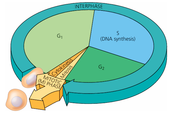
G₁ Phase (Gap 1)

  • Cell grows and increases in size.
  • New organelles and proteins are synthesised.
  • Cell performs normal metabolic activities.
  • Checkpoint ensures the cell is ready for DNA replication.

S Phase (Synthesis)

  • DNA replication occurs – each chromosome is duplicated to form two sister chromatids.
  • Histone proteins are synthesised for packaging DNA.

G₂ Phase (Gap 2)

  • Further cell growth and preparation for mitosis.
  • Microtubule proteins are synthesised for spindle formation.
  • Checkpoint ensures DNA has been correctly replicated.

⚡ 2. Mitosis (Nuclear Division)

  • Division of the nucleus to produce two genetically identical nuclei.
  • Stages: Prophase → Metaphase → Anaphase → Telophase.

✂ 3. Cytokinesis (Cytoplasmic Division)

  • Division of the cytoplasm to form two separate daughter cells.
  • In animal cells: Achieved by a cleavage furrow that pinches the cell in two.
  • In plant cells: A cell plate forms, which develops into a new cell wall.
StageKey EventsOutcome
G₁Cell growth, protein/organelle synthesisPrepares for DNA replication
SDNA replication, histone synthesisEach chromosome becomes two sister chromatids
G₂Cell growth, spindle protein synthesisPrepares for mitosis
MitosisNuclear divisionTwo identical nuclei
CytokinesisCytoplasm dividesTwo identical daughter cells
🧠 Key Takeaways
The cell cycle ensures growth, repair, and genetic stability.
Interphase is not a “resting phase” – it is a period of active preparation.
Mitosis + cytokinesis = one complete cell division.

Role of Telomeres in Protecting Chromosome Ends

📌 What are Telomeres?

  • Repetitive non-coding DNA sequences found at the ends of chromosomes.
  • In humans, the repeated sequence is TTAGGG.
  • Bound by specific protective proteins that maintain chromosome stability.

🔍 Role in DNA Replication

  • DNA polymerase cannot fully replicate the very end of the lagging strand — known as the end-replication problem.
  • Without telomeres, essential gene sequences at chromosome ends would be lost after each cell division.
  • Telomeres act as a protective cap so only telomeric DNA — not essential genes — is lost.
  • Telomerase enzyme (active in germ cells, stem cells, and some cancer cells) extends telomeres, counteracting shortening.

📊 Summary Table

FeatureFunction
StructureRepetitive non-coding DNA + protective proteins
Main RoleProtect genes from loss during DNA replication
MechanismAbsorb the loss of DNA bases from the lagging strand
Enzyme InvolvedTelomerase (adds repeats back)
ImportanceMaintains genetic stability, prevents chromosome degradation
🧠 Key Takeaway: Telomeres work like plastic tips on shoelaces – they prevent chromosome ends from fraying and losing essential genetic information during cell division.

Role of Stem Cells in Cell Replacement and Tissue Repair by Mitosis

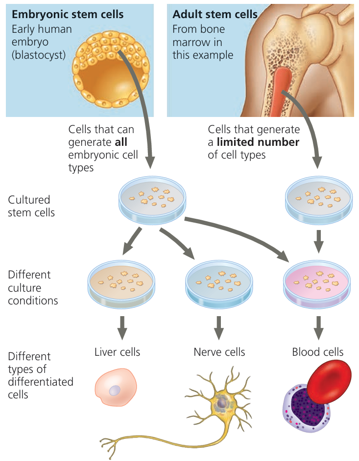
📌 What are Stem Cells?

  • Undifferentiated cells capable of dividing by mitosis to produce more stem cells or specialised cells.
  • Found in both embryonic and adult tissues.

🔬 Role in Cell Replacement & Tissue Repair

  1. Cell Replacement
    • In tissues where cells are regularly lost (e.g., skin, blood, intestinal lining), stem cells divide by mitosis to replace them.
    • Example: Bone marrow stem cells produce new red and white blood cells.
  2. Tissue Repair
    • After injury, stem cells migrate to the damaged area.
    • They divide by mitosis to produce new specialised cells that replace the damaged ones.
    • Example: Muscle stem cells (satellite cells) help repair torn muscle fibres.

📊 Summary Table

Stem Cell TypeLocationRole in Repair & Replacement
Embryonic stem cellsEarly embryoCan differentiate into any cell type (pluripotent)
Adult stem cellsBone marrow, skin, gut lining, etc.Maintain and repair specific tissues
Muscle satellite cellsMuscle tissueRepair and regenerate muscle fibres
🧠 Key Takeaway: Stem cells act as the body’s natural repair system, dividing by mitosis to maintain healthy tissues and replace damaged or lost cells throughout life.

How Uncontrolled Cell Division Can Result in the Formation of a Tumour

🔍 Normal Cell Cycle Control

  • In healthy cells, the cell cycle is tightly regulated by checkpoints and control proteins (e.g., cyclins, tumour suppressor proteins).
  • Cells only divide when needed for growth, repair, or replacement.
  • Damaged cells normally undergo apoptosis (programmed cell death).

❌ What Happens in Uncontrolled Cell Division?

  1. Mutation in DNA
    • Changes in genes that control the cell cycle (e.g., proto-oncogenes or tumour suppressor genes) can disrupt regulation.
    • Causes include radiation, chemicals, viruses, or random DNA errors.
  2. Loss of Regulation
    • Mutated proto-oncogenes become oncogenes → promote excessive cell division.
    • Damaged tumour suppressor genes (e.g., p53) fail to stop division or trigger apoptosis.
  3. Rapid Cell Division
    • Cells divide repeatedly without normal control.
  4. Formation of a Mass of Cells (Tumour)
    • The abnormal, uncontrolled growth forms a tumour.

🧩 Types of Tumours

TypeNatureEffect
BenignCells remain in one location, usually surrounded by a capsuleCan still cause problems by pressing on nearby tissues
MalignantCells invade surrounding tissues and may spread via the blood/lymph (metastasis)Leads to cancer
📌 Summary: Mutations that disrupt normal cell cycle control can cause unchecked mitosis, producing a mass of abnormal cells – a tumour. Malignant tumours can spread and damage vital tissues and organs.
Scroll to Top