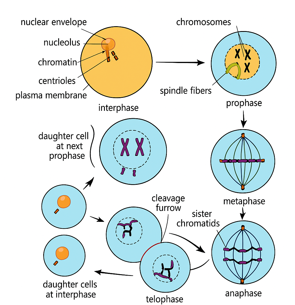
Behaviour of Chromosomes During the Mitotic Cell Cycle in Plant & Animal Cells
1️⃣ Prophase
- Chromosome behaviour:
- Chromatin fibres condense → visible chromosomes (each with two sister chromatids joined at the centromere).
- Sister chromatids are genetically identical (due to DNA replication in S phase).
- Other cell structures:
- Nuclear envelope begins to break down.
- Nucleolus disappears.
- Spindle fibres start forming:
- Animals → from centrosomes; centrioles move to opposite poles.
- Plants → from spindle organising regions; no centrioles.
2️⃣ Metaphase
- Chromosome behaviour:
- Chromosomes align along the cell equator (metaphase plate).
- Centromeres attach to spindle fibres from opposite poles.
- Other cell structures:
- Nuclear envelope completely gone.
- Spindle apparatus fully formed.
3️⃣ Anaphase
- Chromosome behaviour:
- Centromeres split → sister chromatids separate.
- Daughter chromosomes pulled to opposite poles (microtubules depolymerise).
- Other cell structures:
- Animal cells → cleavage furrow formation begins.
- Spindle fibres actively move chromosomes apart.
4️⃣ Telophase
- Chromosome behaviour:
- Daughter chromosomes reach poles and decondense into chromatin.
- Other cell structures:
- Nuclear envelope reforms around each set of chromosomes.
- Nucleolus reappears in each nucleus.
- Spindle fibres break down.
🌱 Cytokinesis (after mitosis)
- Animal cells: Cleavage furrow forms as actin filaments pull the membrane inward until the cell splits.
- Plant cells: Vesicles from the Golgi align at the equator forming a cell plate → becomes a new cell wall.
📌 Summary Table
| Stage | Chromosome Behaviour | Nuclear Envelope | Spindle | Other Notes |
|---|---|---|---|---|
| Prophase | Chromosomes condense, sister chromatids visible | Breaks down | Forms | Nucleolus disappears |
| Metaphase | Chromosomes align at equator | Absent | Fully formed | Centromeres attach to spindle |
| Anaphase | Sister chromatids separate to poles | Absent | Shortens | Animal cell membrane begins constricting |
| Telophase | Chromosomes decondense | Reforms | Breaks down | Nucleolus reappears |
📌 Summary: The mitotic cell cycle ensures equal distribution of identical genetic material into two daughter cells. Although plant and animal cells share the same chromosome behaviour, their spindle formation and cytokinesis differ.
Identifying Stages of Mitosis in Photomicrographs, Diagrams & Slides
1️⃣ Prophase
- Chromosomes appear thick, dark, and distinct under the microscope.

- Each chromosome has two sister chromatids joined at a centromere.
- Nuclear envelope begins breaking down; nucleolus may still be faintly visible.
- In animal cells → centrioles visible at poles (if magnification is high).
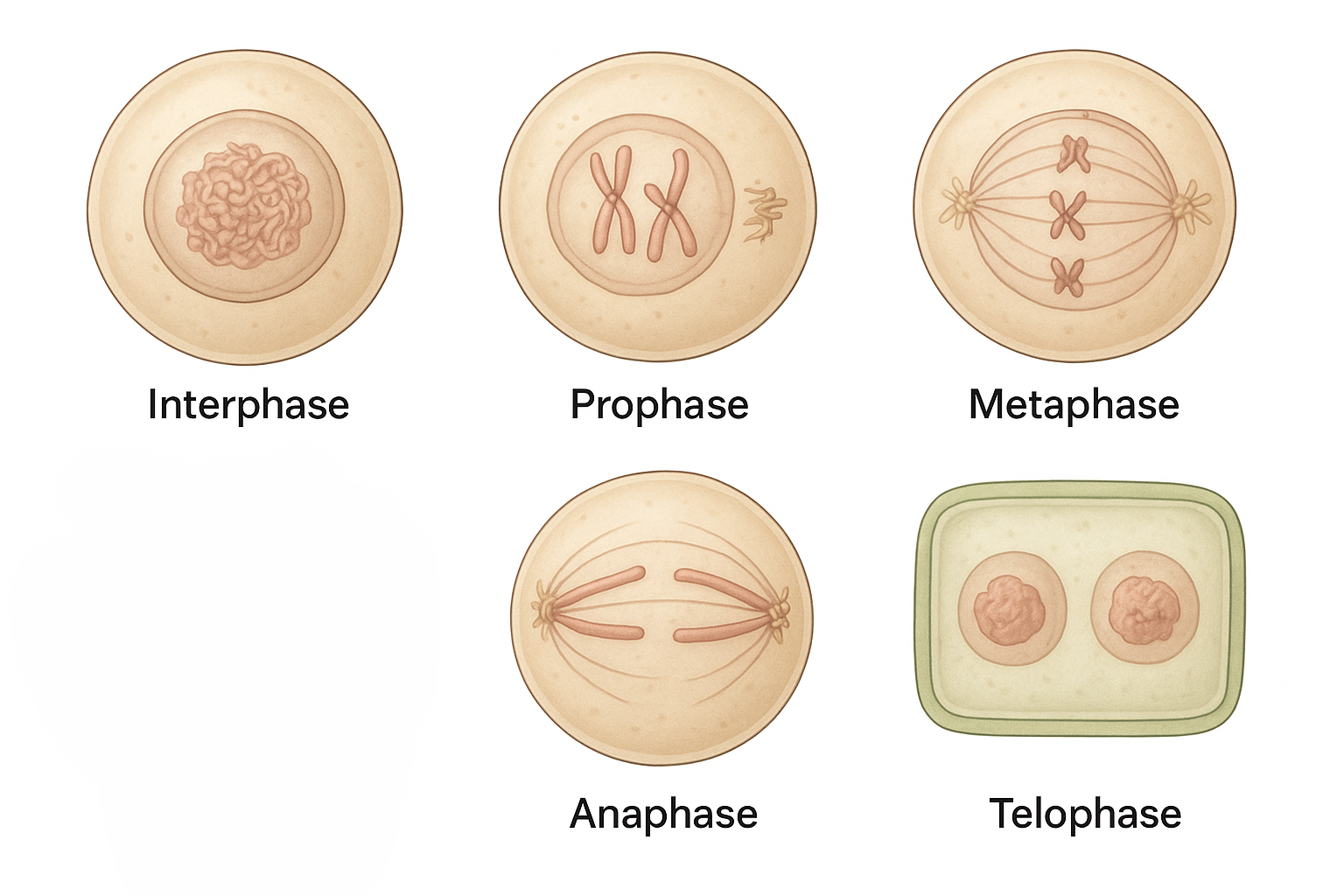
2️⃣ Metaphase
- Chromosomes align along the cell’s equator (metaphase plate).
- No nuclear envelope visible.
- Centromeres attached to spindle fibres from opposite poles.
- Chromosomes are most condensed and distinct → ideal for karyotyping.
3️⃣ Anaphase
- Centromeres split; sister chromatids separate into daughter chromosomes.
- Two V-shaped groups of chromosomes moving toward opposite poles.
- Clear cell equator with no chromosomes present.
- Spindle fibres visible in high-quality micrographs.
4️⃣ Telophase
- Chromosomes reach poles and start decondensing into chromatin.
- Nuclear envelope reforms around each chromosome set.
- Cytokinesis may be visible:
- Animal cells → cleavage furrow forms.
- Plant cells → cell plate forms.
5️⃣ Interphase (Not Mitosis)
- Nucleus intact with diffuse, uncoiled chromatin.
- Nucleolus clearly visible.
- Most cells in a sample are in this stage.
| Stage | Key Features in Photomicrographs |
|---|---|
| Interphase | Large clear nucleus, diffuse chromatin, visible nucleolus. |
| Prophase | Thick, dark chromosomes; nuclear envelope breaking down. |
| Metaphase | Chromosomes aligned at equator; most condensed stage. |
| Anaphase | V-shaped chromatids moving toward poles; clear cell equator. |
| Telophase | Chromosomes decondensing; two nuclei forming; cytokinesis visible. |
🧠 Key Tip: Always scan root tip squash or meristem slides at low power first to locate dividing cells, then zoom in to identify the exact mitotic stage.