Structure of Nucleotides & ATP
🌱 Nucleotides – Basic Unit of Nucleic Acids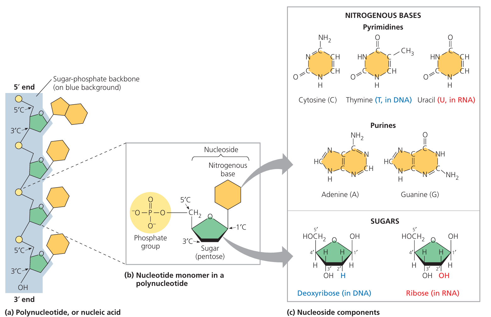
- Definition: Nucleotides are the monomers (building blocks) of nucleic acids (DNA & RNA).
- Each nucleotide is made of three main components:
- Nitrogenous Base
Organic molecule containing nitrogen.
Two types:- Purines (double ring): Adenine (A), Guanine (G)
- Pyrimidines (single ring): Cytosine (C), Thymine (T in DNA), Uracil (U in RNA)
- Pentose Sugar
5-carbon sugar.
Deoxyribose in DNA, Ribose in RNA. - Phosphate Group
One or more phosphate groups attached to carbon 5 of sugar.
Negatively charged → makes nucleotide soluble in water.
- Nitrogenous Base
🔬 Nucleotide Structure Overview
| Component | Function |
|---|---|
| Nitrogenous base | Stores genetic code in sequence |
| Pentose sugar | Forms the backbone with phosphate (sugar-phosphate chain) |
| Phosphate group | Links nucleotides together via phosphodiester bonds |
⚡ Phosphorylated Nucleotides
- Nucleotides can have 1, 2, or 3 phosphate groups:
- Monophosphate – AMP (Adenosine Monophosphate)
- Diphosphate – ADP (Adenosine Diphosphate)
- Triphosphate – ATP (Adenosine Triphosphate)
🔋 ATP – Adenosine Triphosphate
- Structure: Adenine (base) + Ribose (sugar) + Three phosphate groups in a chain.
- High-energy bonds between the last two phosphate groups release energy when broken (hydrolysis).
- Function: Main energy currency of the cell. Hydrolysis of ATP → ADP + Pi + energy for cellular processes.
📊 Comparison Table: Nucleotide vs ATP
| Feature | Nucleotide | ATP |
|---|---|---|
| Components | Base + sugar + phosphate | Base (adenine) + ribose + 3 phosphates |
| Function | DNA/RNA building block | Energy transfer in cells |
| Phosphate groups | 1 | 3 |
| Example | Cytidine monophosphate (CMP) | Adenosine triphosphate (ATP) |
🧠 Key Takeaways:
– Nucleotides are made of a nitrogenous base, a pentose sugar, and phosphate group(s).
– ATP is a special nucleotide with three phosphates, storing energy in its bonds.
– Hydrolysis of ATP releases usable energy for cellular activities.
– Nucleotides are made of a nitrogenous base, a pentose sugar, and phosphate group(s).
– ATP is a special nucleotide with three phosphates, storing energy in its bonds.
– Hydrolysis of ATP releases usable energy for cellular activities.
Structure of the DNA Molecule
🌱 Overall Shape – Double Helix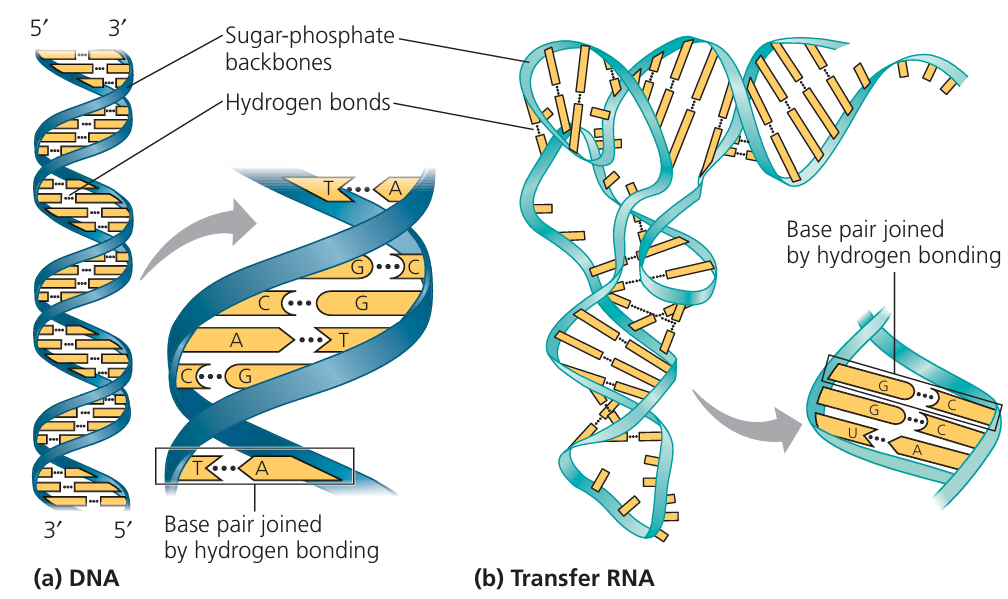
- DNA is a double-stranded molecule twisted into a double helix.
- The two strands run in opposite directions → antiparallel:
- One strand runs 5′ → 3′
- The other runs 3′ → 5′
- Each strand has a sugar-phosphate backbone with nitrogenous bases pointing inward.
🧩 Complementary Base Pairing
- Bases pair through hydrogen bonds:
- Adenine (A) → Thymine (T) → 2 hydrogen bonds
- Cytosine (C) → Guanine (G) → 3 hydrogen bonds
- Purine always pairs with pyrimidine → keeps helix stable and uniform.
- Ensures accurate DNA replication and transcription.
🔗 Phosphodiester Bonds – Backbone Linkage
- Formed between the phosphate group of one nucleotide and the 3′ carbon of the sugar in the next nucleotide.
- Create a sugar-phosphate backbone that is strong and stable.
- Covalent bonds → resistant to breaking.
📊 Summary Table: DNA Structural Features
| Feature | Description | Importance |
|---|---|---|
| Double helix | Two strands twisted | Stability & compact storage |
| Antiparallel strands | 5′ → 3′ and 3′ → 5′ | Correct base pairing & replication |
| Complementary base pairing | A–T (2 bonds), C–G (3 bonds) | Accuracy in copying genetic info |
| Phosphodiester bonds | Sugar–phosphate linkage | Strong backbone support |
🧠 Key Takeaways:
– DNA has two antiparallel strands forming a double helix.
– A–T pairs have 2 hydrogen bonds; C–G pairs have 3, making them stronger.
– Phosphodiester bonds hold nucleotides together in each strand’s backbone.
– Complementary base pairing ensures DNA’s accuracy in replication and transcription.
– DNA has two antiparallel strands forming a double helix.
– A–T pairs have 2 hydrogen bonds; C–G pairs have 3, making them stronger.
– Phosphodiester bonds hold nucleotides together in each strand’s backbone.
– Complementary base pairing ensures DNA’s accuracy in replication and transcription.
Semi-Conservative DNA Replication (S Phase of the Cell Cycle)
🌱 Overview
- Semi-conservative means: after replication, each new DNA molecule has one original (parental) strand and one newly synthesised strand.
- Occurs during the S phase (synthesis phase) of interphase in the cell cycle.
- Ensures accurate copying of genetic material before cell division.
🧬 Step-by-Step Process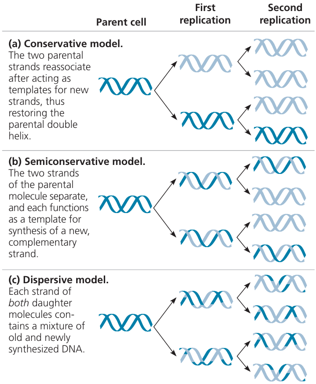
1. Strand Separation
- The two DNA strands unwind and separate (hydrogen bonds between bases break).
- Each parental strand acts as a template for the new strand.
2. Nucleotide Pairing
- Free DNA nucleotides in the nucleus pair with exposed bases on each template strand using complementary base pairing:
- A pairs with T (2 hydrogen bonds)
- C pairs with G (3 hydrogen bonds)
3. Role of DNA Polymerase
- Joins new nucleotides together, forming the sugar-phosphate backbone by phosphodiester bonds.
- Can only add nucleotides in the 5′ → 3′ direction.
4. Leading vs Lagging Strand
- Leading strand: Synthesised continuously in the same direction as the unwinding fork.
- Lagging strand: Synthesised discontinuously in short fragments (Okazaki fragments) because DNA polymerase works only 5′ → 3′.
5. Role of DNA Ligase
- Joins the Okazaki fragments together on the lagging strand to make a complete continuous strand.
📊 Comparison: Leading vs Lagging Strand
| Feature | Leading Strand | Lagging Strand |
|---|---|---|
| Direction relative to fork | Same as fork movement | Opposite to fork movement |
| Synthesis type | Continuous | Discontinuous (Okazaki fragments) |
| Enzyme needed to join fragments | No (polymerase alone) | Yes (DNA ligase) |
| Speed | Faster | Slower |
📌 Why 5′ → 3′ Only?
- DNA polymerase can add nucleotides only to the 3′ end of a growing strand.
- This chemical restriction causes one strand to be continuous and the other discontinuous.
🧠 Key Takeaways:
– DNA replication is semi-conservative: each new molecule has one old and one new strand.
– DNA polymerase builds new DNA only in the 5′ → 3′ direction.
– Leading strand is continuous; lagging strand is discontinuous and joined by DNA ligase.
– Accurate base pairing ensures genetic fidelity.
– DNA replication is semi-conservative: each new molecule has one old and one new strand.
– DNA polymerase builds new DNA only in the 5′ → 3′ direction.
– Leading strand is continuous; lagging strand is discontinuous and joined by DNA ligase.
– Accurate base pairing ensures genetic fidelity.