Functions of Main Blood Vessels in Mammalian Circulation
🌱 Overview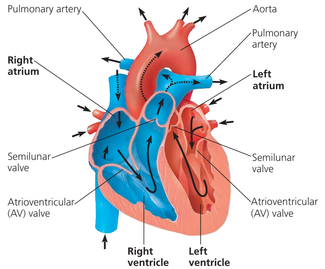
- Mammals have a double circulation system:
- Pulmonary circulation – between heart and lungs
- Systemic circulation – between heart and body
- Key vessels transport oxygenated or deoxygenated blood to maintain efficient circulation.
🔬 Pulmonary Circulation Vessels
| Blood Vessel | Function |
|---|---|
| Pulmonary artery | Carries deoxygenated blood from the right ventricle to the lungs for oxygenation. |
| Pulmonary vein | Carries oxygenated blood from the lungs to the left atrium of the heart. |
🫀 Systemic Circulation Vessels
| Blood Vessel | Function |
|---|---|
| Aorta | Carries oxygenated blood from the left ventricle to all parts of the body. |
| Vena cava | Carries deoxygenated blood from the body back to the right atrium of the heart. |
📊 Quick Reference Table: Blood Flow & Oxygen Status
| Vessel | Carries Blood From → To | Oxygen Status |
|---|---|---|
| Pulmonary artery | Heart → Lungs | Deoxygenated |
| Pulmonary vein | Lungs → Heart | Oxygenated |
| Aorta | Heart → Body | Oxygenated |
| Vena cava | Body → Heart | Deoxygenated |
🧠 Key Points:
– Pulmonary artery is unique as it carries deoxygenated blood away from the heart.
– Pulmonary vein is unique as it carries oxygenated blood towards the heart.
– Aorta distributes oxygen-rich blood to the entire body, while vena cava returns oxygen-poor blood to the heart.
– Together, these vessels maintain efficient double circulation, separating oxygenated and deoxygenated blood.
Recognition and Structure of Blood Vessels
🌱 Overview
- Blood vessels carry blood throughout the body in closed circulation.
- Main types: Arteries, Veins, Capillaries
- Can be identified under light microscope, photomicrographs, or electron micrographs.
- Plan diagrams help illustrate their structural differences.
🔬 Recognition of Blood Vessels
| Vessel Type | Features Under Microscope / Photomicrograph | Key Identifying Traits |
|---|---|---|
| Arteries | Thick muscular wall, narrow lumen, round shape in TS, folds in LS | Thick tunica media, withstands high pressure |
| Veins | Thin wall, wide lumen, often collapsed or irregular in TS | Thin tunica media, contains valves, low pressure |
| Capillaries | Very thin wall (single layer of endothelial cells), very small diameter | Allows exchange of gases and nutrients, only one RBC passes at a time |
🫀 Structural Features of Arteries and Veins
1. Artery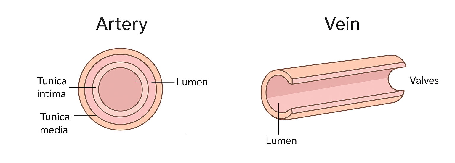
- Transverse Section (TS): Circular with thick wall.
- Longitudinal Section (LS): Tube with prominent layers.
- Layers (from inside out):
- Tunica intima – endothelium (single cell layer)
- Tunica media – thick smooth muscle + elastic fibers
- Tunica externa (adventitia) – connective tissue
2. Vein
- Transverse Section (TS): Wider lumen, thin wall, may appear collapsed.
- Longitudinal Section (LS): Tube with thin walls; valves visible.
- Layers (from inside out):
- Tunica intima – endothelium with valves
- Tunica media – thin smooth muscle, few elastic fibers
- Tunica externa – connective tissue, thicker than tunica media
📊 Comparison Table: Artery vs Vein
| Feature | Artery | Vein |
|---|---|---|
| Wall thickness | Thick | Thin |
| Lumen | Narrow | Wide |
| Shape in TS | Circular | Irregular / collapsed |
| Tunica media | Thick, muscular & elastic | Thin, less muscular |
| Valves | Absent | Present (prevent backflow) |
| Pressure | High | Low |
| Function | Carry blood away from heart | Carry blood toward heart |
🧠 Key Points:
– Arteries are adapted for high pressure, veins for low pressure and valves.
– Capillaries are microscopic and allow exchange, identifiable by single layer of endothelial cells.
Structure-Function Relationship of Blood Vessels
🌱 Overview
- Blood vessels have specialised structures adapted to their roles in circulation.
- Key vessel types: Elastic arteries, muscular arteries, veins, capillaries.
- Structure directly influences blood flow, pressure, and exchange.
🔬 1. Elastic Arteries
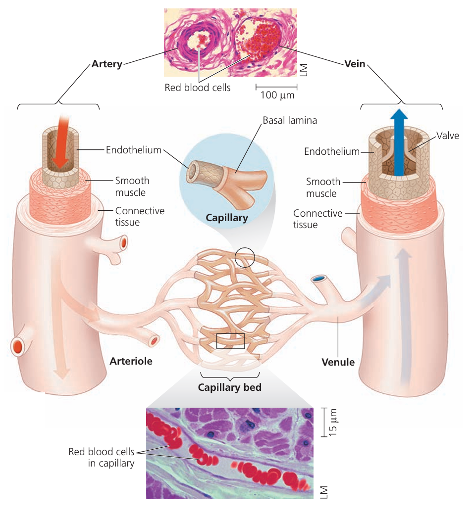
- Examples: Aorta, pulmonary artery
- Structure:
- Thick tunica media with many elastic fibers
- Wide lumen
- Relatively fewer smooth muscle cells than muscular arteries
- Function:
- Elasticity allows stretching during ventricular systole and recoiling during diastole → maintains continuous blood flow despite high pressure.
🔬 2. Muscular Arteries
- Examples: Radial artery, femoral artery
- Structure:
- Thick tunica media rich in smooth muscle
- Fewer elastic fibers compared to elastic arteries
- Narrow lumen
- Function:
- Smooth muscle allows vasoconstriction and vasodilation → regulates blood distribution to different organs.
🔬 3. Veins
- Examples: Superior and inferior vena cava, jugular vein
- Structure:
- Thin tunica media, less smooth muscle
- Wide lumen
- Valves present to prevent backflow
- Tunica externa relatively thick
- Function:
- Thin walls allow accommodation of large blood volumes at low pressure.
- Valves ensure unidirectional blood flow toward the heart.
- Muscle contraction helps push blood back to the heart.
🔬 4. Capillaries
- Examples: Continuous, fenestrated, sinusoidal capillaries
- Structure:
- Wall is one layer of endothelial cells
- Extremely narrow lumen (one RBC fits at a time)
- No muscle or elastic tissue
- Function:
- Thin walls and narrow lumen allow efficient exchange of gases, nutrients, and waste between blood and tissues.
- Slow blood flow increases time for diffusion.
📊 Summary Table: Structure-Function Relationship
| Vessel Type | Key Structural Features | Functional Significance |
|---|---|---|
| Elastic arteries | Many elastic fibers, thick wall, wide lumen | Stretch & recoil; maintains continuous blood flow |
| Muscular arteries | Thick smooth muscle, fewer elastic fibers, narrow lumen | Vasoconstriction & dilation; controls blood distribution |
| Veins | Thin wall, wide lumen, valves, thick tunica externa | Low pressure blood return; prevents backflow |
| Capillaries | Single endothelial layer, very narrow lumen | Exchange of gases, nutrients, and wastes |
– Structure of each vessel type is adapted to its function:
– Elasticity for pressure buffering in elastic arteries
– Muscular control for blood distribution in muscular arteries
– Valves and thin walls for volume accommodation in veins
– Single-cell walls for exchange in capillaries
Recognition and Drawing of Blood Cells
🌱 Overview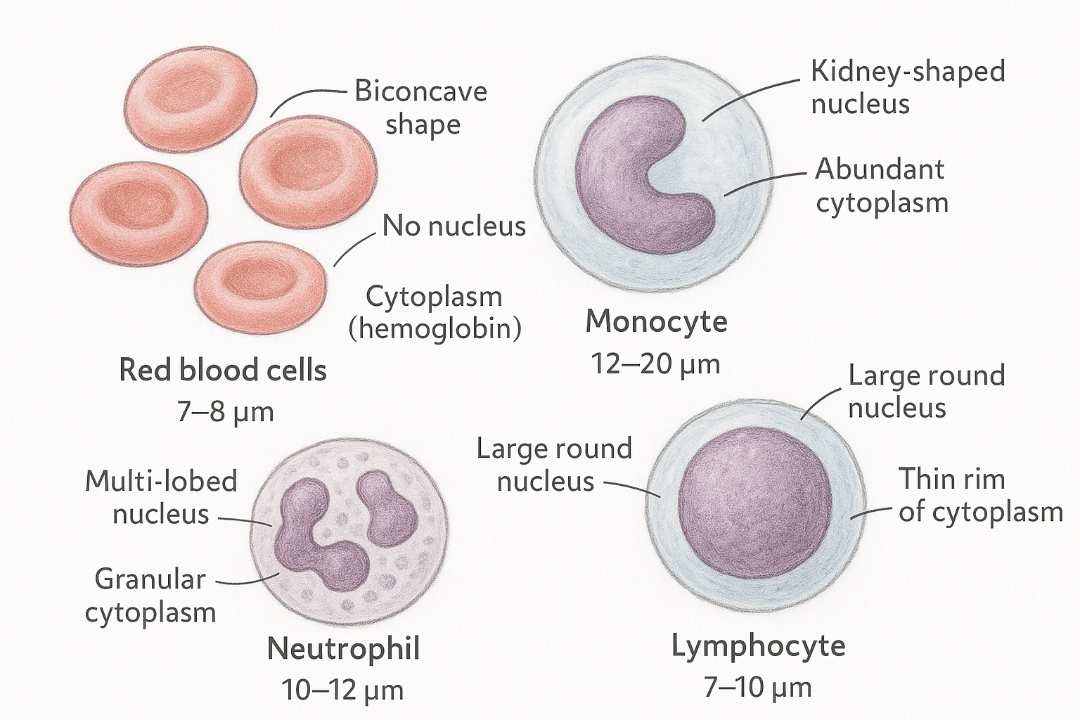
- Blood contains different cell types with specialised functions:
- Red blood cells (RBCs) – oxygen transport
- White blood cells (WBCs) – immune defense
- Monocytes – phagocytosis
- Neutrophils – first line of defense against infections
- Lymphocytes – antibody production and cellular immunity
- Cells can be identified under light microscope, photomicrographs, or electron micrographs.
- Accurate drawings help document cell morphology for study.
🔬 1. Red Blood Cells (Erythrocytes)
- Structure:
- Biconcave disc, no nucleus
- Diameter ~7–8 μm
- Flexible membrane
- Function:
- Transport oxygen and carbon dioxide via hemoglobin
- Recognition:
- Uniform size, central pallor, appear pink with eosin stain
🔬 2. Monocytes
- Structure:
- Largest WBC (12–20 μm)
- Kidney-shaped or horseshoe-shaped nucleus
- Abundant cytoplasm
- Function:
- Develop into macrophages in tissues
- Phagocytose pathogens and debris
- Recognition:
- Large size, pale blue cytoplasm, distinct nucleus
🔬 3. Neutrophils
- Structure:
- 10–12 μm in diameter
- Multi-lobed nucleus (2–5 lobes)
- Granular cytoplasm
- Function:
- First responders to infection
- Phagocytose bacteria and fungi
- Recognition:
- Multi-lobed nucleus, pale pink cytoplasm with fine granules
🔬 4. Lymphocytes
- Structure:
- Small (7–10 μm) or large (10–15 μm) types
- Large round nucleus, occupies most of the cell
- Thin rim of cytoplasm
- Function:
- B cells → produce antibodies
- T cells → cell-mediated immunity
- Recognition:
- Dense round nucleus, scant cytoplasm
📊 Summary Table: Blood Cell Identification
| Cell Type | Size (μm) | Nucleus | Cytoplasm | Function |
|---|---|---|---|---|
| RBC | 7–8 | None | No granules | Transport O₂ & CO₂ |
| Monocyte | 12–20 | Kidney-shaped | Abundant | Phagocytosis, becomes macrophage |
| Neutrophil | 10–12 | Multi-lobed | Granular | Phagocytosis of pathogens |
| Lymphocyte | 7–15 | Large, round | Thin rim | Immune response (B/T cells) |
Tissue Fluid – Formation and Function
🌱 Overview
- Tissue fluid is a watery fluid surrounding body cells, derived from blood plasma.
- It enables exchange of substances between blood and cells.
🔬 Functions of Tissue Fluid
| Function | Description |
|---|---|
| Transport of nutrients | Supplies oxygen, glucose, amino acids, and other nutrients from blood to cells. |
| Removal of waste | Collects carbon dioxide and other metabolic wastes from cells to return to blood. |
| Medium for diffusion | Provides an aqueous environment for substances to diffuse between blood and cells. |
| Homeostasis | Helps maintain stable conditions around cells. |
🔬 Formation of Tissue Fluid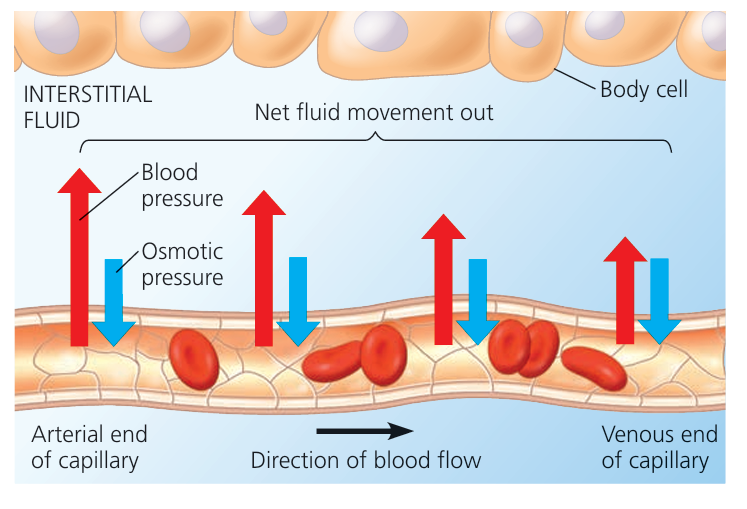
- Arterial End of Capillaries:
- Blood pressure (hydrostatic pressure) inside capillaries is higher than tissue fluid pressure.
- Plasma (except large proteins) is forced out of capillaries into surrounding tissue.
- Exchange of Substances:
- Oxygen, glucose, and nutrients diffuse into cells from tissue fluid.
- Carbon dioxide and metabolic wastes diffuse from cells into tissue fluid.
- Venous End of Capillaries:
- Osmotic pressure due to plasma proteins draws some fluid back into capillaries.
- Remaining tissue fluid drains into lymph vessels.
📊 Summary Table: Tissue Fluid Formation
| Stage | Mechanism/Force | Result |
|---|---|---|
| Arterial end of capillary | High hydrostatic pressure | Plasma forced out → tissue fluid formed |
| Exchange at tissue | Diffusion of nutrients & wastes | Cells receive nutrients, remove wastes |
| Venous end of capillary | Osmotic pressure (from plasma proteins) | Fluid returns to capillaries; excess → lymph |
– Tissue fluid is essential for cell survival, acting as a transport and exchange medium.
– Formation is driven by hydrostatic pressure (outward) and oncotic/osmotic pressure (inward).
– Excess tissue fluid is returned via the lymphatic system, preventing fluid accumulation.