External and Internal Structure of the Mammalian Heart
🌱 Overview
The mammalian heart is a muscular, hollow organ that pumps blood through the double circulatory system. It has four chambers, valves, and is surrounded by protective structures.
1. External Structure of the Heart
| Feature | Description / Function |
|---|---|
| Shape & Size | Cone-shaped, roughly the size of a fist. Apex points downward, forward, and left. |
| Pericardium | Double-layered sac surrounding the heart; protects and reduces friction. |
| Atria (Left & Right) | Upper chambers; receive blood from veins. Thin muscular walls. |
| Ventricles (Left & Right) | Lower chambers; pump blood into arteries. Thick muscular walls, especially the left ventricle. |
| Coronary Blood Vessels | Supply heart muscle with oxygen and nutrients. |
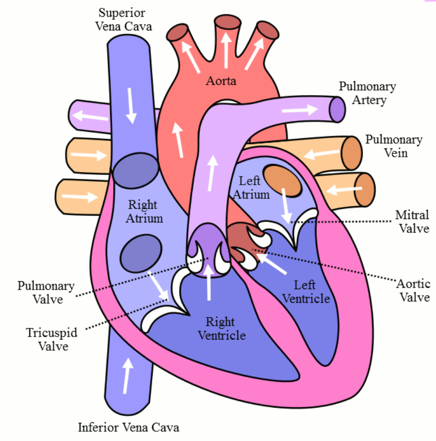
| Major Blood Vessels | – Aorta: carries oxygenated blood to body. – Pulmonary artery: carries deoxygenated blood to lungs. – Pulmonary veins: return oxygenated blood from lungs. – Vena cava (superior & inferior): return deoxygenated blood from body. |
2. Internal Structure of the Heart
| Feature | Description / Function |
|---|---|
| Chambers | – Right atrium: receives deoxygenated blood from body. – Right ventricle: pumps blood to lungs via pulmonary artery. – Left atrium: receives oxygenated blood from lungs. – Left ventricle: pumps oxygenated blood to body via aorta. |
| Atrioventricular (AV) valves | – Tricuspid valve: between right atrium and ventricle. – Bicuspid / mitral valve: between left atrium and ventricle. – Prevent backflow of blood into atria. |
| Semilunar valves | – Pulmonary valve: at pulmonary artery exit. – Aortic valve: at aorta exit. – Prevent backflow into ventricles. |
| Interventricular septum | Muscular wall separating left and right ventricles; prevents mixing of oxygenated and deoxygenated blood. |
| Chordae tendineae & Papillary muscles | Connect AV valves to ventricular walls; prevent valve prolapse during contraction. |
| Endocardium | Smooth inner lining of chambers; reduces friction for blood flow. |
| Myocardium | Thick muscular layer; strongest in left ventricle for systemic circulation. |
| Epicardium | Outer layer of heart wall; forms part of pericardium. |
– Double circulation: Right side pumps to lungs, left side pumps to body.
– Valves ensure unidirectional blood flow.
– Left ventricle is thicker than right due to higher pressure required for systemic circulation.
– Coronary vessels provide oxygen and nutrients to heart muscle itself.
The Cardiac Cycle
🌱 Overview
The cardiac cycle is the sequence of events in one heartbeat: contraction (systole) and relaxation (diastole) of the atria and ventricles. Includes changes in blood pressure, blood flow, and valve movements.
🔬 Phases of the Cardiac Cycle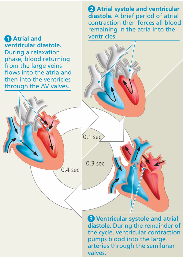
1. Atrial Systole
- What happens: Atria contract, pushing blood into ventricles.
- Valves:
- Atrioventricular valves (tricuspid & mitral) → open
- Semilunar valves (pulmonary & aortic) → closed
- Blood pressure: Slight increase in atrial pressure; ventricular pressure rises slightly as ventricles fill.
2. Ventricular Systole
- What happens: Ventricles contract, pumping blood into arteries.
- Valves:
- Atrioventricular valves → close (prevent backflow, causes first heart sound – “lub”)
- Semilunar valves → open when ventricular pressure > arterial pressure
- Blood pressure: Ventricular pressure rises sharply; atrial pressure low, filling from veins.
3. Diastole (Ventricular and Atrial Relaxation)
- What happens: Heart relaxes, chambers refill with blood.
- Valves:
- Semilunar valves → close (prevent backflow, causes second heart sound – “dub”)
- Atrioventricular valves → open when ventricular pressure < atrial pressure
- Blood pressure: Ventricular pressure falls; blood flows passively from atria to ventricles.
📊 Summary Table: Cardiac Cycle
| Phase | Heart Action | Valves | Blood Pressure Changes |
|---|---|---|---|
| Atrial systole | Atria contract | AV open, SL closed | Atrial pressure ↑, ventricular pressure ↑ slightly |
| Ventricular systole | Ventricles contract | AV closed, SL open | Ventricular pressure ↑ sharply, atrial pressure low |
| Diastole | Heart relaxes, chambers refill | AV open, SL closed | Ventricular pressure ↓, atrial pressure ↑ slightly |
– Valve movements prevent backflow and maintain unidirectional blood flow.
– Systole = contraction, Diastole = relaxation.
– Blood pressure rises during systole and falls during diastole.
– Heart sounds (“lub-dub”) correspond to closure of AV and semilunar valves.
Role of the Sinoatrial Node, Atrioventricular Node, and Purkyne Tissue in the Cardiac Cycle
🌱 Overview
The heart beats rhythmically due to specialized pacemaker and conduction tissues. Key structures: Sinoatrial (SA) node, Atrioventricular (AV) node, Purkyne tissue. This intrinsic system coordinates contraction without nervous or hormonal input.
1. Sinoatrial (SA) Node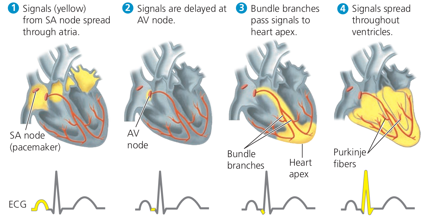
- Location: Wall of the right atrium near the entrance of the superior vena cava.
- Role:
- Acts as the natural pacemaker of the heart.
- Initiates electrical impulses that spread across atria → atrial systole.
- Sets the rate of heartbeat.
2. Atrioventricular (AV) Node
- Location: Between atria and ventricles, near the tricuspid valve.
- Role:
- Receives impulses from SA node.
- Delays transmission to ventricles slightly → allows atria to empty completely before ventricles contract.
3. Purkyne Tissue (Bundle of His and Purkinje Fibres)
- Location:
- Bundle of His runs down interventricular septum.
- Divides into Purkinje fibres spreading into ventricular walls.
- Role:
- Conducts impulses rapidly to ventricular muscle.
- Ensures ventricles contract simultaneously from apex upwards, efficiently pumping blood into arteries.
📊 Summary Table: Pacemaker and Conduction System
| Structure | Location | Function |
|---|---|---|
| SA node | Right atrium wall | Initiates heartbeat; spreads impulse to atria |
| AV node | Between atria and ventricles | Delays impulse; ensures ventricles fill |
| Purkyne tissue | Ventricular walls (via Bundle of His) | Rapid conduction; ventricles contract apex → base |
– SA node = “pacemaker” → sets heartbeat.
– AV node = delays impulse → coordinated atrial and ventricular contraction.
– Purkyne fibres = rapid conduction → synchronized ventricular contraction.
– Ensures efficient pumping of blood during the cardiac cycle.