Structure of the Human Gas Exchange System
🌱 Overview
- The human gas exchange system allows oxygen intake and carbon dioxide removal.
- It consists of airways and lungs that branch into smaller passages ending in alveoli, surrounded by a capillary network for efficient gas exchange.
1. Lungs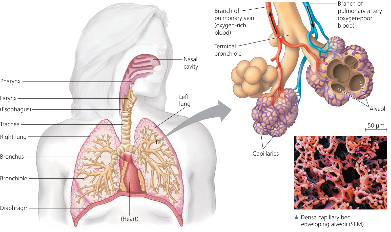
- Paired organs located in the thoracic cavity.
- Spongy texture due to branching airways and alveoli.
- Surrounded by pleural membranes: reduces friction during breathing.
2. Trachea
- Tube connecting larynx to bronchi.
- Structure:
- Supported by C-shaped cartilage rings → prevent collapse.
- Lined with ciliated epithelium and mucus-secreting goblet cells → trap and move dust/microbes.
3. Bronchi
- Two main branches from the trachea (left and right).
- Structure:
- Contain cartilage plates to maintain open passage.
- Lined with ciliated epithelium and goblet cells.
- Function: Conduct air to each lung.
4. Bronchioles
- Smaller branches of bronchi leading to alveoli.
- Structure:
- No cartilage; walls contain smooth muscle → allow constriction/dilation.
- Lined with ciliated epithelium in larger bronchioles; terminal bronchioles lack cilia.
- Function: Regulate airflow to alveoli.
5. Alveoli
- Tiny air sacs at the end of bronchioles (~300 million per lung).
- Structure:
- Single layer of squamous epithelial cells → thin barrier for gas exchange.
- Surrounded by dense capillary network.
- Elastic fibers allow expansion and recoil.
- Lined with surfactant → reduces surface tension, prevents collapse.
- Function: Site of oxygen and carbon dioxide exchange.
6. Capillary Network
- Dense network surrounding alveoli.
- Structure: Walls of endothelial cells → very thin for diffusion.
- Function: Facilitates efficient exchange of O₂ into blood and CO₂ out of blood.
📊 Summary Table: Structure and Function
| Component | Structure | Function |
|---|---|---|
| Lungs | Spongy, paired, pleural-covered | Contain airways and alveoli; gas exchange |
| Trachea | Tube with C-shaped cartilage rings, ciliated epithelium | Conduct air to bronchi, trap particles |
| Bronchi | Cartilage plates, ciliated epithelium | Conduct air to each lung |
| Bronchioles | Smooth muscle walls, small diameter, ciliated (large) | Regulate airflow to alveoli |
| Alveoli | Single-layer squamous epithelium, elastic, surfactant | Site of gas exchange |
| Capillary network | Thin-walled endothelial cells around alveoli | Oxygen and CO₂ exchange with blood |
🧠 Key Points:
– The system is highly branched, increasing surface area for gas exchange.
– Thin barriers and close association with capillaries allow efficient diffusion of gases.
– Elasticity and surfactant prevent alveolar collapse and aid breathing.
– The system is highly branched, increasing surface area for gas exchange.
– Thin barriers and close association with capillaries allow efficient diffusion of gases.
– Elasticity and surfactant prevent alveolar collapse and aid breathing.
Recognition of Key Structures in the Gas Exchange System
🌱 Overview
- In microscopy (light, photomicrograph, or electron micrograph), the airways and alveoli show characteristic structures that can be identified by shape, arrangement, and staining.
- Key structures: cartilage, ciliated epithelium, goblet cells, squamous alveolar epithelium, smooth muscle, capillaries.
1. Cartilage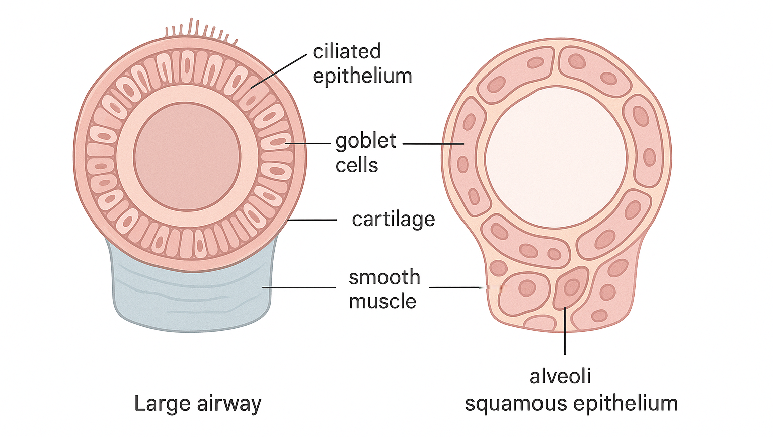
- Appearance: Lightly stained, firm, pale tissue with chondrocytes in lacunae. C-shaped rings in trachea or irregular plates in bronchi.
- Recognition: Under microscope, appears as supportive structure in airway walls.
- Function Reminder: Keeps airways open.
2. Ciliated Epithelium
- Appearance: Columnar cells with hair-like projections (cilia) on the apical surface. Nuclei usually basally located.
- Recognition: Observe the brush border of cilia moving mucus in light micrographs or electron micrographs.
- Function Reminder: Moves mucus and trapped particles.
3. Goblet Cells
- Appearance: Goblet-shaped, pale-staining cytoplasm due to mucus content. Nucleus compressed at the base.
- Recognition: Scattered among ciliated epithelial cells; may appear clear or lightly stained.
- Function Reminder: Secrete mucus to trap dust/microbes.
4. Squamous Epithelium of Alveoli
- Appearance: Flat, thin cells forming alveolar walls. Often closely associated with capillaries.
- Recognition: Single layer of cells; very thin for diffusion.
- Function Reminder: Efficient gas exchange.
5. Smooth Muscle
- Appearance: Spindle-shaped cells with elongated nuclei. Found in walls of bronchioles; no striations.
- Recognition: Look for bundles of elongated cells surrounding airways.
- Function Reminder: Regulates airway diameter.
6. Capillaries
- Appearance: Tiny thin-walled tubes near alveoli. Single layer of endothelial cells; red blood cells may be visible inside.
- Recognition: Seen closely apposed to alveolar squamous epithelium for gas exchange.
- Function Reminder: Transport O₂ and CO₂ between alveoli and blood.
📊 Summary Table:
| Structure | Microscopic Appearance | Function |
|---|---|---|
| Cartilage | Pale, firm, chondrocytes in lacunae | Supports airways |
| Ciliated epithelium | Columnar cells with hair-like cilia | Moves mucus |
| Goblet cells | Goblet-shaped, pale cytoplasm, basal nucleus | Secretes mucus |
| Squamous epithelium (alveoli) | Thin, flat cells forming alveolar walls | Gas exchange |
| Smooth muscle | Spindle-shaped cells, elongated nuclei | Controls bronchiole diameter |
| Capillaries | Thin-walled tubes, sometimes RBCs inside | Gas transport with alveoli |
Recognition of Trachea, Bronchi, Bronchioles, and Alveoli in Microscopy
🌱 Overview
- The airways and alveoli have distinct microscopic features that allow identification in light microscope slides, photomicrographs, and electron micrographs.
- Trachea and bronchi are larger airways; bronchioles are smaller; alveoli are tiny sacs for gas exchange.
1. Trachea
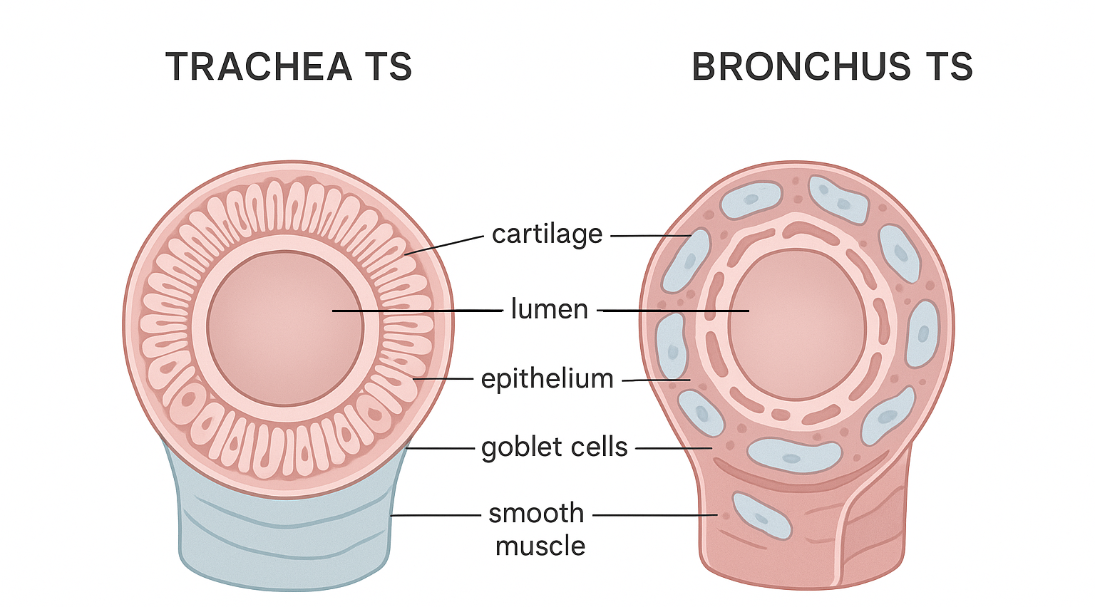
- Appearance: Large tube with C-shaped cartilage rings. Lined with ciliated pseudostratified columnar epithelium. Goblet cells interspersed among epithelial cells.
- Function: Conducts air to bronchi.
2. Bronchi
- Appearance: Similar to trachea but smaller and cartilage forms irregular plates. Lined with ciliated epithelium and goblet cells. Smooth muscle present in wall.
- Function: Distributes air to each lung.
3. Bronchioles
- Appearance: Small, no cartilage. Lined with simple columnar or cuboidal epithelium. Smooth muscle prominent in wall. Terminal bronchioles lack cilia and goblet cells.
- Function: Regulates airflow to alveoli.
4. Alveoli
- Appearance: Tiny sac-like structures. Lined with squamous epithelial cells (type I pneumocytes). Capillaries closely associated. Type II pneumocytes may appear as cuboidal cells producing surfactant.
- Function: Gas exchange (O₂ in, CO₂ out).
📊 Summary Table: Recognition in Microscopy
| Structure | Microscopic Features | Function |
|---|---|---|
| Trachea | C-shaped cartilage rings, ciliated epithelium, goblet cells | Conducts air to bronchi |
| Bronchi | Irregular cartilage plates, ciliated epithelium, smooth muscle | Distributes air to lungs |
| Bronchioles | No cartilage, smooth muscle prominent, cuboidal epithelium | Regulates airflow to alveoli |
| Alveoli | Thin squamous epithelium, associated capillaries | Gas exchange |
🧠 Key Points:
– Cartilage presence decreases from trachea → bronchi → absent in bronchioles.
– Smooth muscle increases in smaller bronchioles for airflow regulation.
– Epithelium changes: ciliated pseudostratified columnar → cuboidal → squamous in alveoli.
– Alveoli are the final site of gas exchange.