AP Biology : 2.4 Plasma Membranes – Exam Style questions with Answer- MCQ

Question

Figure 1. Testosterone movement across the cellular membrane

The figure presents a cellular membrane bilayer. A point between two lipid molecules on the outer surface of the membrane is labeled 1. A protein associated only with the outer surface membrane is labeled 2. A channel protein embedded in the membrane is labeled 3. A solid appearing protein with an extracellular chain of sugars is embedded in the membrane and is labeled 4.
Testosterone is a small steroid hormone that is important in cell signaling. Which of the following describes where testosterone enters a cell and why it is able to cross at that point?
A. 1, testosterone is nonpolar and can diffuse through the membrane.
B. 2, testosterone covalently binds to a surface protein and transports into the cell.
C. 3, testosterone dissolves in water and flows through the channel.

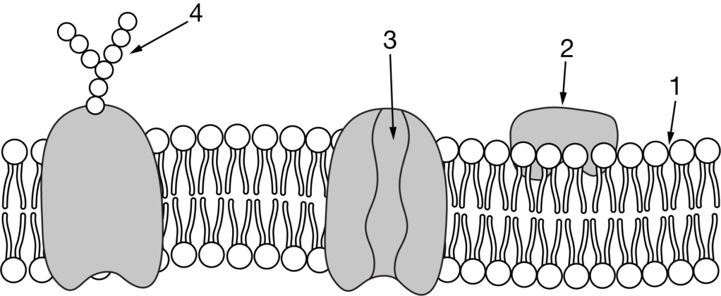
D. 4, testosterone is filtered out of the extracellular fluid and taken into the cell by endocytosis.

▶️Answer/Explanation

Ans: A
Steroids such as testosterone are hydrophobic lipids. Therefore, testosterone can cross the hydrophobic inner region of the phospholipid bilayer.

Question

The figure shows a representation of a protein embedded in a cell membrane. The numbers indicate different structural regions of the protein.

The figure presents a cell membrane lipid bilayer. A protein is embedded in one half of the bilayer. The exposed surface of the protein that protrudes from the membrane is labeled 1, and the part of the protein that associates with the fatty acid tails in the interior of the membrane is labeled 2.
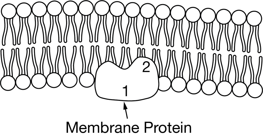
Based on the figure, which of the following statements best describes the relationship between regions 1 and 2 of the protein?
A. Region 1 is hydrophilic because it interacts with the interior of the membrane, whereas region 2 is hydrophobic because it interacts with an aqueous environment.
B. Region 1 is hydrophilic because it interacts with an aqueous environment, whereas region 2 is hydrophobic because it interacts with the interior of the membrane.

C. Region 1 is hydrophobic because it interacts with the interior of the membrane, whereas region 2 is hydrophilic because it interacts with an aqueous environment.
D. Region 1 is hydrophobic because it interacts with an aqueous environment, whereas region 2 is hydrophilic because it interacts with the interior of the membrane.

▶️Answer/Explanation

Ans: B
A cell membrane is a phospholipid bilayer that separates one aqueous environment from another. The interior of a phospholipid bilayer is a hydrophobic environment. Because region 1 interacts with the aqueous environment on one side of the phospholipid bilayer, it is most likely hydrophilic. Because region 2 interacts with the interior of the phospholipid bilayer, it is most likely hydrophobic.

Question

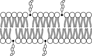
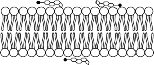
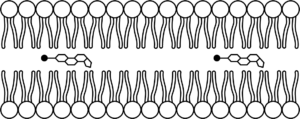
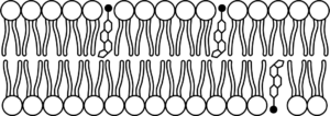
Cholesterol is a naturally occurring substance that helps regulate the fluidity of a cell’s plasma membrane. A cholesterol molecule can be represented as having a polar head and a nonpolar region, as shown in the figure.

The figure presents a cholesterol molecule. A black dot indicates the polar head, which is attached to a nonpolar region that is represented by a sequence of four hexagons or a pentagon, each of which shares one side with the previous and/or next component of the region.
Which of the following models shows how cholesterol molecules most likely interact with the phospholipid bilayer of a cell’s plasma membrane?
 

A.

The figure presents a phospholipid bilayer and cholesterol molecules. The polar head of each cholesterol molecule is situated between the heads of the phospholipids, and the nonpolar region extends out and away from the membrane.

B.

The figure presents a phospholipid bilayer and cholesterol molecules. Each cholesterol molecule lies flat against the outer surface of the membrane so that the polar head and nonpolar region of each molecule are both in contact with the phospholipid heads.

C.

The figure presents a phospholipid bilayer and cholesterol molecules. The cholesterol molecules are located in the interior of the membrane and separate the two layers of the bilayer from each other.

D.

The figure presents a phospholipid bilayer and cholesterol molecules. The polar head of each cholesterol molecule is situated between the heads of the phospholipids, and the nonpolar region extends into the interior of the membrane between the phospholipid tails.

▶️Answer/Explanation

Ans: D
The model correctly shows the polar heads of the cholesterol molecules interacting with the polar heads of the phospholipids. Also, the model correctly shows the nonpolar regions of the cholesterol molecules interacting with the hydrophobic interior of the phospholipid bilayer.

Question

The figure shows a diagram of a portion of a phospholipid bilayer. The upper, middle, and lower areas of the bilayer are labeled 1, 2, and 3, respectively. Areas 1 and 3 show the heads of the phospholipids adjacent to water molecules. Area 2 shows the fatty acid tails of the phospholipids, pointing to the center of the membrane, away from the phospholipid heads at the membrane surfaces.

Which of the following best describes the numbered areas?

A. Areas 1 and 3 are polar, since the membrane molecules are aligned with water molecules.
B. Area 2 is polar, since water has been excluded from this area of the membrane.
C. Areas 1 and 3 are hydrophilic, since membrane molecules formed covalent bonds with water.
D. Area 2 is nonpolar, since hydrogen bonds between the adjacent lipids hold the membrane together.

▶️Answer/Explanation

Ans: A
As indicated in the diagram, the heads of the phospholipids in areas 1 and 3 are adjacent to water molecules, which shows that the phospholipid heads are hydrophilic.

Question

The model below shows the structure of a portion of a plasma membrane in an animal cell.

The model shows the structure of a portion of a plasma membrane in an animal cell. None of the parts are labeled, but the model includes a phospholipid bilayer that contains some embedded proteins. The phospholipids are arranged with their heads oriented toward the external and internal membrane surfaces and their fatty acid tails oriented toward the center of the membrane.

Which statement best explains the orientation of the phospholipid molecules in this model?

A. The nonpolar portions of the phospholipid molecules are attracted to the internal and external environments.
B. The hydrophilic phosphate groups of the phospholipid molecules are attracted to the aqueous internal and external environments.
C. The embedded proteins attract the fatty acid tails of the phospholipids, so the tails point away from the internal and external aqueous environments.
D. The fatty acid tails of the phospholipid molecules are hydrophilic and are repelled by the internal and external aqueous environments.

▶️Answer/Explanation

Ans: B
The hydrophilic polar phosphate groups of the phospholipid molecules orient toward the polar internal and external aqueous environments, forming a bilayer that keeps the hydrophobic portions of the phospholipids away from the polar molecules.

Question

A model of the plasma membrane showing several biological molecules, including a transmembrane protein, is shown in Figure 1.

The figure shows a diagram of a phospholipid bilayer with an embedded transmembrane protein. The protein is coiled into a helical shape where it is buried between the membrane lipids. Polar areas of the protein are labeled on the portions of the protein that stick out from the two membrane surfaces. Nonpolar areas of the protein are labeled on the coiled portion that is buried between the membrane lipids.

Figure 1. Phospholipid bilayer with transmembrane protein

Which statement best explains why correct protein folding is critical in the transmembrane protein shown above?

A. Interactions of the hydrophobic and hydrophilic amino acids help to anchor the protein in the membrane.
B. Interactions of the peptide bonds of the protein with the membrane will affect the rate at which substances can cross the membrane.
C. Interactions of the protein and phospholipids increase membrane fluidity.
D. Interactions of the quaternary structure of the protein will increase hydrogen bonding in the membrane and make the membrane less fluid.

▶️Answer/Explanation

Ans: A
Tertiary structure of the protein resulting from hydrophobic and hydrophilic interactions plays a key role in how the protein interacts with the hydrophobic tails and hydrophilic head of the membrane phospholipids.

Scroll to Top