Question
| Treatment | General Activity \( \left(\text{percent of total time observed, average } \pm 2SE_{\bar{x}} \right) \) | Oriented Activity \( \left(\text{percent of general activity, average } \pm 2SE_{\bar{x}} \right) \) |
|---|---|---|
| Male moths injected with saline (control solution) | \( 95 \pm 5 \) | \( 60 \pm 4 \) |
| Male moths injected with \( \mathrm{siRNAs} \) that inhibit expression of the gene encoding DopEcR | \( 90 \pm 8 \) | \( 25 \pm 6 \) |
(i) Using the template in the space provided for your response, construct an appropriate type of graph that represents the data in Table \( 1 \). Your graph should be appropriately plotted and labeled.
(ii) Ensure that the data and error bars are accurately plotted.
(iii) Ensure that the graph is appropriately labeled.
(iv) Based on the data in Table \( 1 \), determine the type of activity that was affected by inhibiting the expression of the DopEcR receptor.
(i) Based on Table \( 1 \), identify the treatment group in which the oriented activity was greater than \( 50\% \) of the general activity.
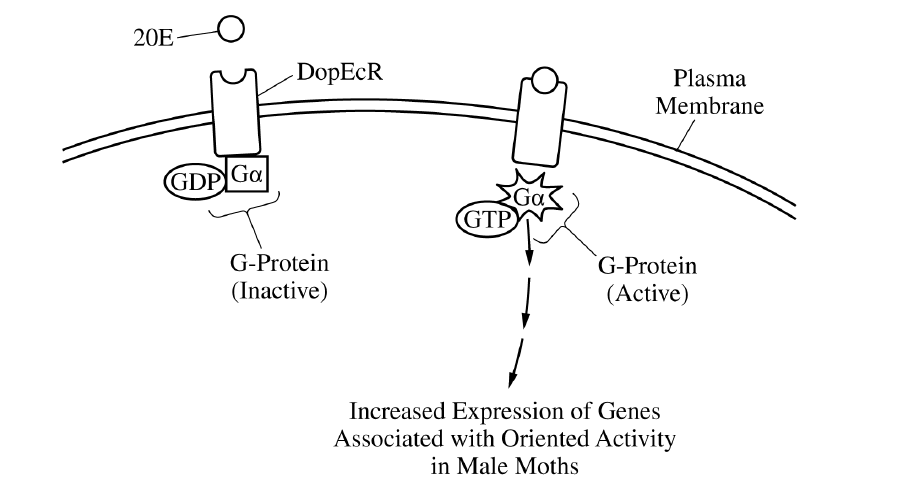
(ii) The scientists studied some moths with a mutation in the gene encoding the G protein. The mutation prevents GTP from displacing the GDP bound to the G protein. Based on Figure \( 1 \), predict the effect of this mutation on the oriented activity in male moths exposed to the pheromone.
(i) Expression of the gene encoding DopEcR is low in male moths during their first few days as adults, when they are sexually immature. Gene expression rapidly increases as the moths reach sexual maturity. The scientists claim that this increase in gene expression increases the likelihood of males finding females with whom to mate. Use evidence from the information provided to support the scientists’ claim.
(ii) Based on Figure \( 1 \), explain how an inhibitor of the DopEcR pathway might serve as an effective chemical to protect crops from moth damage.
Most-appropriate topic codes (AP Biology \( 2025 \)):
• Science Practice \( 4 \) — Representing and Describing Data: Constructing a bar graph, plotting error bars, and labeling axes/legend correctly — parts (B)(i), (B)(ii), (B)(iii)
• Science Practice \( 5 \) — Statistical Tests and Data Analysis: Comparing percentages and identifying which treatment group exceeds \( 50\% \) — parts (B)(iv), (C)(i)
• Topic \( 4.2 \) — Introduction to Signal Transduction: Ligand binding to a receptor, G protein-coupled receptors, and signaling leading to a cellular response — context and part (C)(ii)
• Topic \( 4.3 \) — Signal Transduction Pathways: Predicting how mutation or inhibition changes the signaling pathway and alters the response — parts (C)(ii), (D)(ii)
• Topic \( 6.5 \) — Regulation of Gene Expression: Increased DopEcR gene expression leading to greater pathway activity and a higher likelihood of oriented behavior — part (D)(i)
• Science Practice \( 6 \) — Argumentation: Supporting and explaining claims with evidence from the experiment and model — parts (D)(i), (D)(ii)
▶️ Answer/Explanation
(A)
The portion of the receptor inside the membrane is nonpolar.
It is also hydrophobic.
Summary: because the interior of the plasma membrane is hydrophobic, the membrane-spanning region of the receptor must also be hydrophobic to remain embedded there.
(B)
(i) The data should be represented in a bar graph or modified bar graph.
A correct graph would show the activity values for saline-injected moths and \( \mathrm{siRNA} \)-injected moths using clearly separated bars.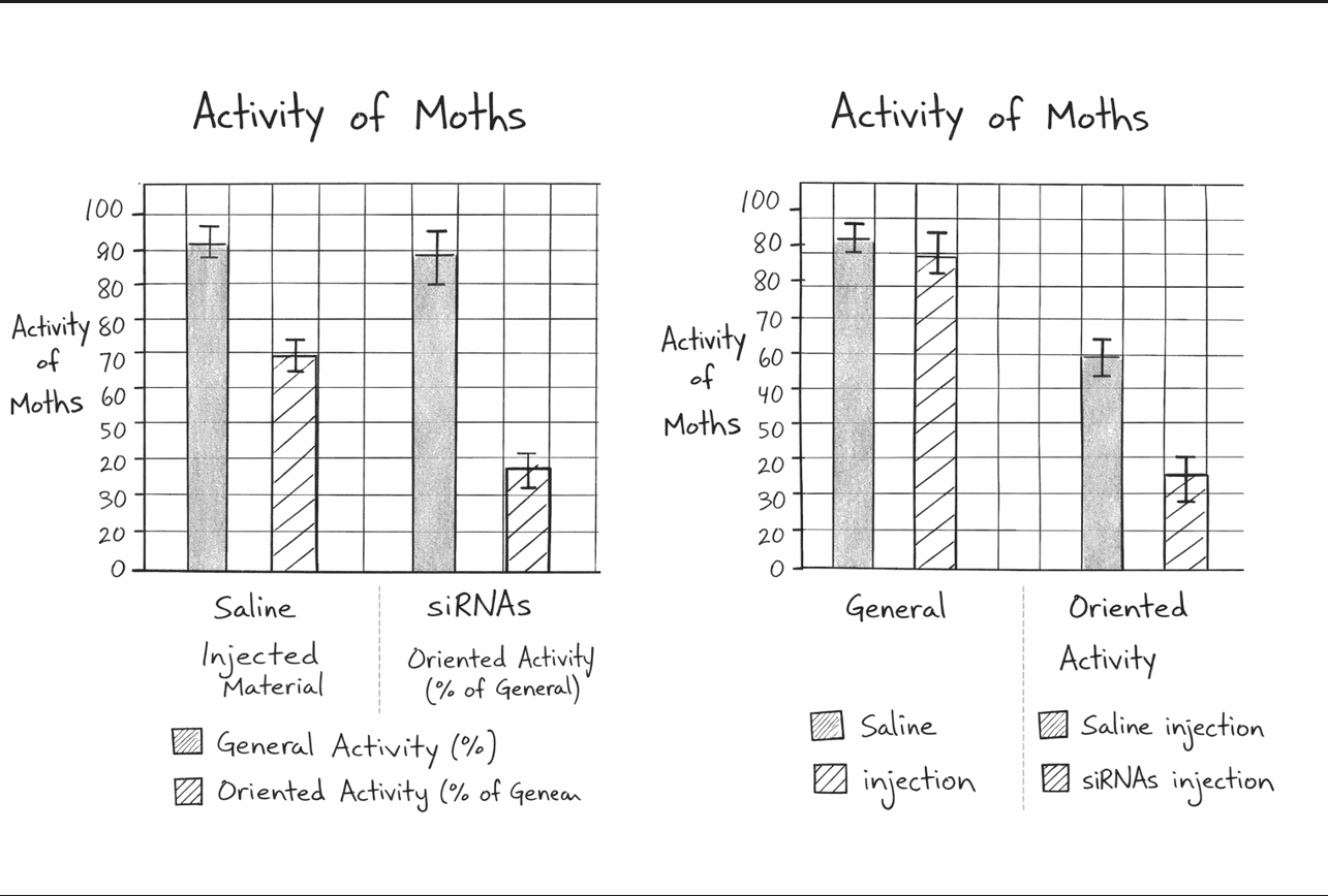
(ii) The data and the error bars should be accurately plotted.
General activity should be shown as approximately \( 95 \pm 5 \) for saline and \( 90 \pm 8 \) for \( \mathrm{siRNA} \).
Oriented activity should be shown as approximately \( 60 \pm 4 \) for saline and \( 25 \pm 6 \) for \( \mathrm{siRNA} \).
(iii) The graph should be appropriately labeled.
Acceptable labels include treatment groups, activity categories, axis titles, and a legend if needed.
Summary: the graph must be the correct type, with the correct values, error bars, and labels.
(B)(iv)
Oriented activity was affected.
It decreased substantially when expression of the DopEcR receptor was inhibited, while general activity changed only slightly.
(C)
(i) The treatment group in which oriented activity was greater than \( 50\% \) of the general activity was the control / saline-treated group.
Male moths injected with saline (control solution) is an acceptable answer.
(ii) The moths will show decreased oriented activity.
Because the mutation prevents GTP from displacing GDP, the G protein would remain inactive, so the signaling pathway would not be properly activated after \( 20E \) binds the receptor.
(D)
(i) Increased expression of DopEcR will increase oriented activity and therefore increase the moths’ ability to find a mate.
An increase in DopEcR expression will enable the binding of \( 20E \) to more receptors.
This will make the males more sensitive to pheromones from the females.
Since oriented activity is movement toward an area of high pheromone concentration, greater receptor expression would increase the likelihood that mature males locate females for mating.
(ii) An inhibitor of the DopEcR pathway will reduce oriented activity and/or reduce the expression of genes associated with oriented activity and therefore decrease mating and population growth.
With fewer successful matings, fewer moths would be produced, so crop damage caused by moth populations would likely decrease.
Summary: blocking the signaling pathway disrupts pheromone-guided behavior and can reduce reproduction.