2.3.A.1 – Phospholipid Structure & Membrane Arrangement
📚 Main Idea:
- Phospholipids have both hydrophilic and hydrophobic regions.
- This dual nature affects how they arrange in the membrane.
🧱 Phospholipid = Amphipathic Molecule
| Region | Type | Faces Toward… |
| Phosphate Head | Hydrophilic 💧 | Water-based areas → outside & inside of cell (cytoplasm + extracellular fluid) |
| Fatty Acid Tails | Hydrophobic 🔥 | Each other → form inner part of membrane |
🧬 Why This Structure Matters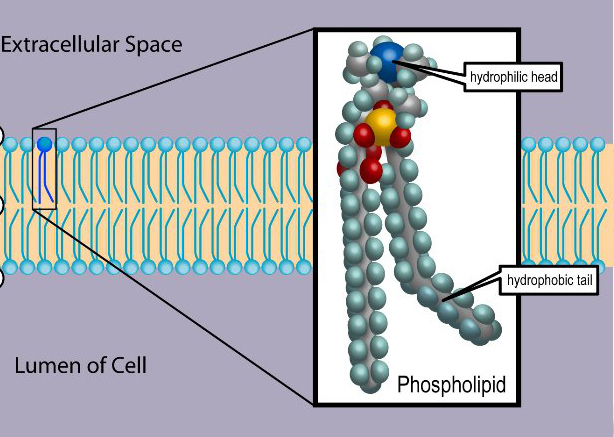
- In water, phospholipids self-arrange into a bilayer
- Heads face watery areas
- Tails hide from water and face inward
- This forms a selectively permeable membrane → only small/nonpolar substances move freely
✅ Summary
- Phospholipids form the basic membrane structure by lining up hydrophilic heads outward and hydrophobic tails inward.
- This creates a stable barrier that protects the cell’s internal environment.
2.3.A.2 – Structure of Embedded Proteins in the Cell Membrane
📚 Main Idea:
- Embedded proteins can be hydrophilic, hydrophobic, or have both regions.
- Their structure determines how they fit and function in the membrane.
🧩 What Are Embedded Proteins?
- Embedded proteins (also called integral proteins) are stuck within the phospholipid bilayer of the cell membrane.
- Some pass all the way through → called transmembrane proteins
- Others are only partially embedded
- Key idea: Their amino acid side chains (R-groups) determine how they sit in the membrane.
💧 Hydrophilic vs. Hydrophobic Protein Regions
| Region Type | Location in Membrane | Why It Works There |
| Hydrophilic | Faces outside (cytoplasm or extracellular fluid) OR hidden inside protein channels | Interacts with water, ions, and polar substances |
| Hydrophobic | Buried in the middle of the bilayer (near fatty acid tails) | Avoids water, interacts with nonpolar lipid interior |
🔬 The way these regions are arranged allows the protein to stay stable in a very unstable (polar + nonpolar) environment.
🌐 How This Affects Function
- Embedded proteins do way more than just sit there. Their hydrophilic/hydrophobic structure is key to their job:
| Protein Function | Role of Hydrophilic/Hydrophobic Parts |
| Transport Proteins | Hydrophilic inside channel to let ions/sugars through, hydrophobic outer part anchors in membrane |
| Receptors | Hydrophilic outer tip binds to signals like hormones |
| Anchors/Linkers | Hydrophobic regions hold protein in membrane; hydrophilic ends connect to cytoskeleton or extracellular matrix |
| Enzymes | Active site (hydrophilic) sticks out to interact with substrates in water-based environments |
✨ Key Insights
- 🌊 Hydrophilic zones often act like “docking bays” for polar molecules – like gates at an airport letting water-loving cargo pass through.
- 🔁 Some proteins flip or rotate slightly in the membrane to activate their function – made possible because of their flexible hydrophobic-hydrophilic regions.
- 🛡️ If protein structure gets disrupted (e.g., due to pH/temp changes), the balance between these regions is thrown off → misfolded proteins can’t stay embedded → diseases like cystic fibrosis.
🧠 Summary:
- Embedded proteins are made of hydrophilic and hydrophobic regions.
- Their structure helps them fit perfectly in the membrane.
- This fit is not random – it’s what lets them transport, signal, or anchor effectively.
- Structure = Function. Always.
2.3.B – Fluid Mosaic Model of cell membrane
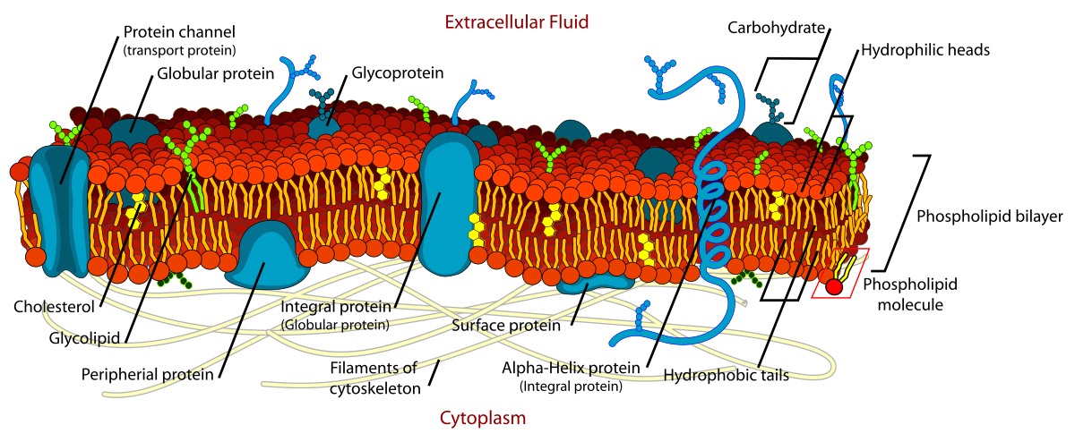
🧩 What Is It?
- The fluid mosaic model is the accepted way of describing the structure of the cell membrane.
- 🔬 “Fluid” = The membrane is flexible; parts can move side to side like a liquid.
- 🧩 “Mosaic” = A patchwork of different molecules (proteins, lipids, carbs) floating in the membrane.
🧱 Main Components
| Component | Role in the Model |
| Phospholipids | Form a flexible bilayer (move freely) |
| Proteins | Float in the bilayer (like boats in a sea); act as transporters, receptors, anchors |
| Cholesterol | Keeps the membrane fluid, stable, and not too rigid or too soft |
| Carbohydrates | Attach to proteins/lipids → help in cell recognition & signaling |
🎯 Why It Matters
- The fluid mosaic model explains how membranes can:
- Let things in/out (selective permeability)
- Move, bend, and reshape
- Support communication and transport
✅ Summary
- The fluid mosaic model shows the membrane as a dynamic, flexible structure.
- It is made of moving lipids and proteins, allowing the cell to function, interact, and protect its internal environment.