Question:
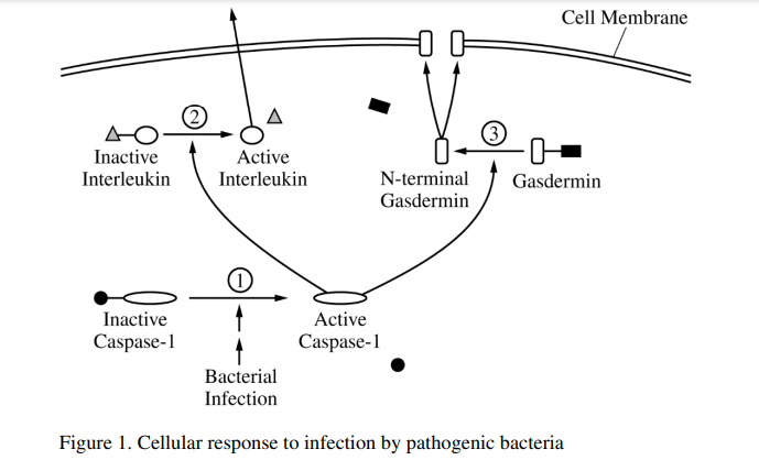
Some pathogenic bacteria enter cells, replicate, and spread to other cells, causing illness in the host organism. Host cells respond to these infections in a number of ways, one of which involves activating particular enzymatic pathways (Figure 1). Cells normally produce a steady supply of inactive caspase-1 protein. In response to intracellular pathogens, the inactive caspase-1 is cleaved and forms an active caspase-1 (step 1). Active caspase-1 can cleave two other proteins. When caspase-1 cleaves an inactive interleukin (step 2), the active portion of the interleukin is released from the cell. An interleukin is a signaling molecule that can activate the immune response. When caspase-1 cleaves gasdermin (step 3), the N-terminal portions of several gasdermin proteins associate in the cell membrane to form large, nonspecific pores. Researchers created the model in Figure 1 using data from cell fractionation studies. In the experiments, various parts of the cell were separated into fractions by mechanical and chemical methods. Specific proteins known to be located in different parts of the cell were used as markers to determine the location of other proteins. The table below shows the presence of known proteins in specific cellular fractions.
CELL FRACTIONS CONTAINING DIFFERENT CELLULAR PROTEINS
| Aconitase (Krebs cycle protein) | DNA polymerase | GAPDH (glycolytic protein) | Sodiumpotassium pump | NF-kB (Immune response protein) | |
| Whole cell sample | + | + | + | + | + |
| Fraction 1 | + | ||||
| Fraction 2 | + | + | |||
| Fraction 3 | + | + | |||
| Fraction 4 | + | ||||
| + = presence of protein | |||||
(a) Describe the effect of inhibiting step 3 on the formation of pores AND on the release of interleukin from the cell.
(b) Make a claim about how cleaving inactive caspase-1 results in activation of caspase-1. A student claims that preinfection production of inactive precursors shortens the response time of a cell to a bacterial infection. Provide ONE reason to support the student’s claim.
(c) A student claims that the NF-kB protein is located in the cytoplasm until the protein is needed for transcription. Justify the student’s claim with evidence. Identify TWO fractions where N-terminal gasdermin would be found in cells infected with pathogenic bacteria.
(d) Describe the most likely effect of gasdermin pore formation on water balance in the cell in a hypotonic environment.
(e) Explain how gasdermin pore formation AND interleukin release contribute to an organism’s defense against a bacterial pathogen.
▶️Answer/Explanation
Ans:
(a) Introducing step 3, the cleavages of separately gasdervaries by Caspar 1. will present the association of gasderamiy proteins and the subsequent formation of poses in the cell membrane. This will not affect the release of interleulax from the cell because outerlacker does not require the poses to lease the cell.
(b) Cleaning inactive caspase-I may alter the interaction of R-groups within the protein, resulting in a slope charge in the cleaned molecule’s tertiary structure that expenses an activate all the cell did not constantly produce inactive caspase-I , the protein would have to be transcribed and translated before performing its function, a process which reprises more engages and more time than a sample clergiable of a polypeptide.
(c) The student’s claim is correct because fraction 2, the nucleus and fraction 3, the equal tested position for RF-kB protein. Fraction 2 is the nucleus be caused contains DNA polymerara, an essays found only in the nucleus where DNA replication occurs and nF -kB would be needed to regulator have secretion. Fraction 3 is the cytoplasm because this fragment tated perstive for glyelytic proteins which and is glycolyass, a process performed in the cytoplasm. It terminal goodermiss would be found in fraction 3 and fraction 4.
(d) IN a hypotonic increment water will more into the cell via arousis through the poses became by debritive, a hypotonic essorounent would have a higher water potential than the cell.
(e) Ucsdermin pere formation cause the flow of water into the cell, which may cause the inflected cell to boost. This would prevent the spread of the infection. because phogocytes could the sleged the conpoxeuts of the cell along with the poctageous. The release of entoslowbix activities the adaptive immence response by stimulating B and J bukecityes to which and produce antibodies or bill infected cells respectively.