Question
In an X-ray tube, electrons are accelerated through a potential difference of \(75\) kV. The electrons then strike a tungsten target of effective mass \(15\) g.
The electron energy is converted into the energy of X-ray photons with an efficiency of \(5.0\) %. The rest of the energy is converted into thermal energy.
(a) The X-ray tube produces an image using a current of \(0.40\) A for a time of \(20\) ms.
The specific heat capacity of tungsten is \(130\, J\, kg^{–1}\, K^{–1}\).
Determine the temperature rise ΔT of the tungsten target.
(b) The linear attenuation coefficient of the X-ray photons in muscle is \(0.22\, cm^{–1}\).
Calculate the thickness t of muscle that will absorb \(80\) % of the incident X-ray intensity.
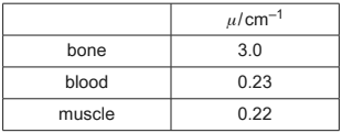
(c) Table 10.1 shows the linear attenuation coefficient μ for the X-ray photons in different tissues.
Table 10.1

Two X-ray images are taken, one of equal thicknesses of bone and muscle and another of equal thicknesses of blood and muscle.
Explain why one of these images has good contrast, but the other does not.
Answer/Explanation
Ans:
(a) energy = mc ΔT
energy = ItV
\((\Delta T =)\frac{0.40 \times 0.020 \times 75\, 000 \times 0.95}{0.015 \times 130}\)
= \(290\) K
(b) \(I\) = \(I_{0}e^{-\mu t}\)
\(0.20\) = \(e^{-0.22t}\)
\(t\) = \(7.3\) cm
(c) either
(linear) attenuation coefficients / μ very different for bone and muscle
(very) different amounts (of X-rays) absorbed so good contrast
or (very) different intensities transmitted so good contrast
or
(linear) attenuation coefficients / μ similar for blood and muscle
similar amounts (of X-rays) absorbed so poor contrast
Question
(a) State, for an X-ray image, what is meant by:
(i) sharpness [1]
(ii) contrast. [1]
(b) A parallel X-ray beam passes through a thickness of 2.3cm of soft body tissue. The intensity of the emerging beam is 12% of the intensity of the incident beam.
Calculate the linear attenuation (absorption) coefficient μ of the soft body tissue. Give a unit with your answer.
μ = …………………………………… unit ………………… [3]
(c) In medical diagnosis, X-rays may be used to produce a single X-ray image or may be used in computed tomography (CT scanning). Suggest an advantage and a disadvantage of CT scanning compared with single X-ray
imaging for diagnosis.
advantage:
disadvantage: [2]
[Total: 7]
Answer/Explanation
Ans
(a) (i) ease with which edges can be distinguished
(a) (ii) difference in degrees of blackening B1
(b) I = I0 exp (–μx) C1
0.12 = exp (–μ × 2.3)
ln 0.12 = –2.3 × μ
μ = 0.92 cm–1
(c) advantage: produces 3-dimensional image
