CIE AS & A Level Physics : 24.3 PET scanning – Exam style question – Paper 4

Question

Positron emission tomography (PET scanning) obtains diagnostic information from a person. The information is used to form an image.

(a)     PET scanning uses a tracer.

Explain what is meant by a tracer.

(b)     PET scanning involves annihilation.

(i)    Explain what is meant by annihilation.

(ii)   State the names of the particles involved in the annihilation process.

(c)      (i)   Calculate the total energy released in one annihilation event in (b).

(ii)   Calculate the wavelength of each gamma photon released.

(d)     Explain how the gamma photons are used to produce an image.

Answer/Explanation

Ans:

(a)     substance containing radioactive nuclei that is introduced into the body or

substance containing radioactive nuclei that is absorbed by the tissue being studied

(b)     (i)      a particle interacting with its antiparticle so that mass is converted into energy

(ii)    electron(s) and positron(s)

(c)     (i)      \(E\) = \(2mc^{2}\)

\(2\times 9.11\times 10^{-31}\times 3.00\times 10^{-82}\)

= \(1.64 \times 10^{-13}\) J

(ii)      \(\lambda\) = \(\frac{2hc}{E}\)

= \(\frac{2\times 6.63\times 10^{-34}\times 3.00\times 10^{8}}{1.64\times 10^{-13}}\)

= \(2.43 \times 10^{-12}\) m

(d)     Any \(3\) from:

• the two gamma photons travel in opposite directions

• gamma photons detected (outside body / by detectors)

• gamma photons arrive (at detector) at different times

• determine location of production (of gamma)


Question

(a)Outline briefly the principles of CT scanning.
…………………………………………………………………………………………………………………………………
…………………………………………………………………………………………………………………………………
…………………………………………………………………………………………………………………………………
…………………………………………………………………………………………………………………………………
…………………………………………………………………………………………………………………………………
…………………………………………………………………………………………………………………………………
…………………………………………………………………………………………………………………………………
…………………………………………………………………………………………………………………………………
…………………………………………………………………………………………………………………………………

(b) In a model for CT scanning, a section is divided into four voxels. The pixel numbers P, Q, R and S of the voxels are shown in Fig. 12.1.

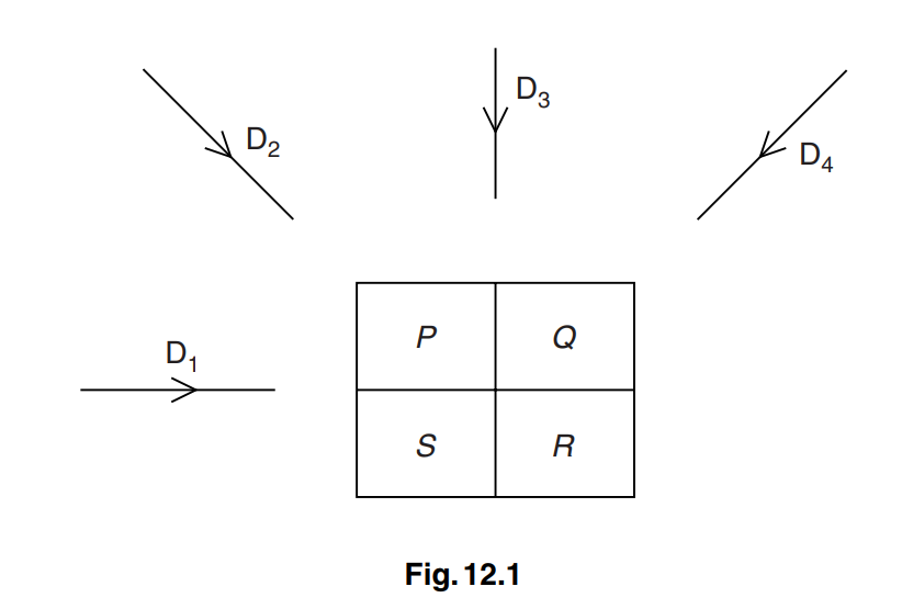
The section is viewed from the four directions D1, D2, D3 and D4.
The detector readings for each direction are noted.
The detector readings are summed as shown in Fig. 12.2.

4961
7355

                  Fig. 12.2
The background reading is 34.
Determine the pixel numbers P, Q, R and S as shown in Fig. 12.3.

PQ
SR

                  Fig. 12.3
P = ……………………………………………………… Q = ………………………………………………………
S = ……………………………………………………… R = ………………………………………………………

Answer/Explanation

(a)

series of X-ray images (for one section/slice)
taken from different angles
to give image of the section/slice
repeated for many slices
to build up three-dimensional image (of whole object)

(b)

deduction of background from readings 
division by three
P = 5 Q = 9 R = 7 S =13

Scroll to Top