Production of X-rays and Minimum Wavelength Calculation
X-rays are generated in an X-ray tube when fast-moving electrons strike a metal target (usually tungsten). This process converts the electrons’ kinetic energy into electromagnetic radiation, including X-rays.
1. How X-rays Are Produced
Electrons are accelerated through a high potential difference (p.d.) and fired at a metal target.
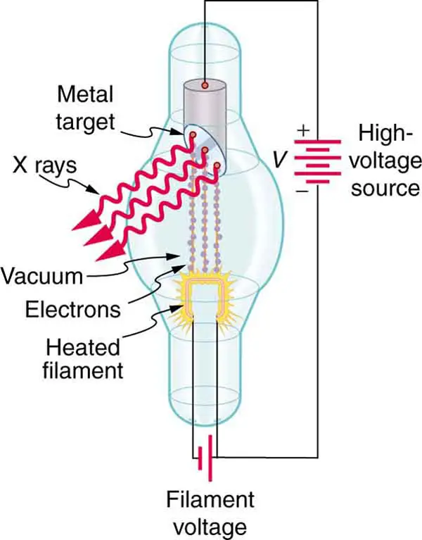
- The electrons gain kinetic energy equal to \( \mathrm{eV} \), where \( \mathrm{V} \) is the accelerating p.d.
- When they hit the metal target, they decelerate suddenly.
- This rapid deceleration emits X-ray photons.
This is known as Bremsstrahlung radiation (“braking radiation”).
Some electrons also knock out inner-shell electrons → characteristic X-rays.
2. Minimum Wavelength of X-rays
The minimum wavelength corresponds to the maximum photon energy, which occurs when all the electron’s kinetic energy is converted into a single photon.
\( \mathrm{eV = hf = \dfrac{hc}{\lambda_{min}}} \)
So:
\( \mathrm{\lambda_{min} = \dfrac{hc}{eV}} \)
- \( \mathrm{h} = 6.63\times10^{-34}\ J\,s \)
- \( \mathrm{c = 3.0\times10^8\ m/s} \)
- \( \mathrm{e = 1.60\times10^{-19}\ C} \)
- \( \mathrm{V} \) = accelerating potential
Example
Electrons are accelerated through \( \mathrm{5.0\ kV} \). Calculate the minimum wavelength of X-rays produced.
▶️ Answer / Explanation
Use:
\( \mathrm{\lambda_{min} = \dfrac{hc}{eV}} \)
Substitute:
\( \mathrm{\lambda_{min} = \dfrac{(6.63\times10^{-34})(3.0\times10^8)}{(1.6\times10^{-19})(5.0\times10^3)}} \)
\( \mathrm{\lambda_{min} = 2.48\times10^{-10}\ m} \)
Minimum wavelength = \( \mathrm{0.248\ nm} \)
Example
An X-ray tube operates at \( \mathrm{20\ kV} \). Find the minimum X-ray wavelength it produces.
▶️ Answer / Explanation
\( \mathrm{\lambda_{min} = \dfrac{hc}{eV}} \)
\( \mathrm{\lambda_{min} = \dfrac{6.63\times10^{-34} \times 3.0\times10^8}{1.6\times10^{-19} \times 2.0\times10^4}} \)
\( \mathrm{\lambda_{min} = 6.2\times10^{-11}\ m} \)
Minimum wavelength = \( \mathrm{0.062\ nm} \)
Example
An electron is accelerated across \( \mathrm{50\ kV} \). What is the minimum wavelength of the X-ray produced? Would this be classified as soft or hard X-rays?
▶️ Answer / Explanation
Step 1: Calculate wavelength
\( \mathrm{\lambda_{min} = \dfrac{hc}{eV}} \)
\( \mathrm{\lambda_{min} = \dfrac{(6.63\times10^{-34})(3.0\times10^8)}{(1.6\times10^{-19})(5.0\times10^4)}} \)
\( \mathrm{\lambda_{min} = 2.48\times10^{-11}\ m} \)
Minimum wavelength = \( \mathrm{0.0248\ nm} \)
Step 2: Classification
- Hard X-rays → shorter wavelengths (≈ 0.01–0.1 nm)
- Soft X-rays → longer wavelengths (≈ 0.1–10 nm)
0.0248 nm → Hard X-rays
Use of X-rays in Imaging Internal Body Structures
X-rays are a powerful diagnostic tool used to visualise internal structures such as bones, lungs, teeth, kidneys, and blood vessels. Their effectiveness relies on how different tissues absorb X-rays differently — a property known as contrast.
1. How X-ray Imaging Works
A beam of X-rays is passed through the body and detected on the other side by a digital detector or photographic plate.
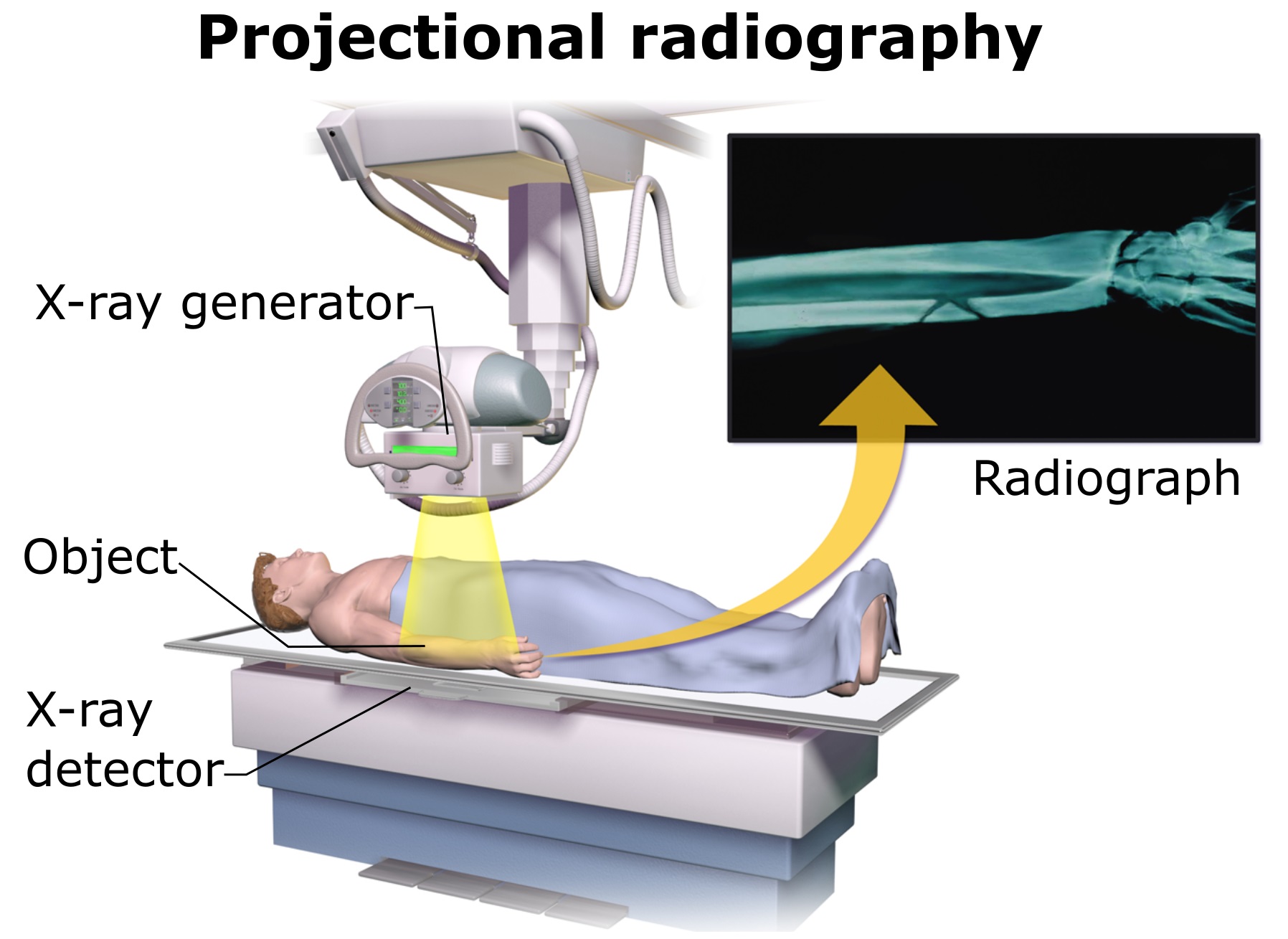
As the X-rays pass through the body:
- Dense materials (e.g., bone) absorb X-rays strongly.
- Less dense tissues (muscle, fat) absorb fewer X-rays.
- Air absorbs very few X-rays (lungs appear dark).
The detector records how much radiation reaches it → forming an image.
2. The Term “Contrast” in X-ray Imaging
Contrast refers to the difference in X-ray absorption between different tissues.
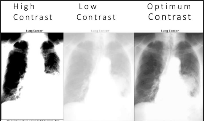
High contrast → easy to distinguish structures
Low contrast → structures look similar or washed out
What determines contrast?
- Density of tissue
- Atomic number of tissue (higher Z absorbs more X-rays)
- Thickness of the tissue
Examples:
- Bones (high calcium content, high Z) → very bright (high absorption)
- Soft organs → grey
- Lungs (air-filled) → dark
3. Improving Contrast Using Contrast Media
Some soft tissues absorb X-rays very weakly. To visualise them, contrast agents are used, such as:
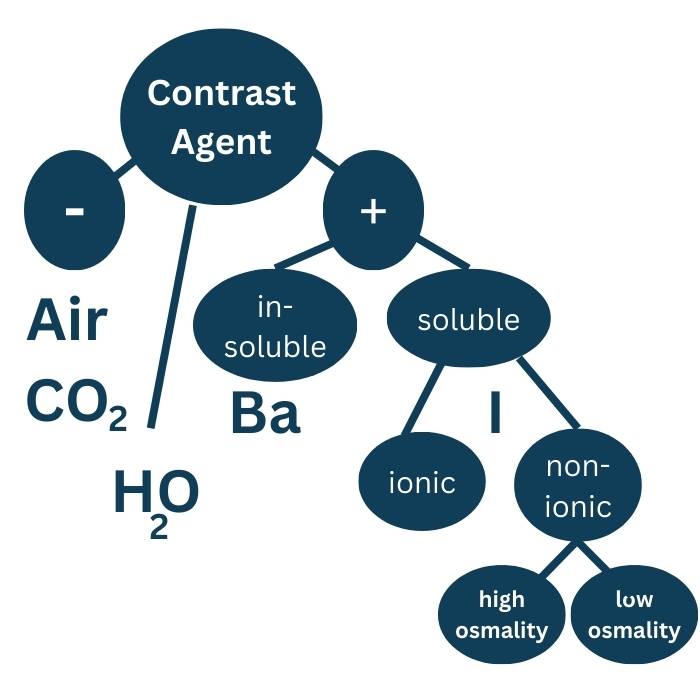
- Barium sulfate for digestive tract imaging
- Iodine compounds for blood vessels, kidneys, bladder
These substances have a high atomic number → absorb X-rays strongly → appear bright.
This enhances contrast so soft structures become visible.
4. Applications of X-ray Imaging
- Bone fractures and alignment
- Dental imaging
- Chest/lung imaging (pneumonia, tumours)
- Mammography
- Contrast-enhanced imaging of blood vessels (angiography)
Example
Why do bones appear white on an X-ray image?
▶️ Answer / Explanation
Bones have high density and high atomic number → they absorb more X-rays → fewer X-rays reach the detector → bones appear bright/white.
Example
Explain why lung tissue produces low absorption and appears dark on an X-ray image.
▶️ Answer / Explanation
Lungs contain air, which has very low density and very low atomic number. X-rays pass through with very little absorption → more X-rays reach the detector → lungs appear dark.
Example
Why is a contrast agent such as iodine needed when imaging blood vessels using X-rays?
▶️ Answer / Explanation
Blood and surrounding soft tissues have similar densities and atomic numbers → low natural contrast. This makes blood vessels difficult to distinguish on a normal X-ray.
Iodine has a high atomic number → absorbs X-rays strongly.
When injected into blood vessels:
- Blood vessels absorb more X-rays
- They appear bright
- Surrounding soft tissue remains darker
This increases contrast and allows clear imaging of arteries and veins.
Attenuation of X-rays in Matter: \( \mathrm{I = I_0 e^{-\mu x}} \)
As X-rays pass through matter, their intensity decreases due to: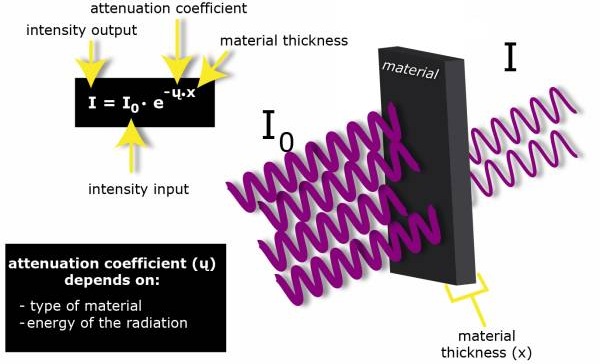
- absorption (mainly by photoelectric effect or Compton scattering)
- scattering of X-rays out of the beam
This decrease in intensity is known as attenuation.
Attenuation Equation
\( \mathrm{I = I_0 e^{-\mu x}} \)
- \( \mathrm{I} \) = intensity after travelling thickness \( \mathrm{x} \)
- \( \mathrm{I_0} \) = initial intensity
- \( \mathrm{\mu} \) = attenuation (or absorption) coefficient (m\(^{-1}\))
- \( \mathrm{x} \) = thickness of material
Meaning:
- Large \( \mathrm{\mu} \): strong absorption (e.g., bone, lead)
- Small \( \mathrm{\mu} \): weak absorption (e.g., soft tissue)
- The decrease is exponential → same mathematical form as radioactive decay.
Example
If the initial X-ray intensity is \( \mathrm{I_0 = 100\ units} \) and the material has \( \mathrm{\mu = 0.30\ cm^{-1}} \), calculate the intensity after the X-rays travel through \( \mathrm{2\ cm} \).
▶️ Answer / Explanation
\( \mathrm{I = I_0 e^{-\mu x}} \)
\( \mathrm{I = 100 \, e^{-0.30 \times 2}} \)
\( \mathrm{I = 100 \, e^{-0.6}} = 100 \times 0.5488 \)
\( \mathrm{I \approx 54.9\ units} \)
Intensity = 54.9 units
Example
X-rays of initial intensity \( \mathrm{500\ units} \) pass through \( \mathrm{5\ cm} \) of a material, emerging with intensity \( \mathrm{150\ units} \). Find the attenuation coefficient \( \mathrm{\mu} \).
▶️ Answer / Explanation
Start with:
\( \mathrm{\frac{I}{I_0} = e^{-\mu x}} \)
\( \mathrm{\frac{150}{500} = e^{-5\mu}} \)
\( \mathrm{0.3 = e^{-5\mu}} \)
Take natural log:
\( \mathrm{\ln(0.3) = -5\mu} \)
\( \mathrm{-1.204 = -5\mu} \)
\( \mathrm{\mu = 0.2408\ cm^{-1}} \)
Attenuation coefficient = \( \mathrm{0.241\ cm^{-1}} \)
Example
An X-ray beam must pass through \( \mathrm{10\ cm} \) of tissue with attenuation coefficient \( \mathrm{\mu = 0.15\ cm^{-1}} \). What percentage of the original intensity reaches the other side?
▶️ Answer / Explanation
Use fractional transmission:
\( \mathrm{\frac{I}{I_0} = e^{-\mu x}} \)
\( \mathrm{\frac{I}{I_0} = e^{-0.15\times10}} = e^{-1.5} \)
\( \mathrm{e^{-1.5} = 0.2231} \)
Therefore 22.3% of the original intensity passes through.
This shows why X-ray beams must be strong enough to penetrate thick tissue.
Computed Tomography (CT) Scanning and 3D Imaging
Computed Tomography (CT) scanning is an advanced medical imaging technique that uses X-rays and computer processing to produce detailed 3D images of internal body structures.
1. How CT Scanning Works
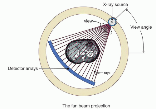
A CT scanner takes many X-ray images (called projections) of the same cross-section of the body from different angles.
Process:
- An X-ray source rotates around the patient.
- Detectors on the opposite side record the transmitted X-rays.
- Hundreds of projections are collected for the same slice.
These projections give information about how much different parts of the section absorb X-rays.
2. Formation of a 2D Cross-Sectional Image

A computer uses mathematical reconstruction algorithms (typically filtered back-projection) to combine all the projections taken from different angles into a single 2D image.
This image represents one “slice” of the body.
Advantages of CT over simple X-ray:
- Much clearer soft-tissue contrast
- No superposition of structures
- Accurate location and shape of abnormalities
3. Building a 3D Image
The scanner then moves slightly along the patient’s body (e.g., head-to-toe direction) and repeats the process for the next slice.
Each slice gives a separate 2D image.
To produce a 3D image:
- Many adjacent 2D slices are taken along the axis
- Computer combines all slices
- A full 3D reconstruction of organs, blood vessels, or bones is created
This allows doctors to “look inside” the body with very high detail.
4. Applications of CT Scans
- Brain imaging (strokes, trauma, tumours)
- Chest and lung imaging
- Abdominal imaging (kidneys, liver, pancreas)
- Blood vessel analysis (CT angiography)
- Bone fracture assessment
Example
Why does a CT scanner take X-ray projections of the same section from many different angles?
▶️ Answer / Explanation
A single X-ray image overlaps many structures and cannot show depth clearly. Multiple angled projections allow a computer to reconstruct a clear 2D slice without overlap.
Example
Explain how a 3D image is produced in CT scanning from a series of 2D slices.
▶️ Answer / Explanation
The scanner moves along the body and acquires many adjacent 2D slices. A computer stacks and combines these slices to create a full 3D reconstruction of the internal structure.
Example
CT scans use a rotating X-ray tube instead of keeping the tube fixed, like in a standard X-ray machine. Explain why this is necessary and how it leads to higher image quality.
▶️ Answer / Explanation
Reason for rotation:
- A fixed X-ray tube gives only one projection → overlapping structures → poor detail.
- Rotating the tube collects hundreds of projections from different angles.
How it improves image quality:
- Reconstruction algorithms combine many projections into a single accurate 2D slice.
- Structures that would overlap in a single X-ray become clearly separated.
- Repeating slices along the axis builds a detailed 3D model.
This results in much better contrast, clarity, and spatial resolution.