
Structure & Function of the Heart
📝 Introduction
- The heart is a muscular organ that pumps blood around the body.
- Works as a double circulatory system → blood passes through the heart twice in one complete circuit.
- Right side → pumps blood to the lungs (pulmonary circulation).
- Left side → pumps blood to the rest of the body (systemic circulation).
📍 Location
- The heart is located in the thorax (chest cavity).
- It lies between the lungs and is slightly towards the left side of the body.
- It is protected by the rib cage.
🫀 Chambers of the Heart
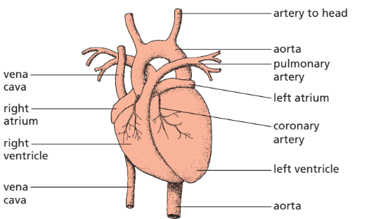
- The heart has four chambers.
- The upper chambers are called atria (singular: atrium).
- The lower chambers are called ventricles.
🔵 Right Atrium
Structure
- Thin-walled upper chamber on the right side of the heart.
Function
- Receives deoxygenated blood returning from the body.
- Blood enters through the vena cava.
- Passes blood into the right ventricle.
🔵 Right Ventricle
Structure
- Lower chamber on the right side.
- Wall is thicker than the atrium but thinner than the left ventricle.
Function
- Pumps deoxygenated blood to the lungs through the pulmonary artery.
- Blood travels to the lungs to collect oxygen and release carbon dioxide.
🔴 Left Atrium
Structure
- Thin-walled upper chamber on the left side.
Function
- Receives oxygenated blood from the lungs.
- Blood enters through the pulmonary vein.
- Passes blood into the left ventricle.
🔴 Left Ventricle
Structure
- Lower chamber on the left side.
- Has the thickest muscular wall of all four chambers.
Function
- Pumps oxygenated blood to all parts of the body through the aorta.
Why is the Wall Thicker?
- Blood must travel to every part of the body.
- A higher pressure is needed to push blood over a much greater distance.
- The thicker muscle produces a more powerful contraction.
🧱 Septum
Structure
- The septum is a thick muscular wall running down the centre of the heart.
Function
- Separates the right and left sides of the heart.
- Prevents oxygenated blood from mixing with deoxygenated blood.
- Ensures body tissues receive fully oxygenated blood.
🚪 Heart Valves
Function of Valves
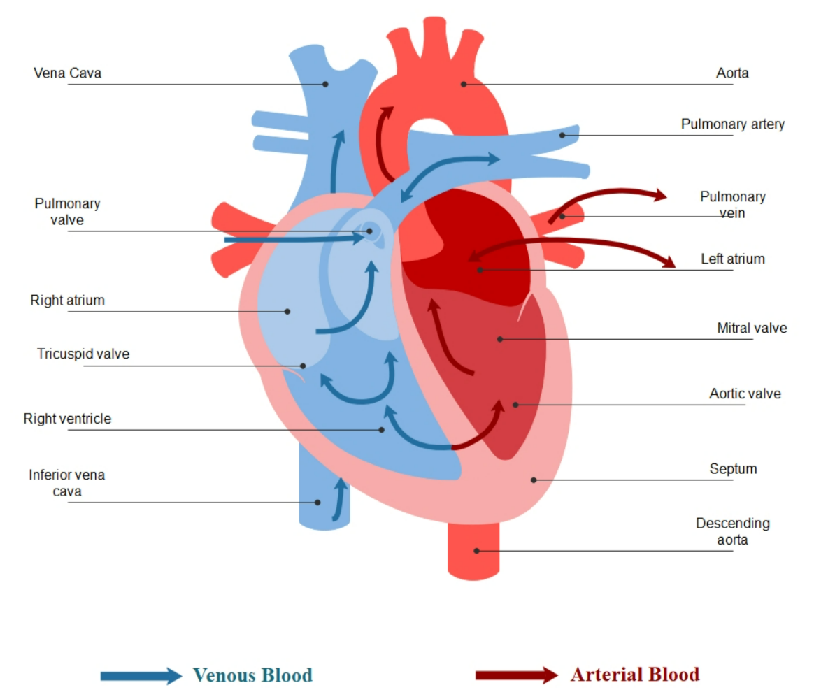
- Valves ensure that blood flows in one direction only.
- They prevent blood from flowing backwards when the heart contracts.
There are two main types of valves.
Atrioventricular (AV) Valves
Location
- Between each atrium and ventricle.
Function
- Open to allow blood to flow from the atria into the ventricles.
- Close when the ventricles contract.
- Prevent blood from flowing back into the atria.
Semilunar Valves
Location
- At the openings of the aorta and pulmonary artery.
Function
- Open when the ventricles pump blood into the arteries.
- Close after contraction.
- Prevent blood from flowing back into the ventricles.
🩸 Major Blood Vessels Connected to the Heart
Vena Cava
- Carries deoxygenated blood from the body to the right atrium.
Pulmonary Artery
- Carries deoxygenated blood from the right ventricle to the lungs.
Pulmonary Vein
- Carries oxygenated blood from the lungs to the left atrium.
Aorta
- Carries oxygenated blood from the left ventricle to the rest of the body.
❤️ Coronary Arteries
Function
- The heart muscle needs a constant supply of oxygen and nutrients because it works continuously.
- Coronary arteries supply oxygenated blood directly to the heart muscle.
- This enables the heart muscle to carry out aerobic respiration and contract continuously.
⚙️ How the Heart Functions
The heart works through a repeating sequence of contraction and relaxation called the cardiac cycle.
Step 1: Atrial Systole
- Both atria contract.
- Blood is pushed into the ventricles through the atrioventricular valves.
- The ventricles fill with blood.
Step 2: Ventricular Systole
- Both ventricles contract.
- The atrioventricular valves close to prevent backflow.
- Blood is pumped:
- From the right ventricle into the pulmonary artery.
- From the left ventricle into the aorta.
- The semilunar valves open to allow blood to leave the heart.
Step 3: Diastole
- The atria and ventricles relax.
- The semilunar valves close to stop blood flowing back into the ventricles.
- Fresh blood enters the atria from the veins.
- The next heartbeat begins.
Blood Flow Through the Heart
🌟 Importance of the Heart
The heart is essential because it:
- Pumps blood continuously around the body.
- Delivers oxygen and nutrients to cells.
- Removes carbon dioxide and other waste products.
- Maintains blood circulation.
- Supplies the heart muscle itself through the coronary arteries.
📊 Summary Table
| Structure | Function |
|---|---|
| Right atrium | Receives deoxygenated blood from the body |
| Right ventricle | Pumps deoxygenated blood to the lungs |
| Left atrium | Receives oxygenated blood from the lungs |
| Left ventricle | Pumps oxygenated blood to the rest of the body |
| Septum | Prevents mixing of oxygenated and deoxygenated blood |
| Atrioventricular valves | Prevent backflow from ventricles to atria |
| Semilunar valves | Prevent backflow from arteries to ventricles |
| Coronary arteries | Supply oxygenated blood to the heart muscle |
| Vena cava | Carries deoxygenated blood to the right atrium |
| Pulmonary artery | Carries deoxygenated blood to the lungs |
| Pulmonary vein | Carries oxygenated blood to the left atrium |
| Aorta | Carries oxygenated blood to the body |
⚡ Quick Recap
Heart = double pump → right side (lungs), left side (body).
Left ventricle thicker → needs more force.
Valves stop backflow → ensure one-way flow.
Cardiac cycle = atrial systole → ventricular systole → diastole.
Coronary arteries keep heart alive.