
Ultrafiltration in the Bowman’s Capsule
📝 Introduction
- The first step in urine formation is called ultrafiltration.
- Ultrafiltration takes place in the renal corpuscle, which consists of the glomerulus and Bowman’s capsule.
- During this process, small molecules are filtered out of the blood, while large molecules remain in the bloodstream.
- The filtered fluid formed is called the glomerular filtrate.
- Ultrafiltration helps remove waste products while allowing useful substances to be reabsorbed later in the nephron.
🩸 What is Ultrafiltration?
Definition
- Ultrafiltration is the process by which small molecules are forced out of the blood in the glomerulus into Bowman’s capsule under high pressure.
It is called “ultra” filtration because only very small molecules can pass through the filtration membrane.
Where Does Ultrafiltration Occur?
Ultrafiltration takes place in two structures:
- Glomerulus – a network of tiny capillaries where blood is filtered.
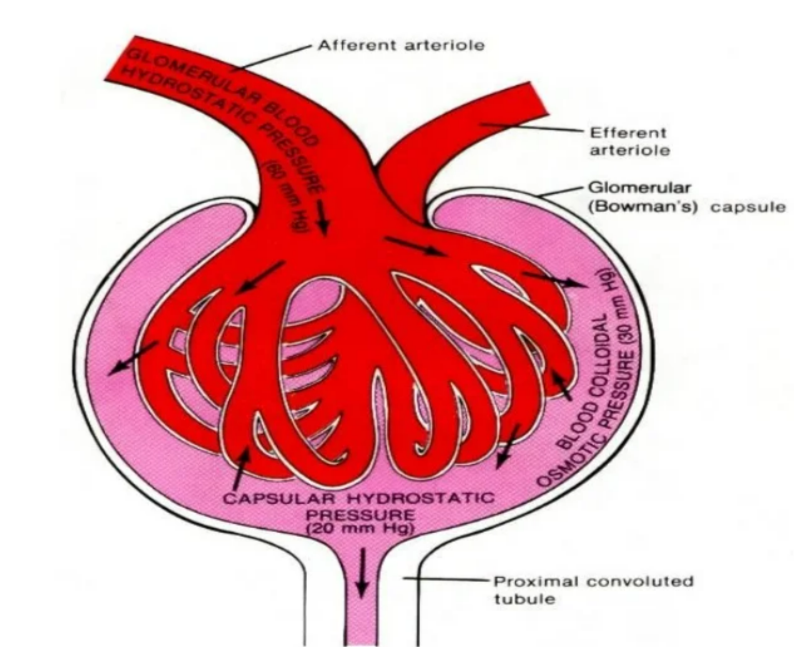
- Bowman’s capsule – a cup-shaped structure that collects the filtered fluid.
Together, they form the renal corpuscle.
🔬 How Does Ultrafiltration Take Place?
Step 1 – Blood Enters the Glomerulus
- Blood enters the glomerulus through the afferent arteriole.
- The afferent arteriole is wider than the efferent arteriole that carries blood away.
- This difference in diameter creates high blood pressure inside the glomerulus.
Why is high pressure important?
- It provides the force needed to push small molecules out of the blood.
Step 2 – Filtration of Small Molecules
- The walls of the glomerular capillaries have tiny pores.
- These pores allow only small molecules to pass through.
- The molecules pass through:
- Capillary wall
- Basement membrane
- Inner wall of Bowman’s capsule
The filtered fluid then enters Bowman’s capsule.
Step 3 – Formation of Glomerular Filtrate
The fluid collected inside Bowman’s capsule is called the glomerular filtrate. It then flows into the proximal convoluted tubule (PCT), where useful substances are reabsorbed.
🌟 Why Don’t Large Molecules Pass Through?
Large substances cannot pass through the filtration membrane because they are too big.
These include:
- Red blood cells
- White blood cells
- Platelets
- Plasma proteins
They remain in the blood and leave the glomerulus through the efferent arteriole. This prevents the loss of important blood components.
🧪 Composition of the Glomerular Filtrate
The glomerular filtrate contains only small molecules.
It contains:
- Water
- Glucose
- Amino acids
- Urea
- Mineral salts (ions)
- Small dissolved substances
It does not contain:
- Red blood cells
- White blood cells
- Platelets
- Plasma proteins
These remain in the bloodstream because they are too large to pass through the filtration membrane.
🔄 What Happens to the Glomerular Filtrate Next?
The filtrate moves into the proximal convoluted tubule (PCT).
Here:
- All glucose is reabsorbed.
- All amino acids are reabsorbed.
- Most water is reabsorbed.
- Most useful mineral salts are reabsorbed.
The remaining fluid eventually becomes urine.
🌟 Importance of Ultrafiltration
Ultrafiltration is important because it:
- Removes waste products such as urea from the blood.
- Begins the process of urine formation.
- Allows useful substances to be reabsorbed later.
- Helps maintain the body’s internal environment (homeostasis).
📊 Stages of Ultrafiltration
| Stage | What Happens? |
|---|---|
| 1. Blood enters glomerulus | Blood enters under high pressure through the afferent arteriole. |
| 2. Filtration | Small molecules pass through the filtration membrane into Bowman’s capsule. |
| 3. Filtrate formed | The filtered fluid is called the glomerular filtrate. |
| 4. Blood leaves | Blood cells and proteins remain in the blood and leave through the efferent arteriole. |
| 5. Filtrate enters PCT | Useful substances are reabsorbed in the next part of the nephron. |
⚡ Quick Recap
Ultrafiltration = blood under pressure filters out small molecules into Bowman’s capsule.
Filtrate = water + glucose + amino acids + salts + urea.
No proteins or blood cells in filtrate.
👉 Think: “Useful + waste small stuff passes, big stuff stays”.
Water Reabsorption from the Collecting Duct
📝 Introduction
- After filtration and selective reabsorption, the kidney still controls the final water content of urine.
- This final adjustment happens in the collecting duct.
- Water is reabsorbed back into the blood depending on how much the body needs.
- This process is controlled by a hormone called ADH (Antidiuretic Hormone).
- It helps maintain water balance in the body (osmoregulation).
💧 What is Water Reabsorption?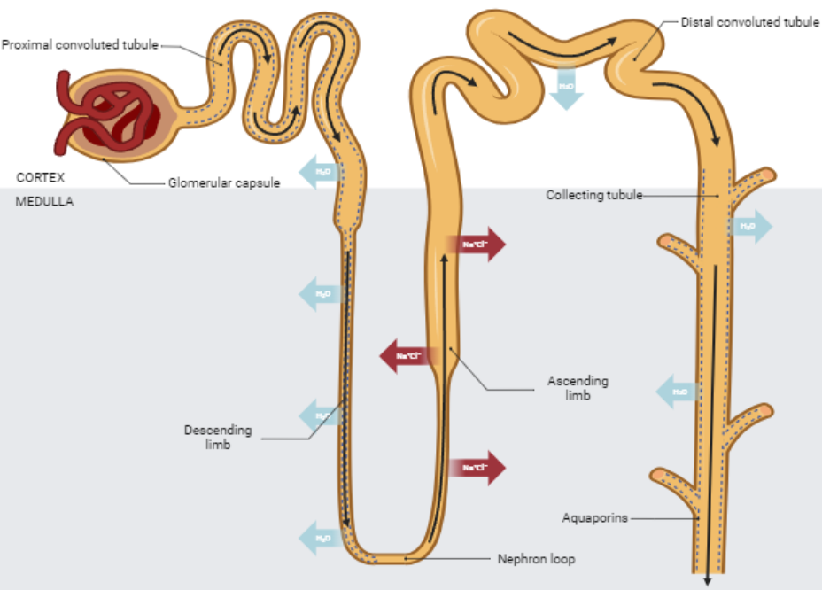
- Water reabsorption means moving water from the kidney tubule back into the blood.
- It mainly happens in the collecting duct.
- The amount of water reabsorbed is not fixed, it changes according to body needs.
🧬 Role of the Collecting Duct
- The collecting duct is the final part of the nephron.
- It receives fluid from many nephrons.
- It decides how much water should be kept in the body and how much should be lost as urine.
🧪 How Water is Reabsorbed
Step 1 – Filtrate reaches collecting duct
- After passing through the nephron, the fluid reaches the collecting duct.
- This fluid still contains:
- Water
- Urea
- Excess salts
Step 2 – Movement of water out of the collecting duct
- Water moves out of the collecting duct into the surrounding blood capillaries.
- This happens by osmosis.
- Water moves from a region of higher water concentration (inside duct) to lower water concentration (blood).
Step 3 – Return to bloodstream
- Reabsorbed water enters nearby capillaries.
- It returns to the bloodstream and is reused by the body.
- This helps maintain blood volume and concentration.
🧬 Role of ADH (Antidiuretic Hormone)
- ADH controls how much water is reabsorbed.
- It is released from the pituitary gland.
When the body is dehydrated (less water in blood)
- More ADH is released.
- Collecting duct walls become more permeable to water.
- More water moves back into blood.
- Urine becomes:
- Small in volume
- Concentrated (dark yellow)
When the body has excess water
- Less ADH is released.
- Collecting duct walls become less permeable.
- Less water is reabsorbed.
- More water stays in urine.
- Urine becomes:
- Large in volume
- Dilute (pale yellow)
🌟 Why Water Reabsorption is Important
- Prevents dehydration.
- Maintains blood volume.
- Keeps concentration of blood constant.
- Helps cells function properly.
- Supports overall homeostasis.
📊 Water Reabsorption in Collecting Duct
| Condition | ADH Level | Water Reabsorbed | Urine Type |
|---|---|---|---|
| Dehydration | High | More water reabsorbed | Small, concentrated urine |
| Normal hydration | Medium | Balanced reabsorption | Normal urine |
| Excess water | Low | Less water reabsorbed | Large, dilute urine |
🔄 Key Idea Flow
- Filtrate enters collecting duct
- Water moves out by osmosis
- ADH controls permeability of duct walls
- Water returns to blood
- Urine concentration is adjusted
⚡ Quick Recap
Collecting duct → water reabsorbed by osmosis into blood capillaries.
Controlled by ADH hormone.
High ADH = concentrated urine.
Low ADH = dilute urine.
👉 Mnemonic: ADH = “Always Drinking Hormone” → tells your kidneys how much water to keep!
Selective Reabsorption of Glucose in the Proximal Convoluted Tubule (PCT)
📝 Introduction
- After ultrafiltration in the glomerulus, the filtrate contains useful substances + waste substances.
- One of the most important useful substances is glucose.
- The body must not lose glucose in urine because it is a vital energy source.
- So, glucose is completely reabsorbed in the proximal convoluted tubule (PCT).
- This process is called selective reabsorption because only useful substances are taken back into the blood.
What is Selective Reabsorption?
- It is the process where useful substances are taken back from the nephron into the blood.
- It happens mainly in the proximal convoluted tubule.
- The word “selective” means only specific substances are absorbed, not everything.
🧪 Why Does Glucose Need to Be Reabsorbed?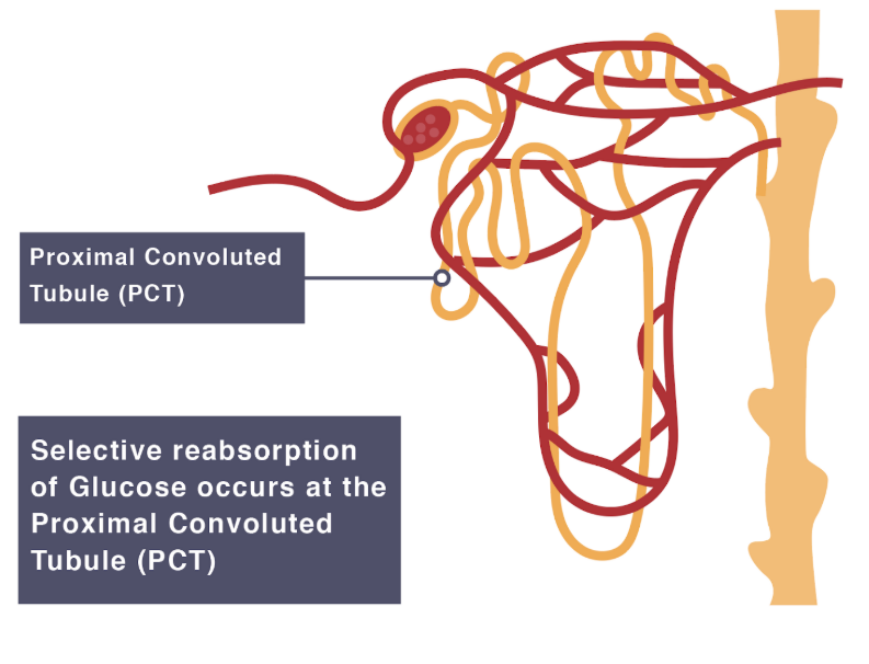
1. Glucose is a major energy source
- Cells use glucose in respiration to release energy (ATP).
- If glucose is lost in urine, the body loses energy supply.
2. Glucose is a useful and essential molecule
- It is not a waste product.
- It is needed for:
- Brain function
- Muscle activity
- Growth and repair
- Continuous energy supply
3. Prevents energy loss
- Losing glucose in urine would mean wasting a valuable nutrient.
- So the kidney ensures all glucose is recovered back into the blood.
Where Does Glucose Reabsorption Happen?
- It occurs in the proximal convoluted tubule (PCT).
- This part of the nephron is specially adapted for absorption.
🧬 How Glucose is Reabsorbed
Step 1 – Glucose enters the PCT
- After filtration, glucose is present in the nephron fluid.
- This fluid flows into the PCT.
Step 2 – Active transport of glucose
- Glucose is moved from the nephron into the blood by active transport.
- Active transport requires energy (ATP) from respiration.
Step 3 – Movement into blood capillaries
- Glucose passes from PCT cells into surrounding blood capillaries.
- Blood then carries glucose to all body cells.
Why is Active Transport Needed?
- Glucose reabsorption cannot rely on diffusion because:
- Glucose concentration in filtrate is already very low at times.
- Blood already contains glucose, so movement is not always down a concentration gradient.
So glucose is moved against the concentration gradient. This is why energy is needed (ATP).
Adaptations of PCT for Glucose Reabsorption
- Many mitochondria → provide energy (ATP) for active transport.
- Microvilli → increase surface area for absorption.
- Thin walls → short distance for movement of substances.
- Good blood supply → maintains concentration gradient.
What Happens After Reabsorption?
- Glucose is completely returned to the blood.
- It is transported to cells for respiration.
- Normally, no glucose should be present in urine.
If glucose is found in urine, it may indicate diabetes mellitus.
📊 Summary of Glucose Reabsorption
| Feature | Explanation |
|---|---|
| Location | Proximal convoluted tubule (PCT) |
| Substance | Glucose |
| Process | Selective reabsorption |
| Method | Active transport |
| Energy source | ATP from respiration |
| Reason | Glucose is needed for respiration and energy |
| Result | All glucose returns to blood (normally no glucose in urine) |
⚡ Quick Recap
Glucose = vital fuel → must be reabsorbed.
PCT is adapted with microvilli + mitochondria.
Process: Active transport + co-transport with sodium ions.
Result: 100% of glucose reabsorbed into blood (normally).
👉 Mnemonic: PCT = Picks up Critical Treasure (glucose)