
Structure and Function of the Eye
📝 Introduction
- The eye is a sense organ that detects light from the environment.
- It works as a receptor for vision, helping us see objects, colours and movement.
- The eye converts light energy into nerve impulses, which are sent to the brain.
- The brain interprets these signals to form an image.
- Different parts of the eye work together to focus light and detect images clearly.
👁️ Structure of the Eye and Their Functions
Cornea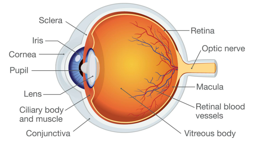
- The cornea is the transparent front part of the eye.
- It is curved and helps to bend (refract) light entering the eye.
- It also acts as a protective layer against dust and injury.
- It is transparent so light can pass through easily.
Aqueous Humour
- A clear fluid found between the cornea and lens.
- Helps to maintain the shape of the eye.
- Refracts light slightly as it passes through.
- Provides oxygen and nutrients to the cornea and lens (as they do not have blood vessels).
Iris
- The iris is the coloured part of the eye.
- It contains muscles that control the size of the pupil.
- It adjusts the amount of light entering the eye.
How it works:
- In bright light → iris contracts → pupil becomes small.
- In dim light → iris relaxes → pupil becomes large.
Pupil
- The pupil is the opening in the centre of the iris.
- It allows light to enter the eye.
- It changes size depending on light intensity (controlled by iris).
Lens
- A transparent, flexible structure behind the pupil.
- It focuses light onto the retina.
- Changes shape to focus on near and far objects.
Accommodation:
- Near objects → lens becomes thicker and more curved.
- Distant objects → lens becomes thin and flatter.
Ciliary Muscles
- Ring-shaped muscles around the lens.
- Control the shape of the lens.
Function:
- Contract → lens becomes thicker (near vision).
- Relax → lens becomes thinner (distant vision).
Suspensory Ligaments
- Thin fibres that hold the lens in place.
- Connect the lens to the ciliary muscles.
- Work with ciliary muscles to change lens shape.
Vitreous Humour
- A clear jelly-like substance inside the main eye cavity.
- Helps maintain the shape of the eyeball.
- Keeps the retina in position.
- Allows light to pass through to the retina.
Retina
- A light-sensitive layer at the back of the eye.
- Contains photoreceptor cells:
- Rod cells (detect dim light, black and white vision)
- Cone cells (detect colour and detail)
Function:
- Converts light into electrical nerve impulses.
- Sends signals to the brain through the optic nerve.
Rods and Cones
Rod Cells
- Work in low light conditions.
- Do not detect colour.
- Provide black and white vision.
- Very sensitive to light.
Cone Cells
- Work in bright light.
- Detect colours (red, green, blue).
- Provide sharp and detailed vision.
Optic Nerve
- Carries nerve impulses from the retina to the brain.
- The brain processes these signals to form a visual image.
- There are no photoreceptors at the optic nerve point → this area is called the blind spot.
Blind Spot
- The point where the optic nerve leaves the eye.
- No rod or cone cells present.
- No image is formed at this point.
🔄 How the Eye Forms an Image
- Light enters through the cornea.
- It passes through aqueous humour and pupil.
- The lens focuses the light onto the retina.
- The retina converts light into nerve impulses.
- The optic nerve sends signals to the brain.
- The brain forms the final image.
📊 Summary Table
| Part | Structure | Function |
|---|---|---|
| Cornea | Transparent curved layer | Refracts and protects |
| Aqueous humour | Clear fluid | Maintains shape, nourishes eye |
| Iris | Pigmented muscle ring | Controls pupil size |
| Pupil | Opening in iris | Allows light entry |
| Lens | Transparent, flexible | Focuses light on retina |
| Ciliary muscles | Circular muscles | Change lens shape |
| Suspensory ligaments | Fibres | Hold lens in place |
| Vitreous humour | Jelly-like fluid | Maintains shape, supports retina |
| Retina | Light-sensitive layer | Converts light to nerve impulses |
| Rods | Photoreceptors | Dim light vision |
| Cones | Photoreceptors | Colour vision |
| Optic nerve | Nerve bundle | Sends signals to brain |
🌟 Why the Eye is a Good Receptor
- Highly sensitive to light.
- Can adjust to different light conditions.
- Produces clear images due to lens focusing.
- Converts light into electrical signals quickly.
- Works continuously without conscious effort.
📌 Quick Recap
Eye = receptor for light.
Detects intensity, colour, and images.
Electrical impulses sent via optic nerve → brain interprets them.
Iris + pupil + lens help control light entry and focus.