▶️ Answer/Explanation
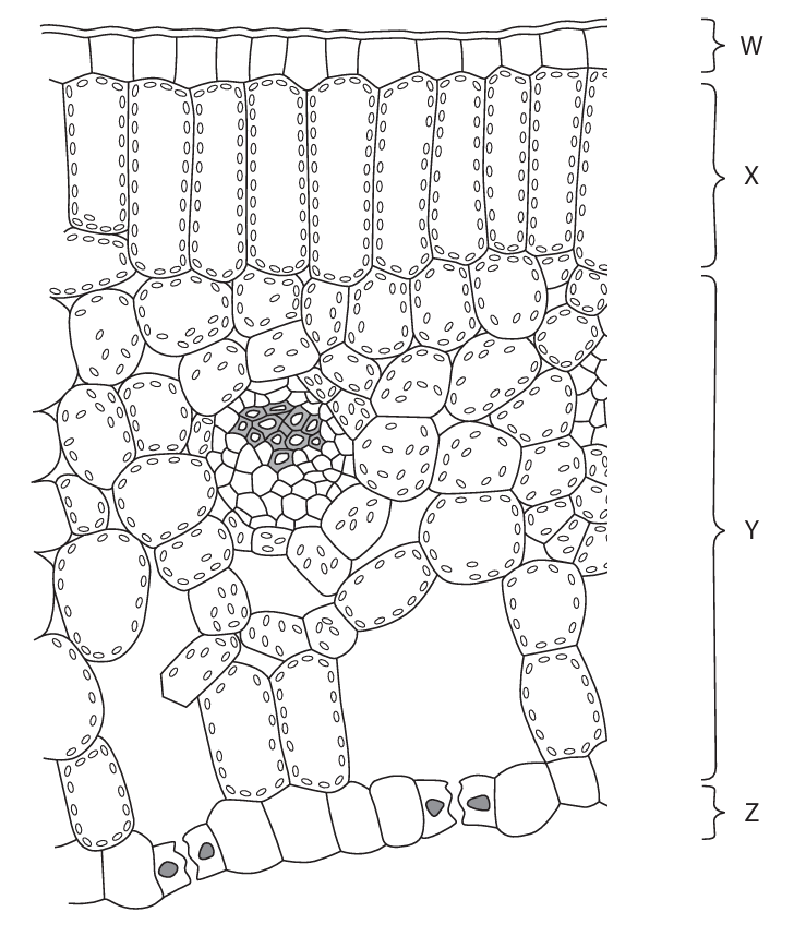
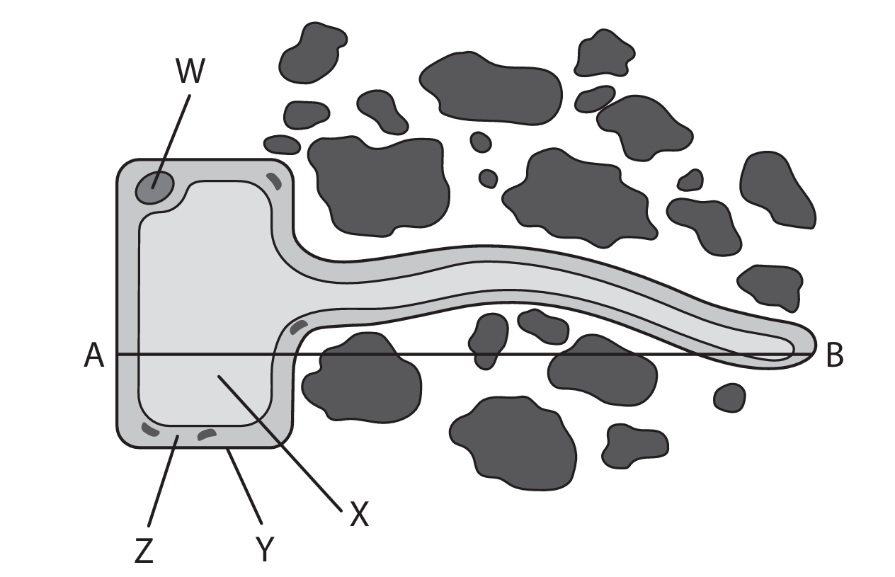
(a) C (Y)
A is not the answer as W does not contain xylem
B is not the answer as X does not contain xylem
D is not the answer as Z does not contain xylem
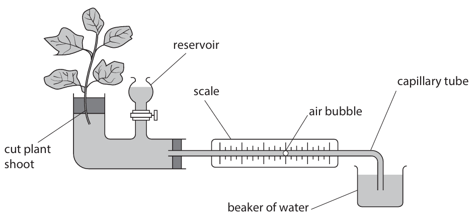
(b)(i) • 8.16(4) (2)
One mark for:
÷ by 5
OR
× 52
OR
× 0.25
OR
0.8(164)
OR
40.82 (allow between 40.82 and 40.85)
Working:
Convert length to mm: \( l = 5.2 \text{ cm} = 52 \text{ mm} \)
Volume = \( \pi r^2 l = 3.14 \times (0.50)^2 \times 52 = 3.14 \times 0.25 \times 52 = 40.82 \text{ mm}^3 \)
Rate = \( 40.82 \div 5 = 8.164 \text{ mm}^3/\text{min} \)
(b)(ii) A description that makes reference to four of the following points:
1. set up potometer underwater / cut stem underwater / dry leaves / eq (1)
2. use a fan at different distances / with and without fans / different fan speeds / eq (1)
3. leave for set time / stated time (1)
4. measure distance bubble moves / distance water moves (on scale) / eq (1)
5. keep other factors constant (1)
6. repeat / reset bubble with reservoir / eq (1)
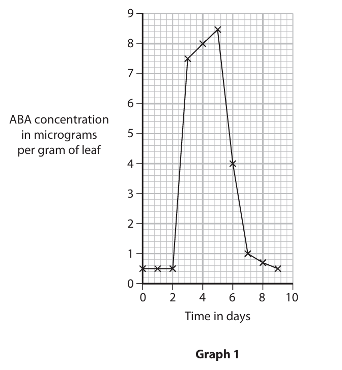
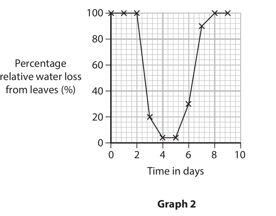
(c) An answer that makes reference to four of the following points:
1. at start / for first two days, ABA is low / is 0.5 OR at start / for first two days water loss is high / is 100 (1)
2. after two days / from three days ABA increases OR after two days / from three days percentage water loss decreases / eq (1)
3. after five days / after watering / ABA decreases OR after five days / after watering water loss increases / eq (1)
4. as ABA increases water loss decreases / inverse relationship / negative correlation / eq (1)
5. ABA closes stomata / stomata open when ABA low / eq (1)
6. stomata close from two days / stomata close from three days / eq (1)
7. stomata closing reduces transpiration / water loss / evaporation / stomata closing prevents wilting / stomata closing prevents loss of turgidity / ABA reduces transpiration / eq (1)
8. light intensity may change / humidity may change / wind may change / eq (1)
▶️ Answer/Explanation
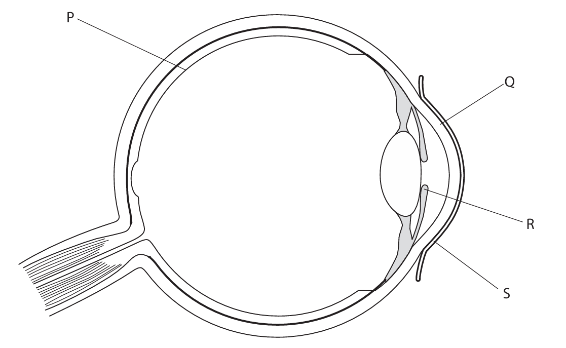
(a) A (P)
B is not the answer as Q is the cornea
C is not the answer as R is the iris
D is not the answer as S is the conjunctiva
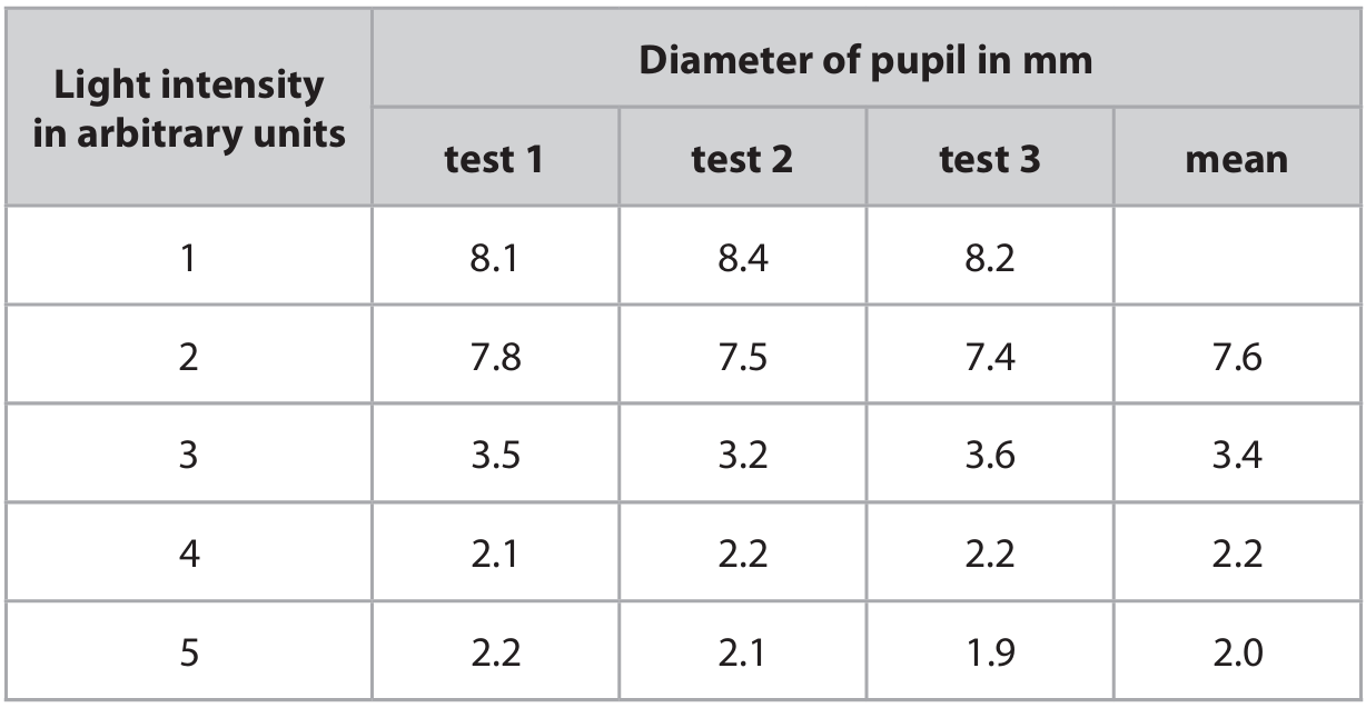
(b)(i) 8.2 (2)
Only one mark for: 24.7 OR division by 3 OR 8.23…
(b)(ii) A description that makes reference to two of the following points:
• (diameter) decreases / pupil smaller / eq (1)
• small decrease between 4(au) and 5(au) / starts to level off after 3(au) / starts to level off from 4(au) / large decrease between 2 and 3 (au) / calculated fall / eq (1)
(b)(iii) An explanation that makes reference to three of the following points:
• less light enters (eye) / passes through pupil / eq (1)
• to prevent damage to retina / eq (1)
• (as pupil narrows) circular muscles (of iris) contract (1)
• (as pupil narrows) radial muscles (of iris) relax (1)
(b)(iv) An explanation that makes reference to one of the following points:
• distance from camera (1)
• same student / person / same eye / left or right eye (1)
• food / drinks / caffeine consumed / eq (1)
• recovery time / time spent with mask on (1)
• noise in room (1)
• other light sources / distance from light (1)
• (type of) mask (1)
• colour of light / wavelength of light (1)
(b)(v) An explanation that makes reference to two of the following points:
1. using a camera (to record) / takes a photograph / uses an image / eq (1)
2. waiting 20 s (each time) / waiting same time / eq (1)
3. pupil is not changing size / is stationary / eye has adjusted / eq (1)
▶️ Answer/Explanation
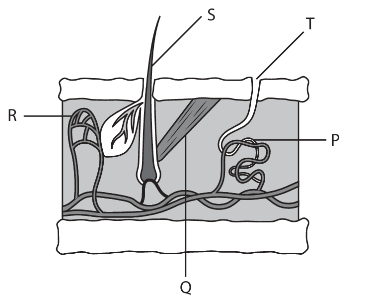
(a)(i) D (T)
A is not correct as Q is not the pore of a sweat gland
B is not correct as R is not the pore of a sweat gland
C is not correct as S is not the pore of a sweat gland
(a)(ii) B (R)
A is not correct as P is not the structure that carries blood
C is not correct as S is not the structure that carries blood
D is not correct as T is not the structure that carries blood
(a)(iii) A description that makes reference to four of the following points:
1. vasoconstriction (1)
2. arterioles supplying capillaries near skin surface constrict / blood vessels supplying skin narrow (1)
3. less blood flows to skin surface (1)
4. hair erector muscle contracts / hair stands up (1)
5. traps air / insulates (1)
6. less heat loss by radiation / convection / evaporation (of sweat) (1)
7. less sweating (1)
(b) An answer that makes reference to five of the following points:
1. (mean) skin temperature is higher in young (1)
2. (mean) skin temperature is higher with heat strain (1)
3. mean sweating rate higher in young (1)
4. mean sweating rate higher in moderate/higher heat strain (1)
5. smaller difference in sweating rate between low and moderate strain in young (1)
6. no information on sweat rate with no heat strain / at rest (1)
7. more sweat glands in young / each gland produces more sweat in young (1)
8. young can reduce (core) body temperature faster / young less likely to overheat (1)
9. numbers very small / not repeated / unreliable / no information on age (1)
10. no information on BMI / fat layers / health / diet (1)
▶️ Answer/Explanation
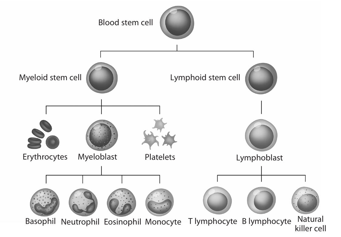
(a) Stem cells are unspecialized/undifferentiated and can divide by mitosis to form other cell types/specialized cells.
Detailed Explanation: Unlike most body cells which are specialized for specific functions (like nerve cells for transmitting signals or muscle cells for contraction), stem cells have not yet undergone differentiation. This means they haven’t developed the specific structures and functions of mature cells. Their key abilities are to continuously divide through mitosis to produce more stem cells (self-renewal) and to differentiate into various specialized cell types when given the right signals. This makes them fundamentally different from the majority of cells in our body that are locked into their specific roles.
(b) Platelets help blood to clot, prevent blood loss, and prevent entry of pathogens.
Detailed Explanation: Platelets are tiny cell fragments that circulate in the blood. When a blood vessel is damaged, they rush to the site and become activated. They change shape, become sticky, and clump together to form a temporary plug that helps stop bleeding. More importantly, they release chemicals that trigger a complex process called the clotting cascade. This process converts a soluble plasma protein called fibrinogen into insoluble strands of fibrin. These fibrin strands form a mesh that traps red blood cells and more platelets, creating a stable blood clot that seals the wound. This not only prevents further blood loss but also creates a barrier that prevents microorganisms like bacteria and viruses from entering the body through the break in the skin.
(c) Vaccination introduces dead/weakened pathogens or their antigens into the body. Lymphocytes respond by producing antibodies and memory cells. If the same pathogen infects the body later, memory cells trigger a faster, stronger secondary immune response with more antibodies produced sooner.
Detailed Explanation: Vaccines work by safely simulating an infection without causing the disease. They contain either killed or greatly weakened (attenuated) pathogens, parts of pathogens (like proteins or sugars which act as antigens), or inactivated toxins. When administered, these antigens are recognized as foreign by the immune system. Specifically, white blood cells called lymphocytes are activated. B lymphocytes produce antibodies that can bind to and neutralize the specific antigens. Additionally, special memory B cells and memory T cells are created during this primary response. These memory cells remain in the body for a long time, sometimes for life. If the person is later exposed to the actual, live pathogen, these memory cells recognize the antigens immediately. They mount a rapid and massive secondary immune response, producing a huge amount of the correct antibodies much faster than the first time. This swift response usually destroys the pathogen before it can multiply to sufficient numbers to cause illness, thus providing protection.
(d) Blood stem cells can differentiate into various blood cells (red blood cells, white blood cells, platelets). They can make red blood cells to transport oxygen and white blood cells to destroy pathogens. They can also make platelets to help blood clotting.
Detailed Explanation: Blood stem cells, also known as hematopoietic stem cells, are multipotent. This means they have the potential to differentiate into all the different types of specialized cells found in blood. This includes red blood cells (erythrocytes) which carry oxygen, the various types of white blood cells (leukocytes like lymphocytes, phagocytes) which fight infection and provide immunity, and platelets (thrombocytes) which are crucial for clotting. In disorders like sickle cell anaemia, the patient produces faulty red blood cells. A transplant of healthy blood stem cells can provide a new population of cells that can differentiate into healthy, oxygen-carrying red blood cells. In leukaemia, a cancer of white blood cells, chemotherapy is often used to destroy the cancerous cells, but this also destroys healthy stem cells. A stem cell transplant then repopulates the bone marrow with healthy stem cells that can differentiate into functional white blood cells, effectively restarting the immune system. Their ability to become any blood cell type makes them a versatile treatment for a wide range of blood disorders.
▶️ Answer/Explanation
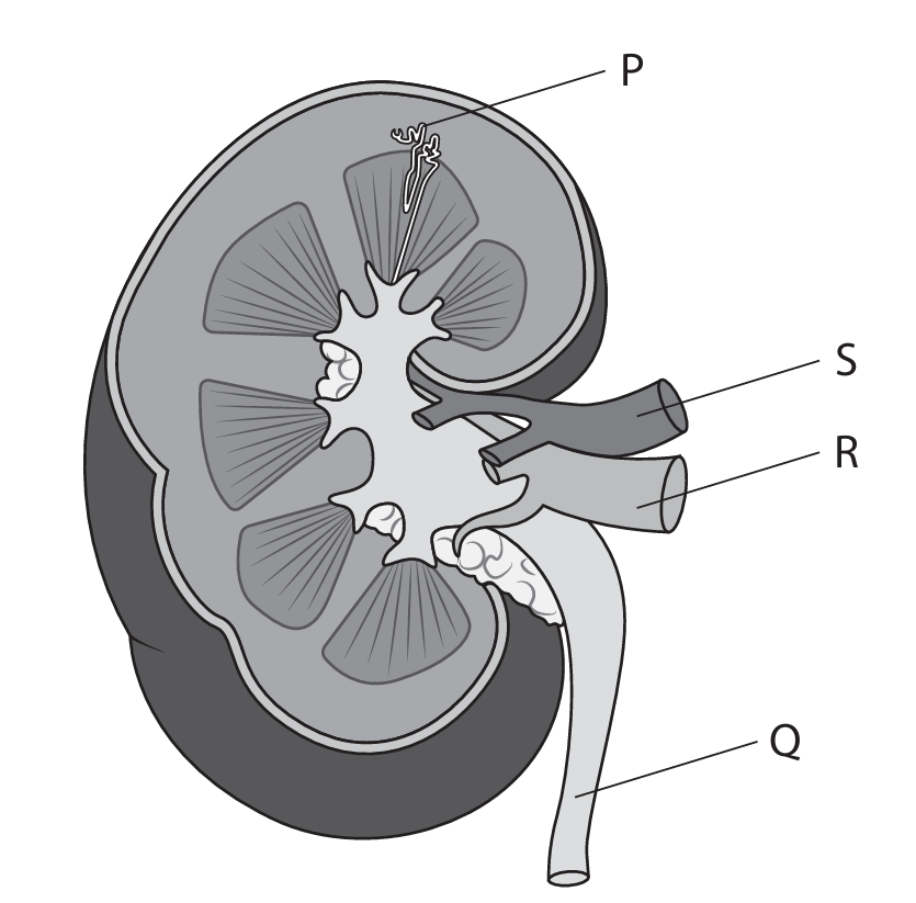
(a)(i) B (nephron)
Explanation: Structure P represents the functional unit of the kidney, which is the nephron.
(a)(ii) B (blood)
Explanation: Tube S carries blood to or from the kidney, specifically it is likely the renal artery or vein.
(a)(iii) C (ureter)
Explanation: Tube Q is the ureter, which transports urine from the kidney to the bladder.
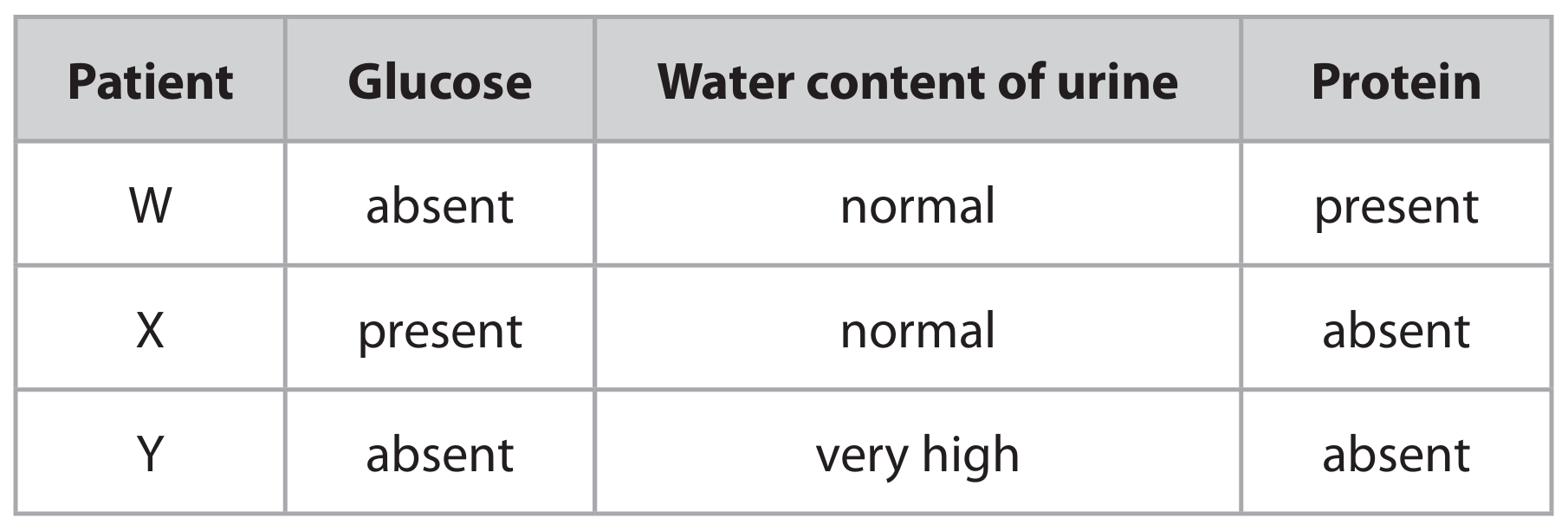
(b)(i) An explanation that makes reference to:
• Patient W: Presence of protein indicates failure of ultrafiltration in the glomerulus/Bowman’s capsule.
• Patient X: Presence of glucose indicates failure of selective reabsorption in the proximal convoluted tubule.
• Patient Y: High water content indicates reduced water reabsorption in the collecting duct, possibly due to low ADH.
(b)(ii) A description that includes:
• Use Benedict’s reagent
• Heat in a water bath
• Observe colour change (green → red indicates glucose)
• Alternatively, use a glucose test strip and compare colour to a chart.
▶️ Answer/Explanation
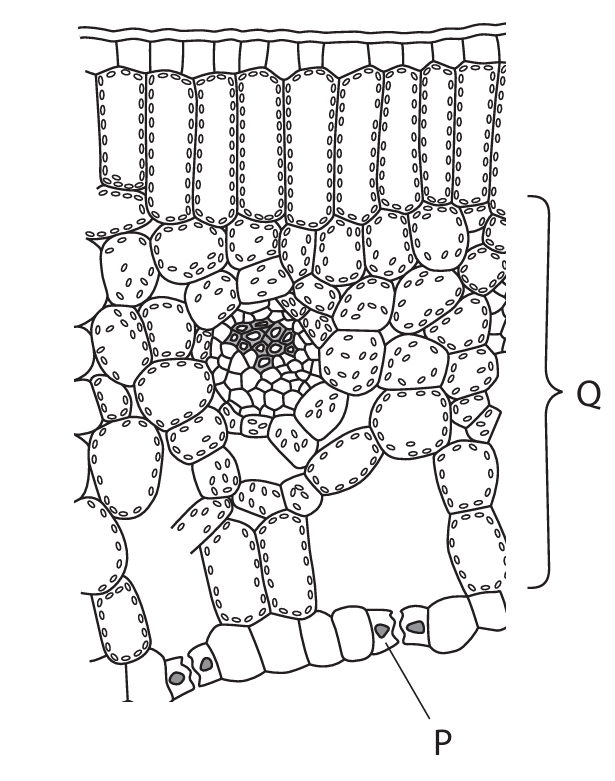
(a)(i) Answer: B (guard)
Explanation: The cell labelled P is a guard cell. Guard cells are specialized cells that surround the stomata (pores) in the leaf epidermis. They control the opening and closing of the stomata, which regulates gas exchange (carbon dioxide in, oxygen out) and water loss through transpiration.
(a)(ii) Answer: Part Q is the spongy mesophyll layer.
Explanation: The spongy mesophyll layer is highly adapted for photosynthesis in several ways. Firstly, it contains numerous air spaces between the cells, which creates a large surface area for the efficient diffusion of gases. Carbon dioxide, which is needed for photosynthesis, can diffuse easily from the stomata through these air spaces to reach the palisade mesophyll cells where most photosynthesis occurs. Similarly, oxygen produced as a byproduct of photosynthesis can diffuse out. Secondly, the cells in the spongy mesophyll contain chloroplasts, although fewer than in the palisade layer, and thus can also carry out photosynthesis. The loose arrangement of cells maximizes the exposure to these gases and facilitates their movement throughout the leaf.
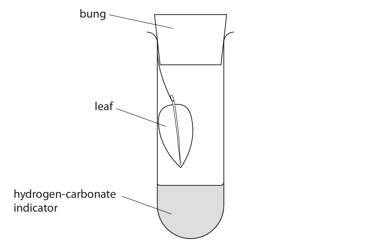
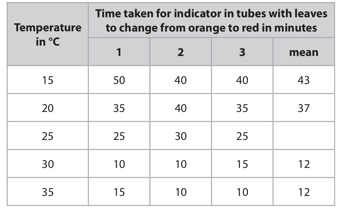
(b)(i) Answer: Temperature (of the water bath)
Explanation: The independent variable is the factor that is deliberately changed or manipulated by the investigator. In this experiment, the student places the tubes into water baths at different temperatures (15°C, 20°C, 25°C, 30°C, 35°C, 40°C). Therefore, temperature is the independent variable.
(b)(ii) Answer: Example factor: Light intensity
Reason: To ensure that light intensity is not a limiting factor for photosynthesis, which would affect the rate of gas exchange and thus the time for the indicator to change color. By keeping it constant and bright, any changes in the rate are due to the temperature and not variations in light.
OR
Answer: Example factor: Volume/concentration of hydrogen-carbonate indicator
Reason: Different volumes or concentrations would absorb or release different amounts of carbon dioxide, which would directly affect the time it takes for the color to change, making comparisons between temperatures invalid.
(b)(iii) Answer: It acts as a control.
Explanation: The tube with no leaf serves as a control experiment. Its purpose is to show that any observed color change in the indicator in the other tubes is due to the presence and activity of the leaf (specifically, its effect on carbon dioxide levels through photosynthesis and respiration) and not due to some other factor, such as the temperature affecting the indicator solution itself.
(c)(i) Answer: 27 minutes
Explanation: To calculate the mean time at 25°C, add the three recorded times together and divide by 3: (25 + 30 + 25) / 3 = 80 / 3 = 26.666… minutes. Rounding this to two significant figures gives 27 minutes.
(c)(ii) Explanation: As the temperature increases from 15°C to 30°C, the mean time taken for the indicator to change color decreases. This indicates that the rate of the process causing the color change (removal of carbon dioxide by photosynthesis) is increasing. This is because temperature increases the kinetic energy of molecules, leading to more frequent collisions between enzymes and substrates involved in photosynthesis, thus speeding up the reaction. However, between 30°C and 35°C, the mean time stops decreasing and remains at 12 minutes. This suggests that the rate of photosynthesis is no longer increasing with temperature, likely because another factor (such as enzyme denaturation or the availability of another substrate like carbon dioxide or light) has become the limiting factor.
(d) Explanation: In the dark, photosynthesis cannot occur as it requires light. However, respiration continues in the leaf cells. Respiration consumes oxygen and produces carbon dioxide. The increase in carbon dioxide concentration in the test tube causes the hydrogen-carbonate indicator to change from orange (at atmospheric CO₂ levels) to yellow (which indicates a high concentration of CO₂). This shows that in the absence of light, the net gas exchange is dominated by the release of carbon dioxide from respiration.
▶️ Answer/Explanation
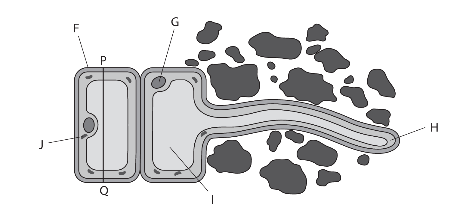
(a)(i) D (I)
A is not the answer as F is the cell wall
B is not the answer as G is the nucleus
C is not the answer as H is the cytoplasm
(a)(ii) B (G)
A is not the answer as F is the cell wall
C is not the answer as H is the cytoplasm
D is not the answer as J is a mitochondrion
(b) Magnification = 475 (or approximately 475×)
The measured length from P to Q on the diagram is approximately 38 mm. Convert this to micrometers: 38 mm × 1000 = 38,000 μm. Using the magnification formula (Magnification = Size of Image / Actual Size): Magnification = 38,000 μm / 80 μm = 475.
(c) An explanation that includes:
• Long root hair / extension increases surface area for absorption of water and mineral ions.
• Thin cell wall reduces distance for diffusion / osmosis.
• Many mitochondria provide ATP / energy for active transport of minerals.
• Large, permanent vacuole maintains water potential gradient for osmosis.
• Cell surface membrane contains transport proteins for selective uptake of ions.
▶️ Answer/Explanation
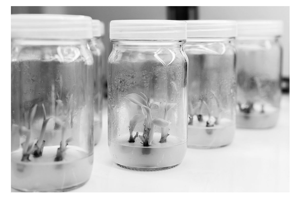
(a) In a test tube / culture dish / jar / glass / petri dish / container / in culture solution / in a lab / outside a living organism.
Explanation: The term “in vitro” literally means “in glass” in Latin, referring to biological processes that are conducted outside of a living organism in an artificial laboratory environment, such as in test tubes or petri dishes. This contrasts with “in vivo” experiments which are conducted within living organisms.
(b) Plant cells can differentiate into all/different types of tissues or specialized cells throughout the plant’s life and can form/regenerate a whole new plant.
Explanation: Plant cells exhibit totipotency, meaning that even mature, differentiated plant cells retain the ability to dedifferentiate and then redifferentiate into any cell type needed to regenerate an entire plant. This is why you can grow a new plant from a cutting. In contrast, human cells have much more limited differentiation capabilities. While stem cells can differentiate into various cell types, most human cells become permanently specialized during development and cannot revert back or form entirely new organisms.
(c)
1. Nitrate – for making amino acids/proteins/DNA/nucleic acids
2. Magnesium – for making chlorophyll/chloroplasts/photosynthesis
Explanation: Plant tissue culture media must contain essential minerals that support plant growth and development. Nitrate is crucial as it provides nitrogen, which is a fundamental component of amino acids, proteins, and nucleic acids (DNA and RNA). Without adequate nitrogen, plants cannot synthesize these essential biomolecules. Magnesium is a central component of the chlorophyll molecule, which is vital for photosynthesis as it captures light energy. Without magnesium, plants cannot produce chlorophyll effectively, leading to chlorosis (yellowing of leaves) and impaired photosynthesis.
(d) Enzymes are affected by pH/ work best at optimum pH. If pH changes, the shape of the active site can change/be denatured so substrates can no longer bind.
Explanation: Maintaining a constant pH is critical because enzymes, which catalyze all biochemical reactions in plant cells, are highly sensitive to pH changes. Each enzyme has an optimal pH range where it functions most efficiently. If the pH deviates from this range, the enzyme’s three-dimensional structure can be altered, changing the shape of its active site. This prevents substrates from binding properly, effectively denaturing the enzyme and halting the metabolic reactions it catalyzes. This would severely disrupt plant growth and development in the culture media.
(e) Place a shoot in light from one side/unidirectional light and another shoot in darkness/light all around. Leave both for a stated time/use shoots of same type/same temperature/other control variable. Observe/measure bending or growing towards light.
Explanation: To demonstrate phototropism (growth response to light), you would set up two identical young plant shoots. One would be placed in a location with light coming from only one direction (e.g., near a window), while the control would be placed in either complete darkness or with light evenly distributed from all sides. Both plants should be kept under the same temperature and watering conditions to ensure any differences are due to light direction only. After a few days, you would observe that the shoot exposed to unilateral light has bent toward the light source. This bending occurs because auxin hormone accumulates on the shaded side of the stem, promoting more cell elongation on that side and causing the stem to curve toward the light.
(f) To maintain biodiversity/reduce damage to ecosystems and to prevent extinction/keep species for future generations/for medicinal properties.
Explanation: Conserving endangered plant species is crucial for several reasons. Firstly, it maintains biodiversity, which ensures ecosystem stability and resilience. Each plant species plays a unique role in its ecosystem, and losing one can disrupt food webs and ecological balance. Secondly, it prevents extinction, preserving genetic diversity that might be valuable for future breeding programs, especially as climate changes. Many plants contain compounds with medicinal properties; for example, aspirin originated from willow bark. By conserving endangered species, we preserve potential future medicines and genetic resources that could be vital for human well-being.
(g) Agitation mixes contents/mixes oxygen/with plant cells. Light is for photosynthesis. Suitable temperature is for enzyme action.
Explanation: Suspension cultures require specific conditions to mimic optimal natural environments. Agitation (shaking or stirring) ensures that cells and nutrients are evenly distributed throughout the liquid media, preventing sedimentation. It also promotes gas exchange, ensuring oxygen (needed for respiration) is available and carbon dioxide (a product of respiration) is removed. Light is essential for photosynthetic plant cells to produce their own energy through photosynthesis. Maintaining a suitable temperature is critical because temperature affects enzyme activity; most plant enzymes function optimally around 25-30°C. Temperatures that are too high can denature enzymes, while temperatures that are too low can slow down metabolic processes to inadequate levels.
▶️ Answer/Explanation
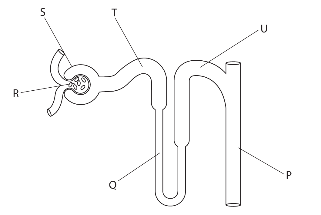
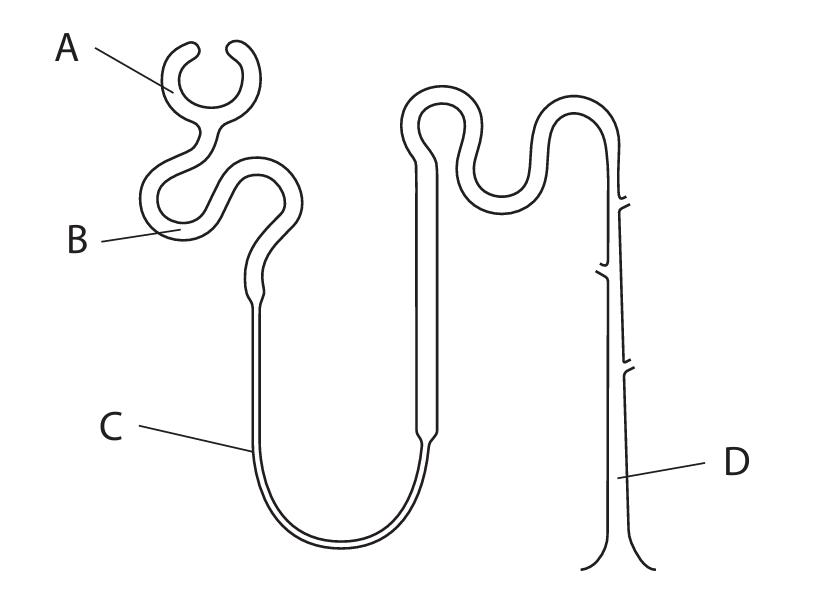
(a)(i) C (S)
Explanation: The Bowman’s capsule (S) is the cup-like sac at the beginning of the nephron that surrounds the glomerulus and receives the filtrate.
(a)(ii) B (Q)
Explanation: The loop of Henle (Q) is a U-shaped tubule that descends into and ascends from the medulla of the kidney. It is crucial for creating a concentration gradient for water reabsorption.
(a)(iii) A (P)
Explanation: ADH (Antidiuretic Hormone) affects the collecting duct (P). ADH increases the permeability of the collecting duct walls to water, allowing more water to be reabsorbed back into the blood, producing more concentrated urine.
(b)(i) An explanation that makes reference to three of the following points:
• Glucose passes from the blood in the glomerulus (R) into the Bowman’s capsule / renal capsule (S) during ultrafiltration. (1)
• (All) glucose is (then) reabsorbed / absorbed back into the blood / eq. (1)
• This reabsorption occurs in the proximal convoluted tubule / PCT (T). (1)
• It is reabsorbed by active transport (which requires energy). (1)
Explanation: During ultrafiltration, small molecules like glucose enter the nephron. The body cannot afford to lose this valuable energy source, so 100% of filtered glucose is normally reclaimed from the filtrate in the proximal convoluted tubule via active transport against its concentration gradient.
(b)(ii) A description that makes reference to two of the following points:
• Add Benedict’s solution to the urine sample (and heat). (1)
• A positive result is indicated by a colour change to green / yellow / orange / brick-red. (1)
Alternative: Use a test strip (e.g., Clinistix) which changes colour (e.g., to brown) in the presence of glucose. (1 each)
Explanation: Benedict’s test is a standard biochemical test for reducing sugars like glucose. Heating with Benedict’s reagent causes a reduction reaction, producing a coloured precipitate of copper(I) oxide.
(c) A description that makes reference to two of the following points:
• Less urine is produced / lower volume. (1)
• The urine becomes more concentrated / contains less water / appears darker in colour. (1)
• (It may contain) a higher concentration of urea / other solutes. (1)
Explanation: Dehydration lowers the water potential of the blood. This is detected by osmoreceptors, leading to increased secretion of ADH. ADH causes more water to be reabsorbed from the collecting duct back into the blood, conserving water. This results in a smaller volume of more concentrated, darker yellow urine.
▶️ Answer/Explanation
(a) Lymphocytes
Explanation: Antibodies are proteins produced by specialized white blood cells called lymphocytes as part of the immune response. When foreign antigens are detected, lymphocytes are activated to produce specific antibodies that bind to and help eliminate the foreign substances.
(b)(i) Both alleles are expressed in the phenotype.
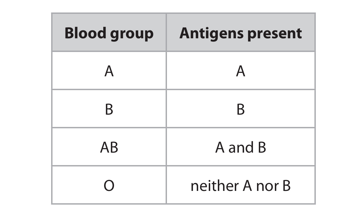
Explanation: Codominance occurs when both alleles in a heterozygous individual are fully expressed, resulting in a phenotype that shows characteristics of both alleles simultaneously. For example, in blood groups, the IA and IB alleles are codominant, meaning an individual with genotype IAIB will have blood group AB, expressing both A and B antigens.
(b)(ii) D (A, B, AB and O)
Explanation: When crossing parents with genotypes IAIO and IBIO, we can create a Punnett square to determine the possible offspring genotypes:
The possible gametes from IAIO parent: IA and IO
The possible gametes from IBIO parent: IB and IO
Possible offspring genotypes: IAIB (blood group AB), IAIO (blood group A), IBIO (blood group B), and IOIO (blood group O). Therefore, all four blood groups are possible.
(c) 4.74 × 10⁷
Explanation: The total global blood donations are 118.5 million, which is 118,500,000. High-income countries contribute 40% of this total. To calculate: 40% of 118,500,000 = 0.40 × 118,500,000 = 47,400,000. In standard form, this is written as 4.74 × 10⁷.
(d) Artificial spherical red blood cells have a smaller surface area to volume ratio compared to normal biconcave red blood cells, reducing oxygen diffusion efficiency.
Explanation: Normal human red blood cells have a unique biconcave disc shape that maximizes their surface area to volume ratio. This specialized shape allows for more efficient gas exchange as it provides a larger surface area for oxygen to diffuse across. Spherical artificial cells have a lower surface area to volume ratio, meaning less surface is available for oxygen binding and release. Additionally, the biconcave shape of natural red blood cells allows them to flow more easily through narrow capillaries, while spherical cells might not navigate the circulatory system as efficiently.
(e) Artificial blood lacks platelets and clotting factors.
Explanation: Natural blood contains platelets and various clotting factors that work together to form clots when bleeding occurs. Artificial blood, being designed primarily for oxygen transport, doesn’t include these components. Without platelets and clotting factors like fibrinogen, the coagulation cascade cannot be initiated, preventing clot formation during storage.
(f) Sodium chloride solution maintains osmotic balance, preventing cell bursting or shrinkage.
Explanation: If artificial red blood cells were suspended in pure water, water would enter the cells by osmosis due to the higher solute concentration inside the cells. This would cause the cells to swell and potentially burst (hemolysis). Sodium chloride solution is isotonic, meaning it has the same solute concentration as the interior of the cells. This prevents net movement of water across the cell membrane, maintaining cell integrity and function during storage.
(g)(i) Stem cells can undergo mitosis to produce more cells and differentiate into specialized cell types.
Explanation: Stem cells are undifferentiated cells with two key properties: self-renewal (ability to divide and produce more stem cells through mitosis) and differentiation (ability to develop into specialized cell types). By controlling the growth conditions and providing specific signals, scientists can direct stem cells to differentiate exclusively into red blood cells, allowing for large-scale production in laboratory settings.
(g)(ii) Blood group O lacks A and B antigens, making it universally compatible.
Explanation: Blood group O red blood cells don’t have A or B antigens on their surface. This means they won’t trigger an immune response when transfused into recipients with any blood type (A, B, AB, or O). People with other blood types don’t have pre-formed antibodies against O blood cells. This universal compatibility makes blood group O particularly valuable in emergency situations when there might not be time to determine the recipient’s blood type.
(h) Any two from: urea, carbon dioxide, hormones, mineral ions, vitamins, proteins (antibodies/clotting factors), digested food (glucose/amino acids)
Explanation: Natural blood plasma contains numerous substances that artificial blood lacks. Urea is a waste product transported to the kidneys for excretion. Carbon dioxide is carried from tissues to the lungs. Hormones act as chemical messengers throughout the body. Mineral ions and vitamins serve various metabolic functions. Proteins include antibodies for immunity and clotting factors for wound healing. Digested nutrients like glucose and amino acids are transported to cells for energy and growth.
▶️ Answer/Explanation
(a) Salt concentration / concentration of salt solution / sodium chloride concentration / percentage sodium chloride / water potential of solution
Explanation: The independent variable is the factor that is deliberately changed by the investigator. In this experiment, the teacher is testing different concentrations of salt solution (water, 1%, and 5% sodium chloride) to see their effect on the red blood cells.
(b) Volume of (diluted) blood / volume of solution added / time solution left for / concentration of (diluted) blood sample
Explanation: A controlled variable is one that is kept constant to ensure a fair test. The teacher controls several factors, such as using the same volume of diluted blood in each tube (1 cm³), adding the same volume of different solutions (10 cm³), and leaving all tubes for the same duration (5 minutes).
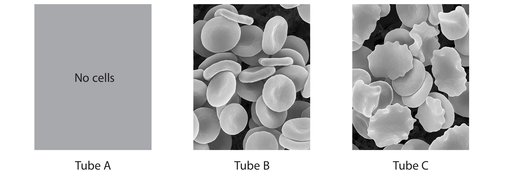
(c)(i) In tube A (water), the cells burst (lyse) and release haemoglobin, creating a clear red solution. In tubes B and C (salt solutions), the cells remain mostly intact, creating a cloudy suspension.
Explanation: The cloudiness indicates the presence of intact cells scattering light. In tube A, distilled water is hypotonic relative to the red blood cells. Water enters the cells by osmosis, causing them to swell and burst (haemolysis), releasing haemoglobin into the solution and making it clear. In tubes B and C, the salt solutions are closer to isotonic (B) or hypertonic (C), so the cells do not burst and remain in suspension, making the solution cloudy.
(c)(ii) In tube A (water), water enters the red blood cells by osmosis, causing them to swell and burst, so no intact cells are seen. In tube B (1% salt), the solution is isotonic, so water enters and leaves at equal rates, and normal biconcave cells are seen. In tube C (5% salt), the solution is hypertonic, so water leaves the cells by osmosis, causing them to shrink and develop cremated (shrunken) edges.
Explanation: The differences are due to osmosis, the movement of water across the cell membrane from a region of higher water potential to a region of lower water potential. In tube A, the external water potential is higher than inside the cell, so water rushes in, bursting the cell. In tube B, the water potential is balanced, so the cell shape is maintained. In tube C, the external water potential is lower (due to high salt), so water leaves the cell, causing it to shrink.
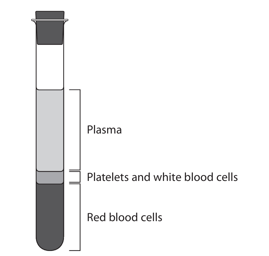
(d) Tube A would show no red cell layer (the red color would be distributed throughout the tube). Tubes B and C would show normal layers, but the red cell layer in C might be slightly smaller.
Explanation: Centrifugation separates blood components based on density. In a normal blood sample, red blood cells form the bottom layer. In tube A, the cells have burst, so there are no intact cells to form a pellet; the haemoglobin is dissolved in the solution. In tubes B and C, the cells are intact (though shrunken in C) and will form a red cell layer at the bottom. The layer in C might be smaller if the cells have lost water and become denser.
▶️ Answer/Explanation
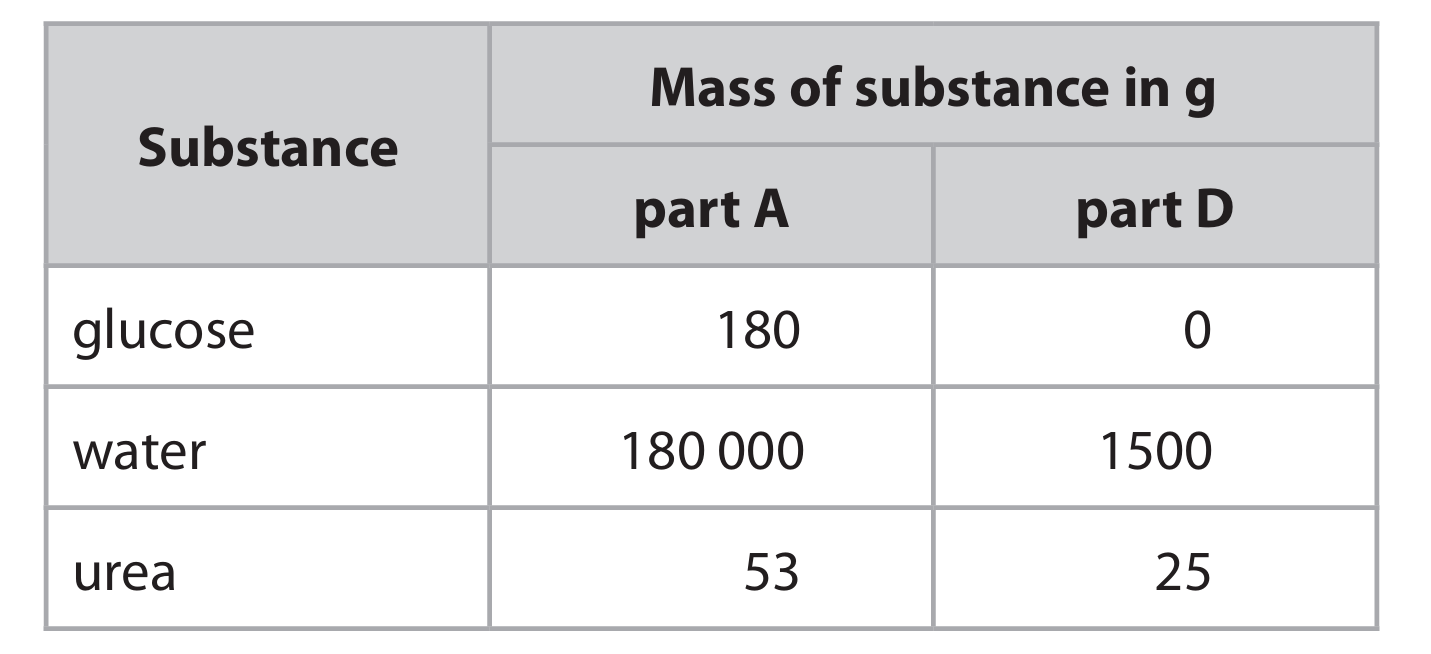
(a)
W: Nucleus
X: Vacuole (or cell sap)
Y: Cell Wall (specifically cellulose cell wall)
Z: Cytoplasm
Explanation: In a typical plant cell, the nucleus (W) contains the genetic material and controls cell activities. The vacuole (X) is a large, fluid-filled sac that stores water, nutrients, and waste, helping maintain turgor pressure. The cell wall (Y) is a rigid outer layer made of cellulose that provides structural support and protection. The cytoplasm (Z) is the gel-like substance inside the cell where most cellular activities occur.
(b)
magnification = 80 (accept values in the range 79-82)
Explanation: To calculate magnification, we use the formula:
\[ \text{Magnification} = \frac{\text{Size of Image}}{\text{Actual Size}} \]
First, measure the length between A and B in the drawing. Let’s assume this measures 80 mm (or 8.0 cm).
Convert this measurement to micrometers (μm) to match the units of the actual size. Since 1 mm = 1000 μm, 80 mm = 80,000 μm.
The actual size is given as 1000 μm.
Now, plug the values into the formula:
\[ \text{Magnification} = \frac{80,000 \ \mu m}{1,000 \ \mu m} = 80 \]
So, the drawing is magnified 80 times.
(c) (i)
Explanation: Root hair cells are specially adapted for absorbing water from the soil. Their long, hair-like projection significantly increases the surface area of the root, allowing it to absorb more water. Water enters the root hair cell from the soil via osmosis. This process occurs because the water potential inside the root hair cell is lower (meaning it has a higher concentration of solutes like minerals) than the water potential in the soil (which is generally higher, or more dilute). Water molecules naturally move from an area of high water potential (soil) to an area of low water potential (root hair cell) across the partially permeable cell membrane. This movement of water is often driven by a water potential gradient set up in the plant as water is lost through transpiration from the leaves. The absorbed water is essential for various plant functions, including photosynthesis and maintaining turgor pressure, which keeps the plant upright.
(c) (ii)
Explanation: When too much water is added to the soil, it fills the air spaces that normally contain oxygen. Plant roots, like all living cells, require oxygen for respiration to release energy. This energy is crucial for active transport, the process by which roots absorb essential mineral ions (like nitrates and magnesium) from the soil against a concentration gradient. If the soil becomes waterlogged and oxygen is depleted, root respiration is severely reduced. Consequently, active transport cannot occur effectively, leading to a decreased uptake of vital minerals. Without sufficient nitrates, the plant cannot synthesize amino acids and proteins properly, and without magnesium, it cannot produce chlorophyll, which is essential for photosynthesis. This overall lack of energy and essential nutrients causes the plant to fail to grow properly, leading to stunted growth, yellowing leaves, and potentially plant death.
▶️ Answer/Explanation
(a) (i) D (protoctists)
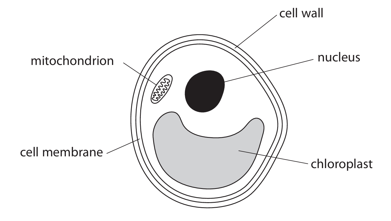
Explanation: Chlorella is a single-celled, photosynthetic organism with a nucleus and chloroplasts. It is not an animal (A) because it has chloroplasts and a cell wall. It is not a bacterium (B) because it has a true nucleus and membrane-bound organelles like chloroplasts and mitochondria. It is not a plant (C) because it is unicellular, whereas plants are multicellular. Therefore, it belongs to the kingdom Protoctista (D), which contains various unicellular and simple multicellular eukaryotes, including algae.
(a) (ii) B (cell membrane and mitochondrion)
Explanation: Animal cells have a cell membrane and mitochondria. They do not have chloroplasts (so A and C are incorrect) and they do not have a cell wall (so D is incorrect). Both animal cells and Chlorella require mitochondria for respiration to release energy.
(b) 6CO2 + 6H2O → C6H12O6 + 6O2
Explanation: The balanced equation for photosynthesis shows that six molecules of carbon dioxide (6CO2) and six molecules of water (6H2O), in the presence of light energy and chlorophyll, react to produce one molecule of glucose (C6H12O6) and six molecules of oxygen (6O2). The reactants must be placed on the left-hand side of the arrow.
(c) (i)
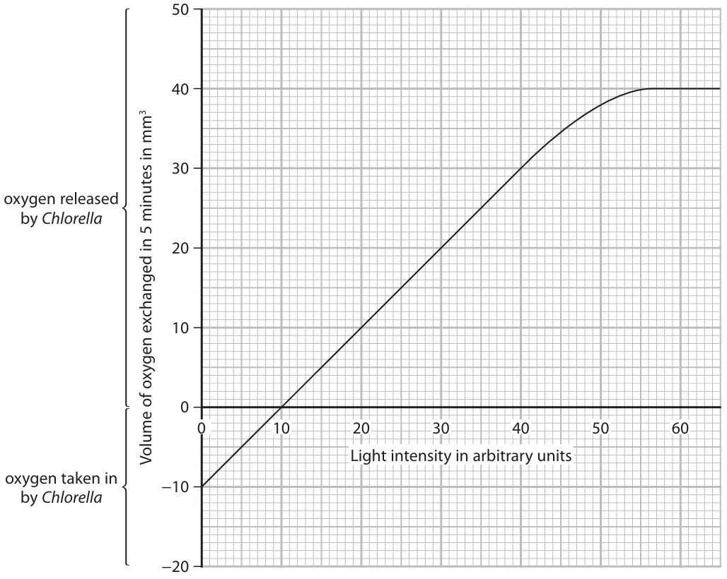
Explanation: At low light intensities (below 10 arbitrary units), the rate of photosynthesis is very low because there is insufficient light energy. However, respiration continues at all times to release energy for cell processes. Therefore, the oxygen produced by photosynthesis is less than the oxygen consumed by respiration. This results in a net uptake of oxygen from the surroundings, which is why the graph shows a negative value for oxygen exchange (indicating net intake).
(c) (ii)
Explanation: As light intensity increases from 10 arbitrary units, the rate of photosynthesis also increases because light is a key factor for the light-dependent reactions. At 10 arbitrary units, the compensation point is reached where the rate of photosynthesis equals the rate of respiration, so there is no net gas exchange. Above this point, the rate of photosynthesis becomes greater than the rate of respiration. This means more oxygen is produced by photosynthesis than is consumed by respiration, leading to a net release of oxygen, which is shown by the positive values on the graph. The curve eventually levels off because another factor, such as carbon dioxide concentration or temperature, becomes limiting and prevents the rate of photosynthesis from increasing further, even with more light.
(c) (iii) 48 mm3
Explanation: The graph shows the net oxygen released, which is the oxygen from photosynthesis minus the oxygen used in respiration. At 50 arbitrary units, the net oxygen released is approximately 38 mm³. We are told that the oxygen taken in (used in respiration) is 10 mm³ (this value is consistent across light intensities as respiration rate is relatively constant). To find the gross oxygen produced by photosynthesis, we add the oxygen used in respiration to the net oxygen released: 38 mm³ + 10 mm³ = 48 mm³.
(d)
Explanation: To investigate the effect of light intensity on carbon dioxide exchange, you could set up the following experiment. Place equal volumes or masses of Chlorella in several test tubes containing the same volume of hydrogen-carbonate indicator solution. Seal the tubes. Hydrogen-carbonate indicator changes color with carbon dioxide concentration: it turns yellow when carbon dioxide levels are high, red at atmospheric levels, and purple when carbon dioxide levels are low. You would then place the tubes at different distances from a light source to create different light intensities (e.g., 10 cm, 20 cm, 30 cm away). A control tube with no Chlorella should be set up to show that any color change is due to the organism. You would also need to control other variables, such as temperature and the initial concentration of the algae and indicator. After leaving the tubes for a set period, you would observe and record the final color of the indicator in each tube. In high light, photosynthesis would be high, so carbon dioxide would be absorbed, and the indicator would turn purple. In low light or darkness, respiration would dominate, releasing carbon dioxide, and the indicator would turn yellow.
▶️ Answer/Explanation
(a) (i) C
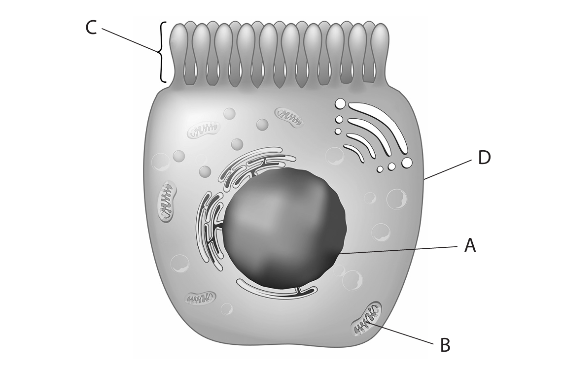
Explanation: Microvilli are tiny, finger-like projections on the surface of some cells, especially those involved in absorption, like the cells lining the small intestine. They greatly increase the surface area of the cell membrane, which allows for more efficient absorption of nutrients. In the diagram, structure C is correctly identified as the microvillus.
(a) (ii) B
Explanation: ATP (Adenosine Triphosphate) is the main energy currency of the cell, produced during cellular respiration. The organelles responsible for this process are the mitochondria. In the diagram, structure B represents a mitochondrion, which is often described as the “powerhouse” of the cell because it generates most of the cell’s supply of ATP.
(b) The small intestine has several structural adaptations for efficient absorption:
- It is very long, providing a large surface area over which absorption can occur.
- The inner lining is folded, and these folds are covered in tiny finger-like projections called villi. The cells on the surface of the villi themselves have microvilli, forming a “brush border”. Both villi and microvilli massively increase the surface area for absorption.
- Each villus contains a network of blood capillaries that absorb and transport products of digestion like glucose and amino acids. Good blood flow in these capillaries helps maintain a steep concentration gradient for rapid diffusion.
- Each villus also contains a lacteal, which is a lymphatic vessel that absorbs fatty acids and glycerol.
- The walls of the villi are only one cell thick, creating a very short diffusion distance for nutrients to pass from the gut into the blood.
(c) The human placenta is a vital organ that forms during pregnancy and has several key roles:
- It allows for the exchange of materials between the mother’s blood and the foetus’s blood without the two blood supplies mixing. Oxygen and digested food nutrients (like glucose, amino acids, and minerals) diffuse from the mother’s blood into the foetal blood.
- It removes waste products from the foetus, such as carbon dioxide and urea, which then pass into the mother’s blood for her to excrete.
- The placenta acts as a barrier against some harmful substances, like certain bacteria, although some viruses and drugs can cross it.
- It produces important hormones, such as progesterone, which helps to maintain the pregnancy.
- Towards the end of pregnancy, the placenta passes antibodies from the mother to the foetus, providing the baby with passive immunity for the first few months after birth.
▶️ Answer/Explanation
(a) in glass / in test tube / in (Petri) dish
Explanation: The term “in vitro” is a Latin phrase that literally means “in glass.” In biology, it refers to experiments or processes that are conducted outside of a living organism, in an artificial environment controlled by a scientist, such as a test tube, Petri dish, or flask. This is the opposite of “in vivo,” which means experiments conducted within a living organism.
(b)(i)
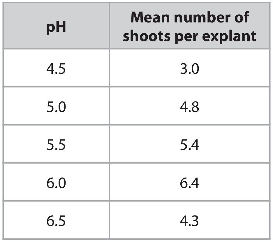
Answer: The mean number of shoots increases as the pH increases from 4.5 to 6.0, and then it decreases at pH 6.5. This pattern occurs due to the effect of pH on enzyme activity, with an optimum pH around 6.0 for the enzymes involved in shoot growth.
Explanation: When we look at the data, we can see a clear trend. At a very acidic pH of 4.5, the mean number of shoots is low (3.0). As the pH becomes less acidic and moves towards neutral, the number of shoots increases, reaching a maximum of 6.4 at pH 6.0. However, when the pH is increased further to 6.5, the number of shoots drops to 4.3. This pattern is classic for enzyme-controlled processes. The enzymes responsible for cell division and growth in the plant tissues have an optimal pH at which they work most efficiently, which appears to be pH 6.0 in this case. At pH values above and below this optimum, the enzymes become less active (they may denature at extremes), leading to reduced shoot formation.
(b)(ii)
Answer: The student should use sterile techniques to obtain and prepare the explants. Small pieces of plant tissue (explants) are cut from a parent plant using a sterilized scalpel. These explants are then surface-sterilized by washing them in a disinfectant like bleach or ethanol to kill any microorganisms. The sterile explants are placed on a nutrient agar gel in Petri dishes or test tubes. The agar contains essential nutrients, minerals, and plant hormones to support growth. The student would prepare several identical agar plates, each buffered to a specific pH (4.5, 5.0, 5.5, 6.0, 6.5). Multiple explants are placed in each pH condition to ensure the results are reliable. All plates are kept in a controlled environment (e.g., constant temperature and light) for a set period. After this time, the number of new shoots on each explant is counted, and a mean is calculated for each pH level.
Explanation: To get valid and reliable results, the procedure must be very careful and controlled. The key steps involve:
- Obtaining Explants: Using a sterile tool like a scalpel or forceps to take small tissue samples from the same part of the same plant species to keep the starting material consistent.
- Sterilization: Washing the explants in a disinfectant is crucial. Any bacteria or fungi present would contaminate the nutrient agar and compete with the plant tissue, ruining the experiment.
- Growing Medium: The explants are placed on a solid agar medium. This agar is not just a solidifier; it’s a “growth cocktail” containing sugars for energy, mineral ions, vitamins, and plant growth regulators (hormones) like auxins and cytokinins that stimulate shoot and root development.
- Controlling Variables: The pH is the independent variable, so it is deliberately changed for each set of plates. All other factors that could affect growth, such as temperature, light intensity, and the composition of the agar (except for pH), must be kept the same for all explants. Using multiple explants (repeats) at each pH allows the student to calculate a mean, making the results more trustworthy and accounting for natural variation between individual explants.
- Data Collection: After a predetermined growth period, the student counts the number of shoots that have developed from each explant and calculates the average for each pH group.
(c)
Answer:
- It produces genetically identical plants (clones), ensuring desirable characteristics from the parent plant are preserved.
- It allows for the rapid production of a large number of plants in a relatively short time, independent of seasonal constraints.
Explanation:
Benefit 1: Genetic Uniformity When you grow plants from seeds, the offspring show genetic variation due to sexual reproduction (cross-pollination). This means the new plants might not have the exact same desirable traits as the parent plant, such as specific flower color, fruit taste, or disease resistance. Micropropagation, however, is an asexual process. All the new plants are clones, meaning they are genetically identical to the original parent plant. This is extremely valuable for farmers and horticulturists who want to propagate a specific cultivar with known, superior qualities reliably.
Benefit 2: Speed and Season Independence Micropropagation can produce a vast number of plants from a single piece of tissue much faster than waiting for seeds to germinate and grow. A single explant can be induced to produce multiple shoots, and each of those shoots can then be divided and cultured again, leading to an exponential increase in plant numbers. Furthermore, this process is carried out in a lab under controlled conditions, meaning it is not dependent on seasons or weather. Plants can be produced all year round, which is not always possible with seeds that may have specific germination requirements.
Other potential benefits include: producing plants that are difficult to grow from seed, and conserving rare or endangered plant species.
▶️ Answer/Explanation
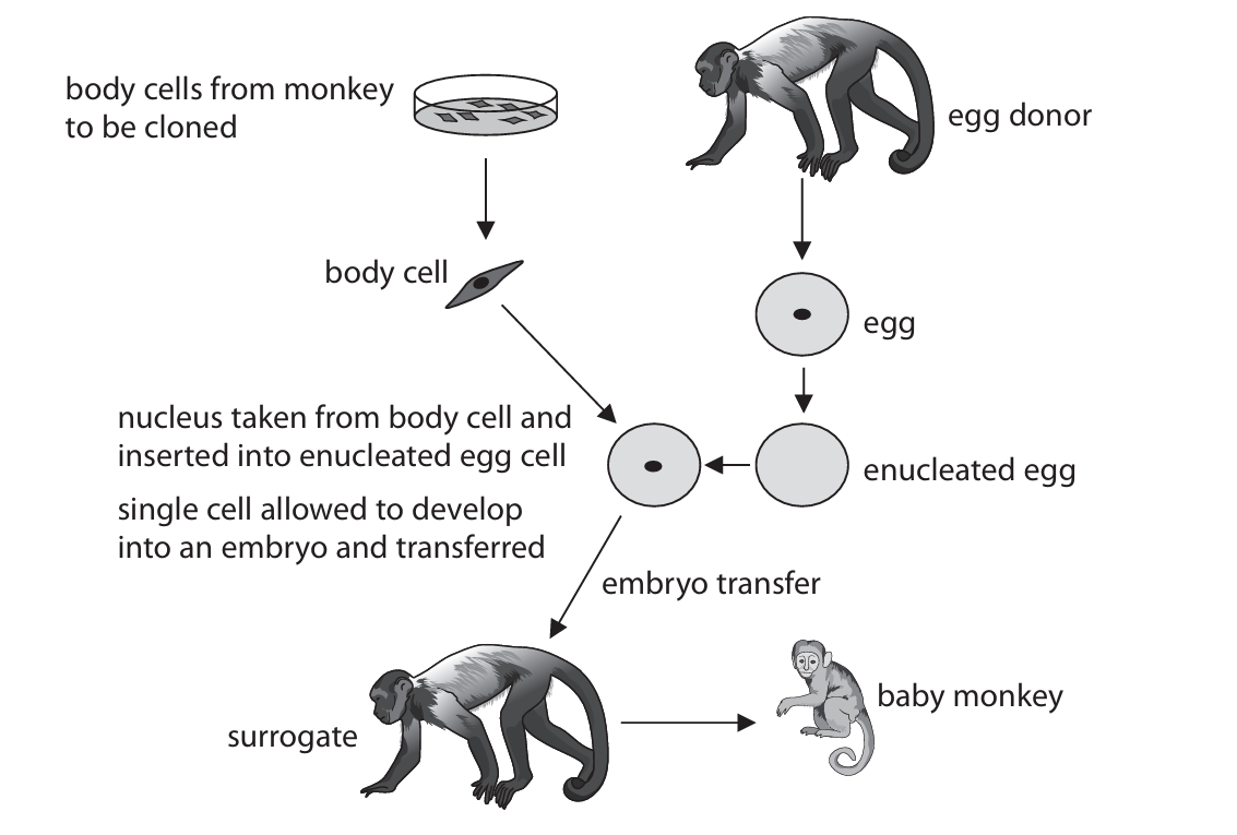
(a) (i) The nucleus is removed.
Explanation: In the context of cloning, “enucleated” specifically refers to an egg cell that has had its nucleus carefully removed. This is a crucial first step in the somatic cell nuclear transfer (SCNT) process, as it creates a vacant cellular environment ready to receive the nucleus from the donor body cell.
(a) (ii) The single cell divides many times by mitosis, producing a ball of cells, and then the cells differentiate.
Explanation: After the donor nucleus is inserted into the enucleated egg cell and stimulated, the newly formed single cell begins a process of rapid, successive divisions. This type of cell division is called mitosis, which produces genetically identical daughter cells. These divisions lead to the formation of a solid ball of cells. Following this, the process of differentiation begins, where the initially identical cells start to specialize, taking on different structures and functions to form the various tissues that will make up the embryo.
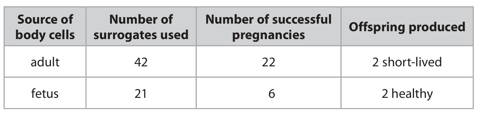
(b) Fetal body cells are more successful.
Explanation: When evaluating the data, we need to look at both the efficiency of the process and the health of the resulting offspring.
Looking at the success rates: For adult cells, 22 out of 42 surrogates resulted in a pregnancy, which is a success rate of approximately 52%. For fetal cells, 6 out of 21 surrogates resulted in a pregnancy, which is a lower success rate of about 29%. Based purely on the number of successful pregnancies, adult cells seem more efficient.
However, the critical factor is the outcome for the offspring. The two offspring produced from adult body cells were “short-lived,” indicating significant health problems. In contrast, the two offspring produced from fetal body cells were “healthy.”
Therefore, while using fetal cells led to fewer successful pregnancies, it resulted in viable, healthy clones. The ultimate goal of cloning is to produce a healthy organism, which makes fetal cells the more successful source in this investigation, despite the lower pregnancy rate.
▶️ Answer/Explanation
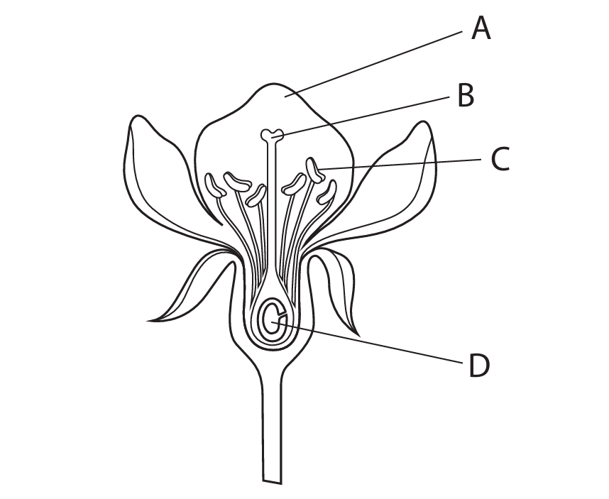
(a)(i) C
Explanation: The part labelled C is the anther, which is the male reproductive organ of the flower responsible for producing and releasing pollen grains.
(a)(ii) B
Explanation: The stigma is the sticky, receptive tip of the female reproductive structure (carpel) that captures pollen grains. In the diagram, this is represented by label B.
(b)
Explanation: The pollen tube plays a crucial role in plant fertilization. After a pollen grain lands on the stigma, it germinates and grows a pollen tube. This tube extends down through the style, which is the long stalk connecting the stigma to the ovary. The pollen tube acts as a conduit, transporting the male gametes (sperm cells) from the pollen grain to the ovary. Once the pollen tube reaches the ovary, it enters an ovule through a small opening called the micropyle. This allows the male gametes to fertilize the female gamete (egg cell) inside the ovule, leading to the formation of a seed.
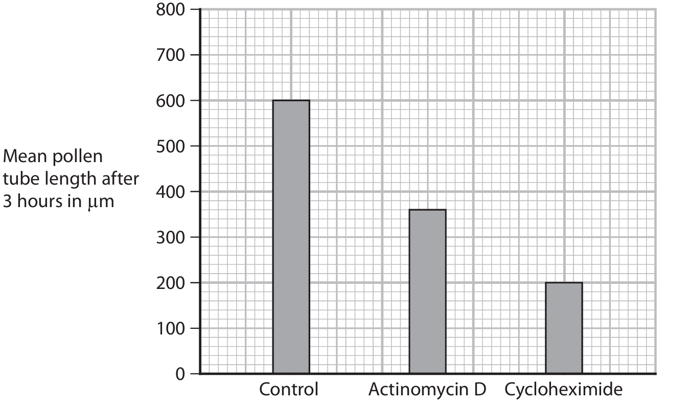
(c)(i) 80 µm per hour
Explanation: To calculate the growth rates, we first determine the rate for each solution:
Control: 600 µm ÷ 3 hours = 200 µm/hour
Actinomycin D: 360 µm ÷ 3 hours = 120 µm/hour
Difference: 200 µm/hour – 120 µm/hour = 80 µm/hour
Alternatively: (600 µm – 360 µm) = 240 µm total difference over 3 hours, so 240 µm ÷ 3 hours = 80 µm/hour.
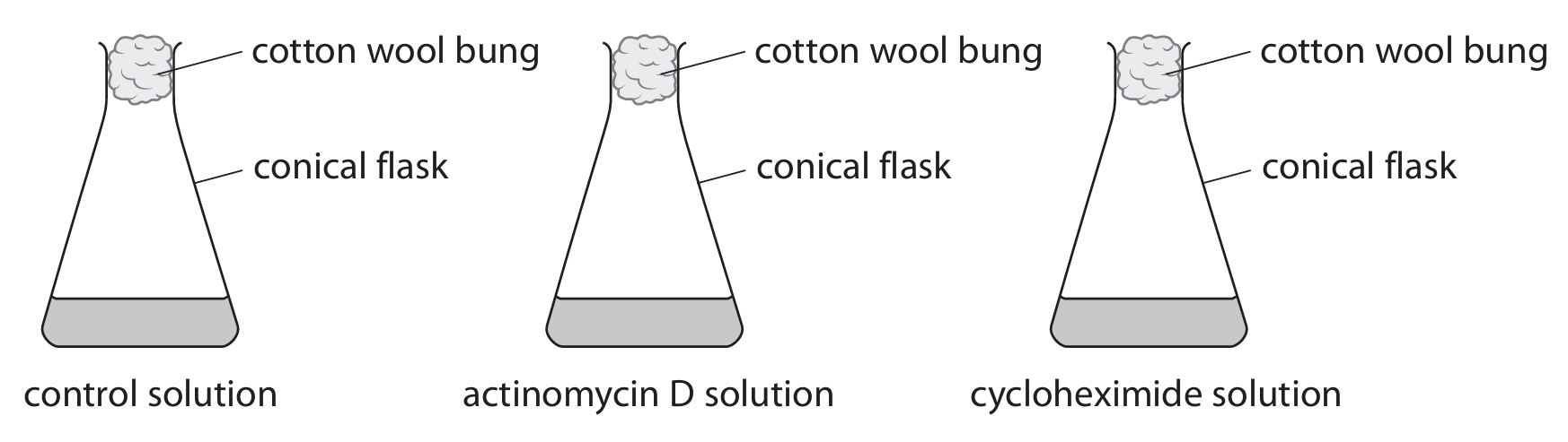
(c)(ii)
Explanation: Both chemicals inhibit pollen tube growth because they disrupt protein synthesis, which is essential for growth. Actinomycin D prevents transcription, the process where DNA is copied into mRNA. Without mRNA, the instructions for making proteins cannot reach the ribosomes. Cycloheximide prevents translation, where mRNA is read by ribosomes to assemble amino acids into proteins. Since both processes are blocked, protein synthesis decreases, slowing down pollen tube growth. The growth with actinomycin D is higher than with cycloheximide because some mRNA molecules might already be present in the pollen grain and can still be translated until they degrade, allowing limited protein production. With cycloheximide, translation is directly blocked, so even existing mRNA cannot be used, resulting in more severe growth inhibition.
(d)
Explanation: To observe pollen grains effectively, the scientist should use microscopy. First, place a pollen grain sample on a clean glass slide. Add a drop of water or a suitable stain (like methylene blue) to make the structures more visible. Carefully lower a cover slip over the sample to avoid air bubbles. Then, examine the slide under a light microscope, starting with low magnification to locate the pollen grains and switching to higher magnification to observe detailed structures such as the shape, size, and surface patterns of the pollen grains.