▶️ Answer/Explanation
(a)(i) B (carbon dioxide and water)
A is not the answer as lungs do not excrete urea
C is not the answer as lungs do not excrete urea
D is not the answer as lungs do not excrete urea
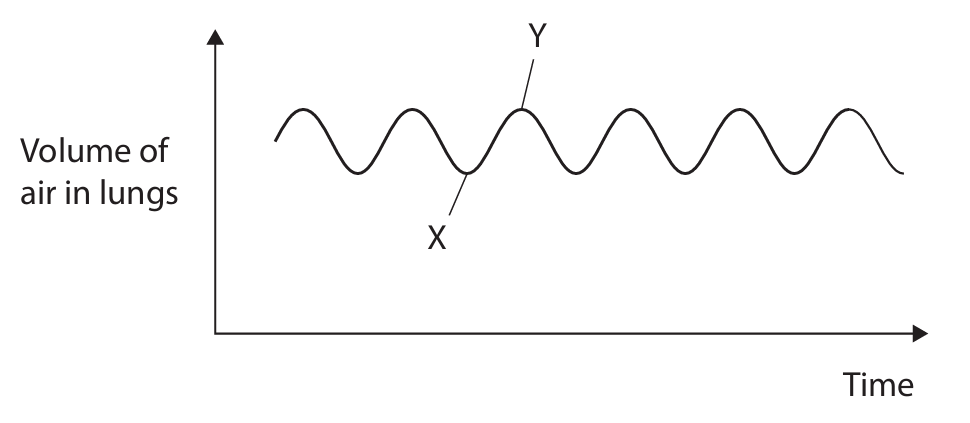
(a)(ii) An explanation that makes reference to three of the following points:
- volume increases / inhalation occurs / air drawn in (1)
- diaphragm / intercostal muscles contract (1)
- diaphragm moves down / flattens (1)
- ribcage expands (1)
- pressure decreases (inside thorax / lungs) (1)
Accept: internal intercostal muscles relax; ribs move up / move out; thorax / chest expands; pressure higher outside
(b) An explanation that makes reference to three of the following points:
- glucose in urine (1)
- glucose released by ultrafiltration (into filtrate) (1)
- glucose not reabsorbed / too much glucose (in filtrate) to reabsorb (1)
- in the proximal convoluted tubule / PCT / first convoluted tubule (1)
- by active transport (1)
Accept: glucose not absorbed into blood; some glucose not reabsorbed
Reject: if active transport pumping glucose into filtrate
▶️ Answer/Explanation
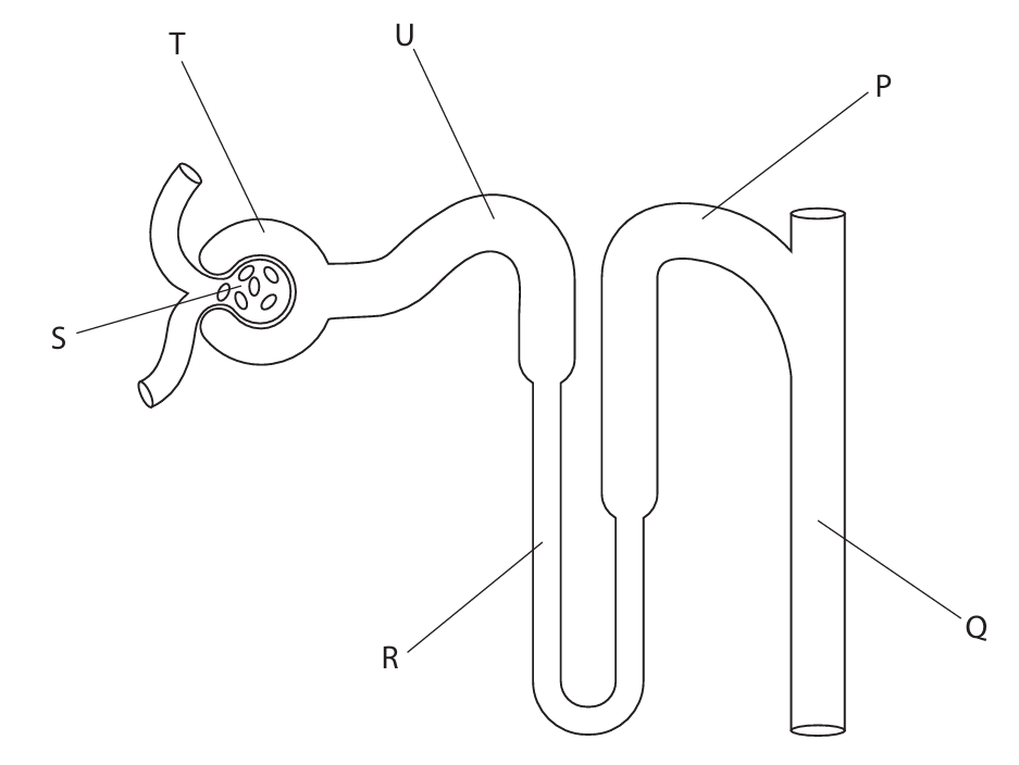
(a)(i) D (proximal convoluted tubule)
A is not correct as U is not the Bowman’s capsule
B is not correct as U is not the collecting duct
C is not correct as U is not the loop of Henle
(a)(ii) D (ultrafiltration)
A is not correct as it is not ADH production
B is not correct as it is not selective reabsorption
C is not correct as it is not transpiration
(a)(iii) B (Q)
A is not correct as P is the distal convoluted tubule
C is not correct as S is the glomerulus
D is not correct as T is the Bowman’s capsule
(b)(i) Calculation:
Concentration of glucose in blood plasma = 0.1 g/100 cm³
5 dm³ = 5000 cm³
Mass of glucose = (0.1 g/100 cm³) × 5000 cm³ = 5 grams
(b)(ii) An explanation that makes reference to two of the following:
• protein molecules too large / large mass / too heavy (1)
• cannot pass out of glomerulus / into Bowman’s capsule / into nephron / into tubules / through basement membrane (1)
• so stay in blood / not in filtrate (1)
(b)(iii) An explanation that makes reference to two of the following:
• glucose passes out of glomerulus / into Bowman’s capsule / into nephron / into tubules / through basement membrane (1)
• (then) reabsorbed / back into blood (1)
• by active transport (1)
• in proximal convoluted tubule / PCT (1)
▶️ Answer/Explanation
(a) D (homeostasis)
A is not correct as it is not absorption
B is not correct as it is not diffusion
C is not correct as it is not egestion
(b)(i) Calculation method:
Insensible loss = gas exchange + sweating = \(0.4 + 0.5 = 0.9\) litres
Total water loss = urine + gas exchange + sweating + faeces = \(1.5 + 0.4 + 0.5 + 0.1 = 2.5\) litres
Percentage insensible loss = \(\frac{0.9}{2.5} \times 100 = 36\%\)
(b)(ii) Calculation method:
Water input from food for 70 kg person = 0.5 litres
Water input per kg = \(\frac{0.5}{70} = 0.007142857\) litres/kg
For 110 kg person: \(0.007142857 \times 110 = 0.7857\) litres ≈ \(0.79\) litres (accept \(0.77–0.80\))
(b)(iii) An explanation that makes reference to four of the following points:
1. more water lost / less water in body /eq (1)
2. water / liquid lost in vomiting / faeces / diarrhoea / eq (1)
3. (more) sweating / eq (1)
4. concentration of blood increases / water potential lowered / eq (1)
5. (more) ADH produced / secreted / eq (1)
6. by hypothalamus / pituitary (1)
7. permeability of collecting duct increases / eq (1)
8. more water reabsorbed / less water lost in urine / more concentrated urine / less urine / eq (1)
(c) An answer that makes reference to four of the following points:
In winter:
1. body mass lower / eq (1)
2. as fewer plants available / less food / hibernating / eq (1)
3. less sunlight / lower temp / less photosynthesis / eq (1)
4. (much) more water intake / eq (1)
5. as more rainfall/ eq (1)
6. less concentrated urine in winter / more urine / eq (1)
7. (so) more water lost in urine / eq (1)
8. Ref to data for urine 3× less concentrated in winter / water input 4 × higher in winter / eq (1)
In summer: converse of above points applies.
▶️ Answer/Explanation
(a) B (bacteria, fungi, and protoctists)
Explanation: Pathogens are disease-causing microorganisms. While viruses like rabies are one type, other major groups also contain pathogenic species. Bacteria include pathogens like those causing tuberculosis and cholera. Fungi include pathogens responsible for athlete’s foot and ringworm. Protoctists (protists) include pathogenic organisms like Plasmodium which causes malaria and Entamoeba which causes dysentery. Therefore, all three groups – bacteria, fungi, and protoctists – include pathogens.
(b)(i)
Explanation: The rabies vaccine contains weakened or inactivated forms of the rabies virus or its antigens. When this vaccine is administered to dogs, it stimulates their immune system without causing the actual disease. The immune system recognizes these viral antigens as foreign invaders and produces specific antibodies against them. Specialized white blood cells called lymphocytes are activated during this process. Some of these lymphocytes develop into memory cells that remain in the body long-term. If the vaccinated dog is later exposed to the actual rabies virus, these memory cells recognize the pathogen immediately and trigger a rapid, strong immune response. This secondary response produces antibodies much faster and in greater quantities, effectively neutralizing the virus before it can establish an infection and cause disease.
(b)(ii)
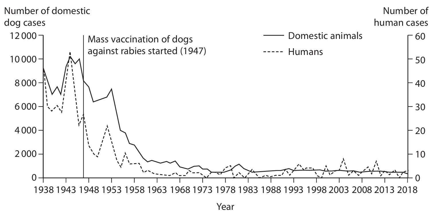
Explanation: The graph shows a clear correlation between the vaccination of domestic dogs and the decline in human rabies cases. Before mass vaccination began in 1947, both dog and human rabies cases were relatively high. After vaccination programs were implemented, we observe a continuous and dramatic decline in rabies cases in both dogs and humans throughout the 1950s and beyond. This strong correlation suggests that most human rabies cases were originating from infected domestic dogs rather than wild animals. The data shows that as dog vaccination reduced the reservoir of infection in the canine population, human cases consequently decreased. However, the graph also indicates that human rabies cases didn’t disappear completely but plateaued at low levels, suggesting that some transmission still occurs from wild animals or unvaccinated dogs. This demonstrates that while vaccinating domestic dogs significantly reduces human rabies risk, it doesn’t completely eliminate it due to other potential sources of infection.
(b)(iii)
Explanation: When the RNA vaccine is injected, cells take up the RNA molecules that code for parts of the rabies virus protein coat. Inside the cell, these RNA molecules move to the ribosomes, which are the cellular structures responsible for protein synthesis. The process of translation then occurs, where the genetic code on the RNA is read and converted into a sequence of amino acids. Transfer RNA (tRNA) molecules with specific anticodons bind to complementary codons on the mRNA, each bringing with it a specific amino acid. As the ribosome moves along the mRNA strand, it facilitates the formation of peptide bonds between adjacent amino acids, creating a growing polypeptide chain. This chain folds into the three-dimensional structure of the viral protein. These viral proteins then act as antigens, stimulating the immune system to produce antibodies against rabies without exposure to the actual virus, thus providing protection against future infection.
▶️ Answer/Explanation
(a) Answer: 9.5 × 10⁸
Explanation: To calculate the number of people with chronic kidney disease, we multiply the world population by 12% (or 0.12).
Calculation: 7,900,000,000 × 0.12 = 948,000,000
In standard form, this is 9.48 × 10⁸, which rounds to 9.5 × 10⁸ when expressed to two significant figures as appropriate for the percentage given (12%).
(b) Answer: Carbon dioxide / CO2 or Water (vapour) / H2O
Explanation: The lungs are responsible for excreting carbon dioxide, which is a waste product of cellular respiration. Water vapor is also excreted through the lungs during exhalation, especially in humid environments.
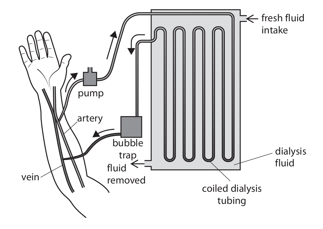
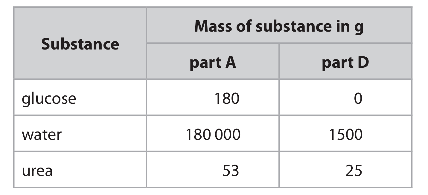
(c) Answer: The dialyser is designed in two key ways to increase urea removal:
1. The temperature is maintained at 40°C, which is slightly higher than normal body temperature. This increases the kinetic energy of urea molecules, making them move faster and diffuse more rapidly across the partially permeable membrane.
2. The dialysis fluid contains no urea, creating a steep concentration gradient between the blood and the dialysis fluid. This maximizes the rate of diffusion of urea from the blood into the dialysis fluid, following the principle of moving from an area of high concentration to an area of low concentration.
Additional design features include the long, coiled tubing which provides a large surface area for diffusion, and the thin partially permeable membrane which shortens the diffusion pathway.
(d)(i) Answer: A. Bowman’s capsule
Explanation: Ultrafiltration occurs in the Bowman’s capsule of the nephron, where high pressure forces small molecules like water, glucose, urea, and salts out of the blood and into the nephron tubule, while larger molecules like proteins and blood cells remain in the blood.
(d)(ii) Answer: Glucose is reabsorbed from the filtrate in the nephron through selective reabsorption in the proximal convoluted tubule. This process involves active transport, which requires energy (ATP) to move glucose molecules against their concentration gradient from the tubule back into the blood capillaries. Specialized carrier proteins in the cells lining the tubule facilitate this transport.
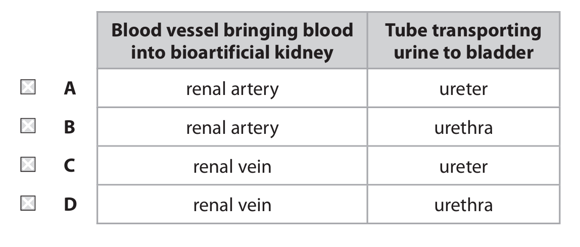
(d)(iii) Answer: A (renal artery / ureter)
Explanation: The renal artery brings oxygenated blood into the kidney for filtration, while the ureter is the tube that transports urine from the kidney to the bladder. The urethra is not correct as it transports urine from the bladder out of the body, not from the kidney to the bladder.
(e) Answer: In a dehydrated patient:
1. Osmoreceptors in the hypothalamus detect the increased solute concentration (decreased water potential) in the blood.
2. The hypothalamus stimulates the pituitary gland to release more antidiuretic hormone (ADH).
3. ADH travels through the bloodstream to the kidneys (or bioreactor nephron cells).
4. ADH makes the walls of the collecting duct (or bioreactor membrane) more permeable to water.
5. More water is reabsorbed from the filtrate back into the blood, resulting in a smaller volume of more concentrated urine.
This negative feedback mechanism helps conserve water in the body and restore normal blood concentration.
▶️ Answer/Explanation
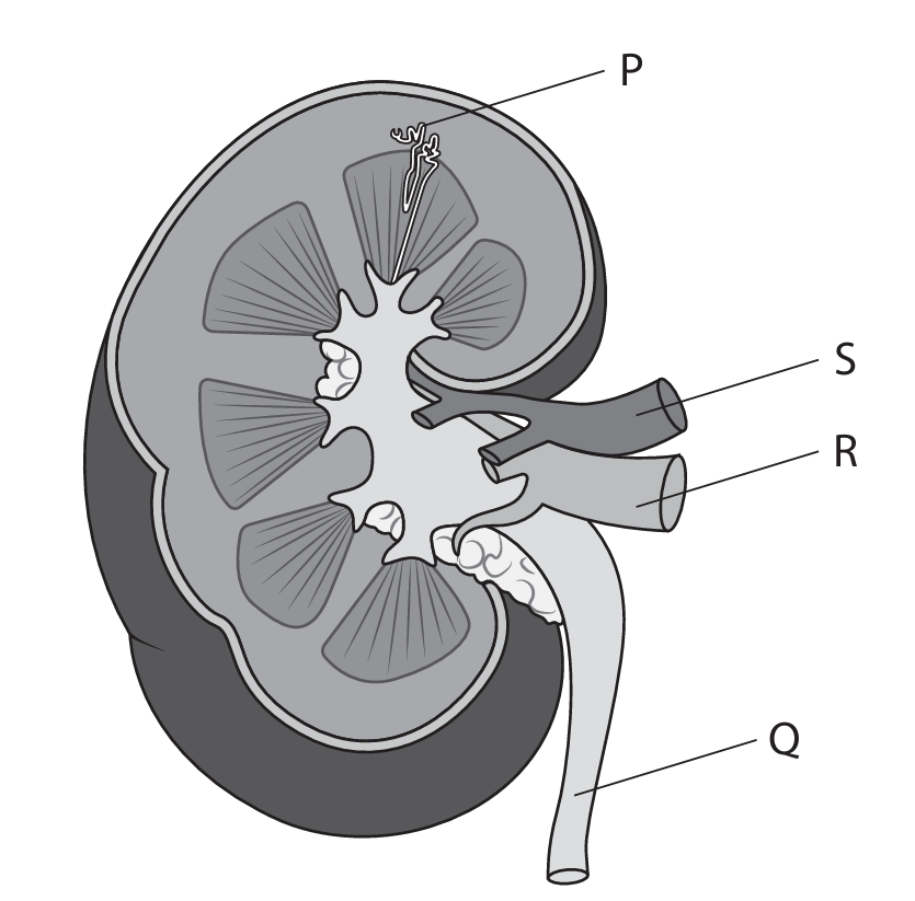
(a)(i) B (nephron)
Explanation: Structure P represents the functional unit of the kidney, which is the nephron.
(a)(ii) B (blood)
Explanation: Tube S carries blood to or from the kidney, specifically it is likely the renal artery or vein.
(a)(iii) C (ureter)
Explanation: Tube Q is the ureter, which transports urine from the kidney to the bladder.
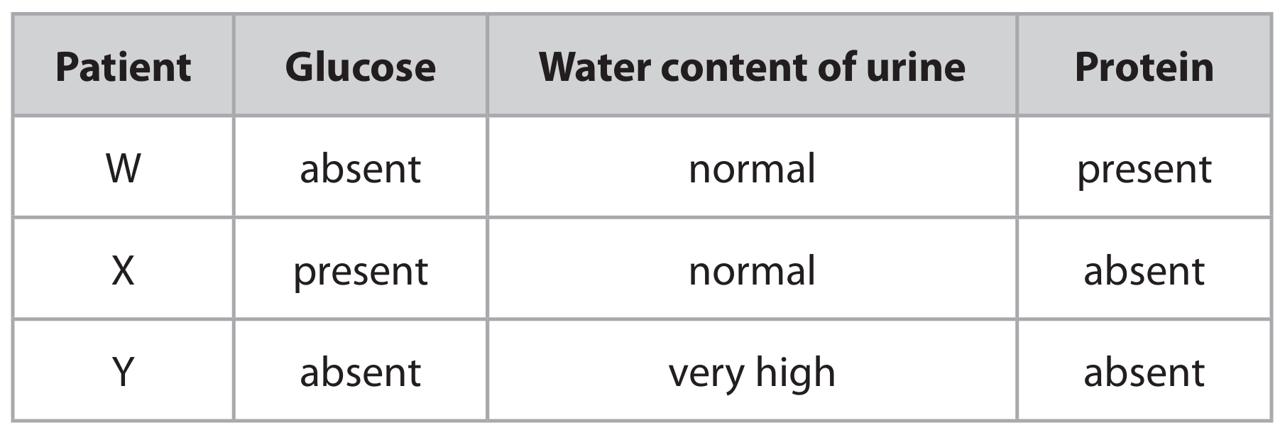
(b)(i) An explanation that makes reference to:
• Patient W: Presence of protein indicates failure of ultrafiltration in the glomerulus/Bowman’s capsule.
• Patient X: Presence of glucose indicates failure of selective reabsorption in the proximal convoluted tubule.
• Patient Y: High water content indicates reduced water reabsorption in the collecting duct, possibly due to low ADH.
(b)(ii) A description that includes:
• Use Benedict’s reagent
• Heat in a water bath
• Observe colour change (green → red indicates glucose)
• Alternatively, use a glucose test strip and compare colour to a chart.
▶️ Answer/Explanation
(a) A
Explanation: The proximal convoluted tubule is the first part of the tubule system in the nephron, located right after the Bowman’s capsule. In a standard nephron diagram, it’s often the first coiled section labelled. Option B is the distal convoluted tubule (later section), C is the collecting duct (where urine is collected), and D is the loop of Henle (the U-shaped part that dips into the medulla).
(b)(i) 0.0314 mm²
Explanation: The radius is 100 μm. First, convert this to millimetres: 100 μm = 100/1000 mm = 0.1 mm. Now, use the area formula: Area = π × (radius)² = 3.14 × (0.1 mm)² = 3.14 × 0.01 mm² = 0.0314 mm².
(b)(ii)
Explanation: Blood vessel X is the afferent arteriole (wider lumen), and blood vessel Y is the efferent arteriole (narrower lumen). This size difference is crucial for creating high hydrostatic pressure within the glomerulus. The wider afferent arteriole allows a large volume of blood to flow into the glomerulus. The narrower efferent arteriole restricts the outflow of this blood. This “bottleneck” effect significantly increases the blood pressure inside the glomerular capillaries. This high pressure is the driving force for ultrafiltration, where water, ions, glucose, and urea are forced out of the blood and into the Bowman’s capsule to form the glomerular filtrate.
(c)
Explanation: The standard test for protein in urine is the Biuret test. Pour a small sample of urine into a test tube. Add an equal volume of sodium hydroxide (NaOH) solution and mix. Then, add a few drops of very dilute copper(II) sulfate (CuSO₄) solution. Do not shake. If protein is present, the solution will turn a lilac or purple colour. Alternatively, a urine test strip (dipstick) can be used. The strip is dipped into the urine sample, and after the specified time, the colour change on the protein pad is compared to a chart on the bottle.
▶️ Answer/Explanation
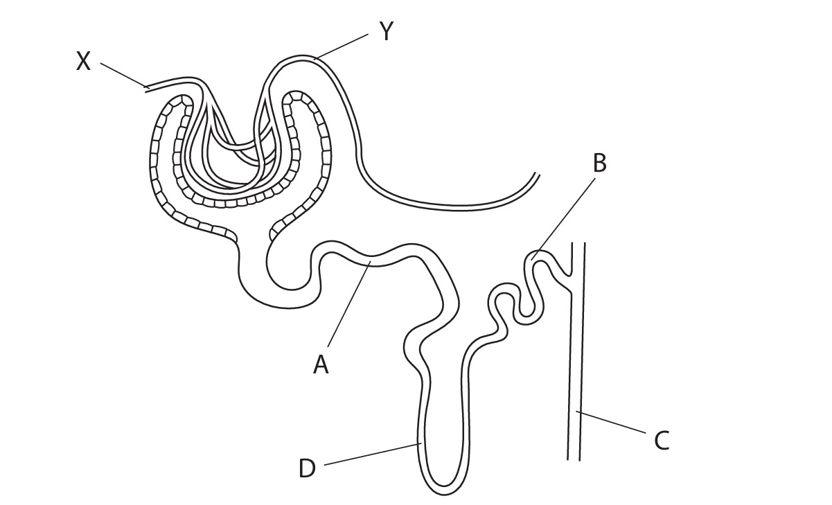
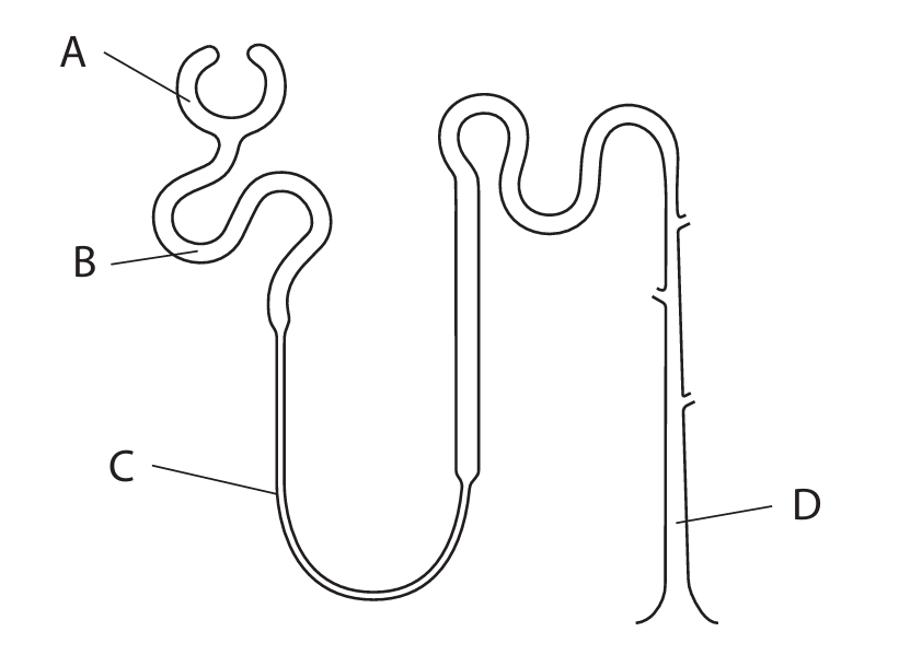
(a)(i) C (S)
Explanation: The Bowman’s capsule (S) is the cup-like sac at the beginning of the nephron that surrounds the glomerulus and receives the filtrate.
(a)(ii) B (Q)
Explanation: The loop of Henle (Q) is a U-shaped tubule that descends into and ascends from the medulla of the kidney. It is crucial for creating a concentration gradient for water reabsorption.
(a)(iii) A (P)
Explanation: ADH (Antidiuretic Hormone) affects the collecting duct (P). ADH increases the permeability of the collecting duct walls to water, allowing more water to be reabsorbed back into the blood, producing more concentrated urine.
(b)(i) An explanation that makes reference to three of the following points:
• Glucose passes from the blood in the glomerulus (R) into the Bowman’s capsule / renal capsule (S) during ultrafiltration. (1)
• (All) glucose is (then) reabsorbed / absorbed back into the blood / eq. (1)
• This reabsorption occurs in the proximal convoluted tubule / PCT (T). (1)
• It is reabsorbed by active transport (which requires energy). (1)
Explanation: During ultrafiltration, small molecules like glucose enter the nephron. The body cannot afford to lose this valuable energy source, so 100% of filtered glucose is normally reclaimed from the filtrate in the proximal convoluted tubule via active transport against its concentration gradient.
(b)(ii) A description that makes reference to two of the following points:
• Add Benedict’s solution to the urine sample (and heat). (1)
• A positive result is indicated by a colour change to green / yellow / orange / brick-red. (1)
Alternative: Use a test strip (e.g., Clinistix) which changes colour (e.g., to brown) in the presence of glucose. (1 each)
Explanation: Benedict’s test is a standard biochemical test for reducing sugars like glucose. Heating with Benedict’s reagent causes a reduction reaction, producing a coloured precipitate of copper(I) oxide.
(c) A description that makes reference to two of the following points:
• Less urine is produced / lower volume. (1)
• The urine becomes more concentrated / contains less water / appears darker in colour. (1)
• (It may contain) a higher concentration of urea / other solutes. (1)
Explanation: Dehydration lowers the water potential of the blood. This is detected by osmoreceptors, leading to increased secretion of ADH. ADH causes more water to be reabsorbed from the collecting duct back into the blood, conserving water. This results in a smaller volume of more concentrated, darker yellow urine.
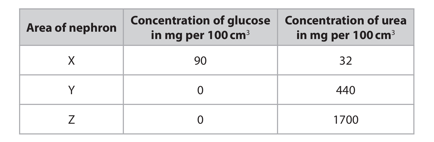
▶️ Answer/Explanation
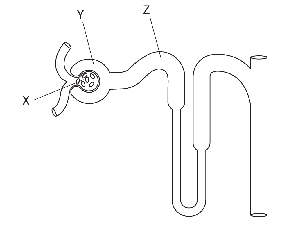
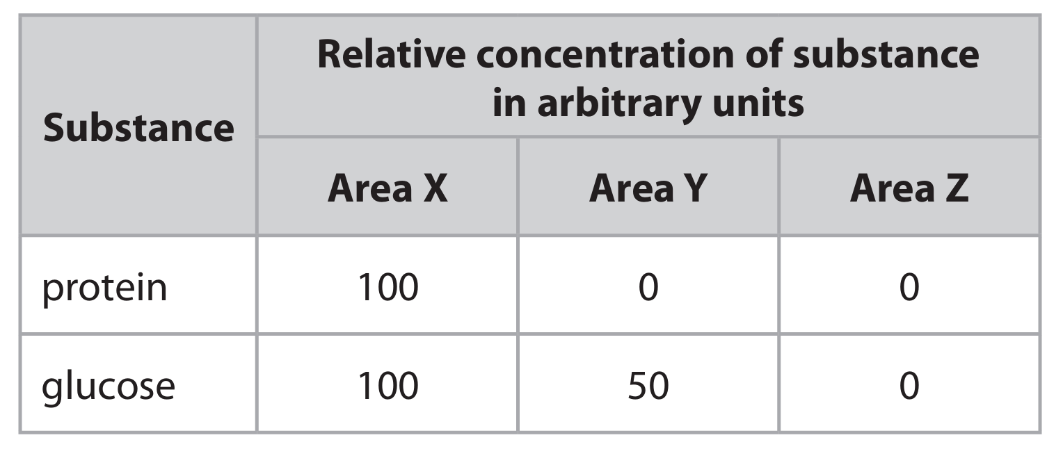
(a)
Explanation: The difference in glucose concentration between area X and area Y occurs due to selective reabsorption in the nephron. Area X represents the filtrate in the Bowman’s capsule, which contains glucose at the same concentration as blood plasma. As the filtrate moves through the proximal convoluted tubule (area Y), all glucose is actively reabsorbed back into the blood. This process requires energy (ATP) and specific carrier proteins to transport glucose against its concentration gradient. Since glucose is a valuable nutrient that the body needs to conserve, it is completely removed from the filtrate in the proximal convoluted tubule, resulting in zero glucose concentration in area Y.
(b)(i) 3.9
Explanation: To calculate how many times more concentrated urea is in area Z compared to area Y, we divide the concentration in Z by the concentration in Y: 1700 ÷ 440 = 3.8636… Rounded to two significant figures, this gives 3.9. This means urea is approximately 3.9 times more concentrated in area Z than in area Y.
(b)(ii)
Explanation: The difference in urea concentration between area Y and area Z occurs primarily due to water reabsorption. As the filtrate moves through the distal convoluted tubule and collecting duct (area Z), water is reabsorbed back into the blood by osmosis. This process is regulated by the hormone ADH (antidiuretic hormone). While urea itself is not actively reabsorbed in significant amounts, the removal of water from the filtrate causes the remaining substances, including urea, to become more concentrated. Therefore, the urea concentration increases significantly from area Y to area Z as water is removed from the filtrate.
(c)(i)
Explanation: To test for protein in urine, a common method is to use urine test strips (dipsticks) that contain a chemical indicator. The strip is dipped into the urine sample for a specified time, then removed. The test pad on the strip will change color if protein is present – typically turning green or blue depending on the concentration. Alternatively, a chemical test can be performed using reagents like Biuret solution, which would change from blue to purple in the presence of protein. The intensity of the color change corresponds to the amount of protein present in the sample.
(c)(ii)
Explanation: High blood pressure during pregnancy can cause protein to appear in urine because the increased pressure can damage the filtration barrier in the glomerulus. Normally, the glomerular filter prevents large molecules like proteins from passing through into the filtrate. However, when blood pressure is excessively high, it creates increased mechanical stress on the glomerular capillaries and basement membrane. This can cause the filtration pores to enlarge or the membrane to become more permeable, allowing proteins (which are large molecules) to be forced through into the filtrate. Once in the filtrate, proteins are too large to be reabsorbed effectively by the nephron, so they remain and are excreted in the urine.

▶️ Answer/Explanation
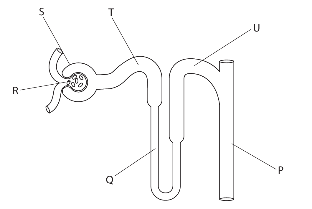
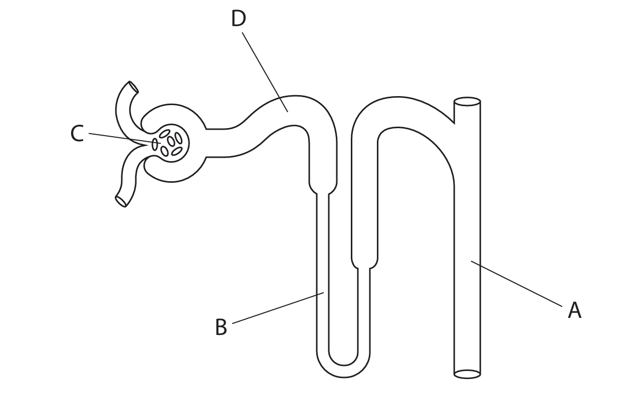
(a)(i) D S
Explanation: The structure labelled S is the glomerulus, which is a network of capillaries where ultrafiltration occurs. Blood pressure forces water, ions, and small molecules out of the blood and into the Bowman’s capsule, forming the filtrate.
(a)(ii) D U
Explanation: The structure labelled U is the proximal convoluted tubule (PCT). This is where the majority of glucose reabsorption takes place through active transport, returning this valuable nutrient to the bloodstream.
(a)(iii) B R
Explanation: The structure labelled R is the loop of Henle. This hairpin-shaped section of the nephron creates a concentration gradient in the kidney medulla, which is essential for water reabsorption and urine concentration.
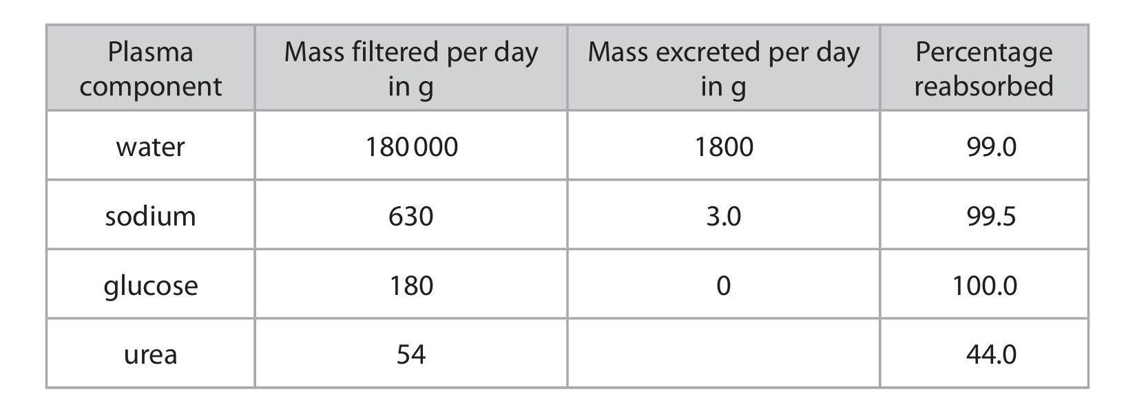
(b)(i) Substances/chemicals present in solution/dissolved/carried in the liquid part of the blood/plasma.
Explanation: Plasma components refer to the various dissolved substances found in blood plasma, which is the liquid matrix of blood. These include water, electrolytes (like sodium), nutrients (like glucose), waste products (like urea), hormones, and proteins.
(b)(ii) 30.24 g
Explanation: The calculation is based on the percentage reabsorbed. If 44% of urea is reabsorbed, then 56% is excreted. The mass excreted per day is therefore 56% of the mass filtered: \( \frac{56}{100} \times 54 = 30.24 \) g.
(b)(iii) To prevent glucose from being excreted/lost from the body and to maintain blood glucose levels for respiration/energy release.
Explanation: Glucose is a vital energy source for cells. The body carefully regulates blood glucose levels. Reabsorbing glucose in the nephron ensures that this important fuel is not wasted in urine and is kept in the bloodstream to be used for cellular respiration, which releases the energy needed for all bodily functions.
(b)(iv)
Explanation: A high-protein, high-salt meal with low water intake would have several effects. The breakdown of excess protein increases urea production, leading to a higher concentration of urea in the plasma and subsequently more urea being filtered and excreted. The high salt intake increases the sodium concentration in the blood, lowering its water potential. This is detected by osmoreceptors, which signal the pituitary gland to release more Anti-Diuretic Hormone (ADH). ADH makes the walls of the collecting duct more permeable to water. As a result, more water is reabsorbed back into the blood from the filtrate, producing a smaller volume of more concentrated urine to conserve water and excrete the excess salts and urea.
▶️ Answer/Explanation
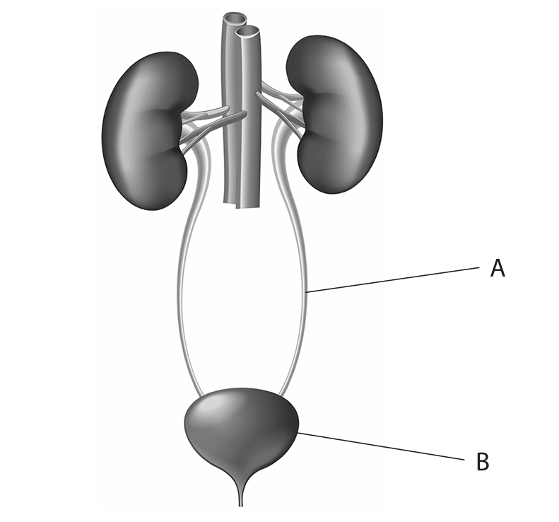
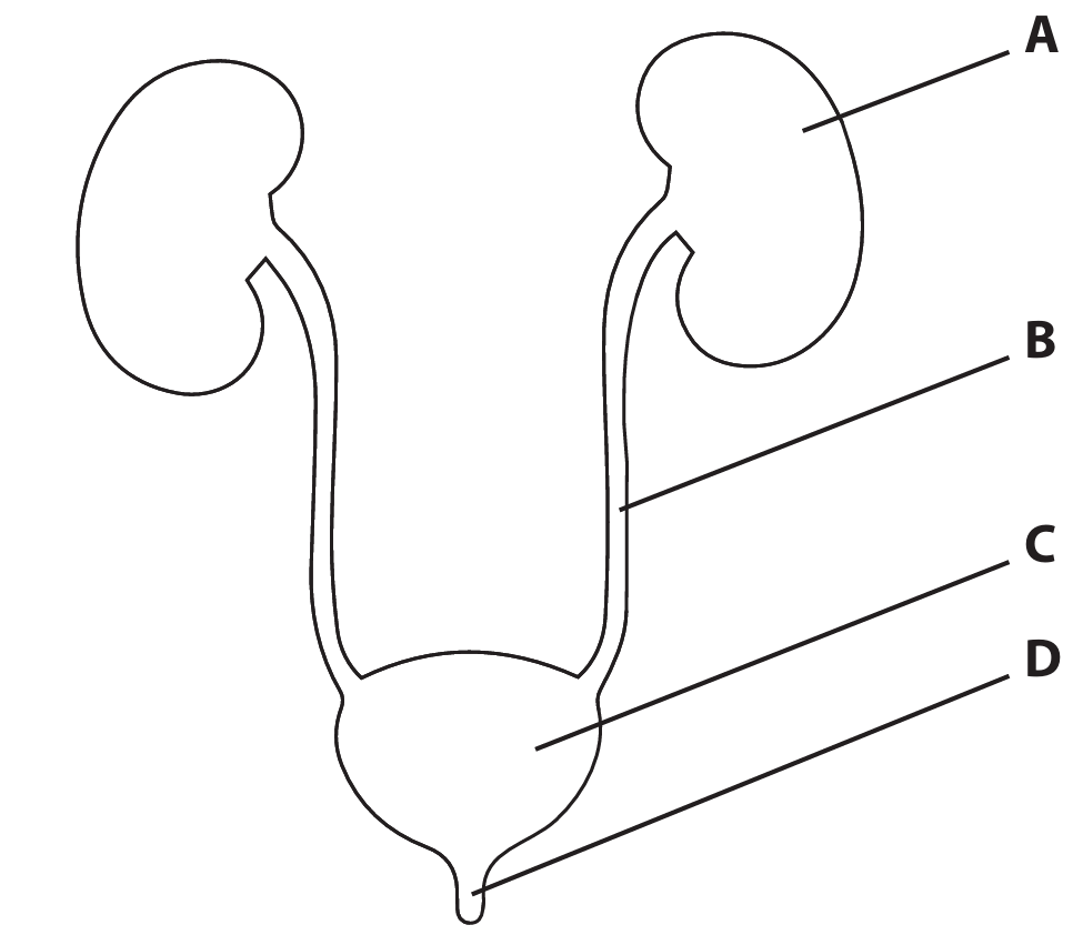
(a)
A: Ureter
B: Bladder
Explanation: The ureter is the tube that carries urine from the kidney to the bladder. The bladder is the muscular sac that stores urine until it is expelled from the body. These are two key components of the urinary system’s transport and storage pathway.
(b)(i)
Explanation: The concentration of protein drops from 100 arbitrary units in area X to 0 in area Y because proteins are too large to pass through the filtering system of the glomerulus. The glomerulus acts like a sieve, allowing small molecules like water, salts, glucose, and urea to pass into the Bowman’s capsule (area Y), while large protein molecules remain in the blood (area X). This is a crucial filtration mechanism that prevents essential proteins from being lost in urine.
(b)(ii)
Explanation: The concentration of glucose decreases from 50 arbitrary units in area Y to 0 in area Z because glucose is actively reabsorbed back into the blood from the filtrate. This process occurs primarily in the proximal convoluted tubule. The body recognizes glucose as a valuable energy source and uses active transport, which requires energy, to reclaim it from the filtrate and return it to the bloodstream, ensuring no glucose is wasted in the urine under normal conditions.
(c)
Explanation: When the body is dehydrated, the blood becomes more concentrated (has a lower water potential). This is detected by osmoreceptors in the hypothalamus of the brain. In response, the pituitary gland releases more Antidiuretic Hormone (ADH) into the bloodstream. ADH travels to the kidneys and makes the walls of the collecting ducts more permeable to water. As a result, more water is reabsorbed from the filtrate back into the blood by osmosis. This process conserves water for the body, produces a smaller volume of more concentrated urine, and helps to raise the blood water potential back towards normal levels.
▶️ Answer/Explanation
(a) A
Explanation: The nephrons are the functional units of the kidney responsible for filtration, reabsorption, and secretion. In the urinary system diagram, part A represents the kidney, which is where the nephrons are located. The other parts (B, C, D) represent other structures like the ureter, bladder, and urethra, which do not contain nephrons.
(b) Water / Urea / Ions (e.g., sodium, chloride) / Salts
Explanation: Urine is composed of water and various waste products that the body needs to excrete. The primary component is water, which acts as a solvent. Urea is a major nitrogenous waste product from the breakdown of proteins. Ions such as sodium, potassium, chloride, and other salts are also found in urine, their concentrations varying based on the body’s needs to maintain homeostasis.
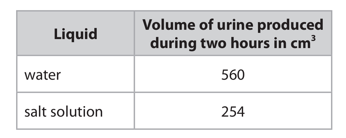
(c)(i)
Explanation: The scientist’s results show that drinking water leads to a much higher volume of urine produced (560 cm³) compared to drinking a salt solution (254 cm³). This happens due to the body’s osmoregulatory mechanisms. When pure water is consumed, the blood becomes more dilute (its water potential increases). This change is detected by osmoreceptors in the hypothalamus. In response, the pituitary gland releases less Antidiuretic Hormone (ADH). With less ADH, the walls of the kidney’s collecting ducts become less permeable to water. This means less water is reabsorbed back into the blood, resulting in a larger volume of dilute urine being produced. Conversely, when a salt solution is consumed, the blood becomes more concentrated (its water potential decreases). This stimulates the release of more ADH. More ADH makes the collecting ducts more permeable, allowing more water to be reabsorbed into the blood to correct the concentration. Consequently, a smaller volume of more concentrated urine is produced.
(c)(ii)
1. Exercise / Physical activity
2. Temperature / Environmental temperature
Explanation: Several other factors can influence urine volume. Exercise or physical activity causes sweating, which loses water from the body. To conserve fluid, the body produces less urine. The temperature of the environment also plays a role; in hot conditions, the body sweats more to cool down, leading to reduced urine production. In cooler temperatures, less sweating occurs, potentially resulting in higher urine output. Other factors not listed here could include the volume of other fluids consumed before the test, certain foods in the diet (like those with high water or salt content), and the intake of diuretics such as caffeine.
▶️ Answer/Explanation
(a)
(i) C
Explanation: Ultrafiltration occurs in the Bowman’s capsule, which is labelled as structure C in the diagram. This is where high pressure forces small molecules like water, glucose, and urea out of the blood and into the nephron, forming the filtrate.
(ii) D
Explanation: Glucose is reabsorbed back into the blood in the proximal convoluted tubule, labelled as structure D. This process, called selective reabsorption, uses active transport to move useful substances like glucose from the filtrate back into the bloodstream.
(b)
(i) Osmoregulation
Explanation: The control of blood concentration, specifically water and salt balance, is known as osmoregulation. The kidneys play a vital role in this by adjusting the amount of water reabsorbed or excreted.
(ii) The removal of metabolic waste / waste from chemical reactions (from cells).
Explanation: Excretion is the biological process of removing waste products generated by metabolic activities within cells. In the context of the kidney, this primarily involves removing urea, a toxic substance produced from the breakdown of excess proteins in the liver.
(c)
Explanation: ADH (Antidiuretic Hormone) makes the walls of the collecting duct more permeable to water. In diabetes insipidus, no ADH is produced. This means the collecting duct walls remain impermeable to water. As a result, very little water is reabsorbed from the filtrate back into the blood in this final part of the nephron. This leads to the production of a large volume of very dilute urine. Consequently, the body loses excessive amounts of water, which can lead to dehydration and an increase in blood concentration (it becomes more concentrated).
(d)
(i) It increases the permeability of the collecting duct (wall).
Explanation: Desmopressin acts as a substitute for ADH. Its primary effect on the nephron is to bind to the cells of the collecting duct and cause the insertion of aquaporins (water channels) into their walls. This makes the walls permeable to water, allowing water to be reabsorbed.
(ii) The drug would decrease the volume of urine produced and increase the concentration of the urine.
Explanation: By making the collecting duct permeable, Desmopressin allows more water to be reabsorbed from the filtrate back into the blood. This means less water remains in the tubule to be excreted, so the volume of urine decreases. As more water leaves the filtrate, the remaining substances (like urea) become more concentrated, resulting in the production of more concentrated urine.
▶️ Answer/Explanation
(a) C (ultrafiltration)
A is incorrect because digestion is not a process in the kidneys.
B is incorrect because mutation is not a process in the kidneys.
D is incorrect because vaccination is not a process in the kidneys.
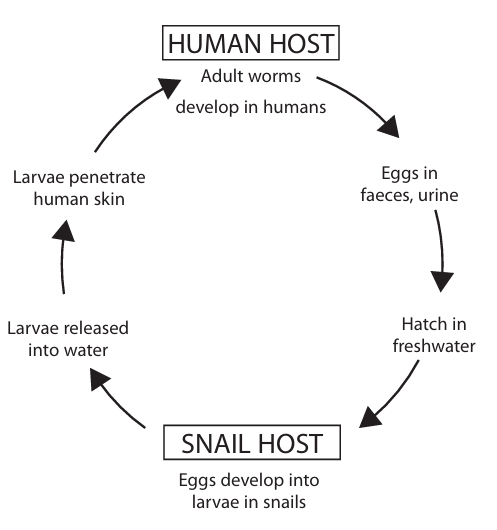
(b) An answer that makes reference to three of the following points:
• treat drinking water / boil water (before drinking) / do not drink water / drink bottled water / eq (1)
• sanitation / no faeces in water / no urine in water / eq (1)
• remove snails / eq (1)
• vaccination (1)
Additional guidance: Allow “do not go in infected rivers or lakes / cover skin when in water / avoid contact with affected water / only wash in clean water”.
(c) An answer that makes reference to two of the following points:
• red blood cells / rbc (1)
• white blood cells / wbc (1)
• lymphocytes (1)
• phagocytes / macrophages (1)
(d) D (4800 mg)
Calculation: \(120\ \text{kg} \times 0.040\ \text{g/kg} = 4.8\ \text{g} = 4800\ \text{mg}\)
A is incorrect because it is the wrong value.
B is incorrect because it is the wrong value.
C is incorrect because it is the wrong value.
(e) \(1920\) people
Working:
• \(8 \times 10^{-4}\% = 0.0008\%\)
• \(0.0008\% \text{ of } 240,000,000 = 0.000008 \times 240,000,000 = 1920\)
Allow 1 mark for: \(19200000 / 1920000 / 192000 / 192200 / 192 / 19.2 / 1.92 / 0.192 / 0.0192\)
Award full marks for correct numerical answer without working.
(f) A (a circle of DNA)
B is incorrect because it is not RNA.
C is incorrect because it is not a protein.
D is incorrect because it is not RNA.
(g) An explanation that makes reference to three of the following points:
• antigen (1)
• memory cells / lymphocytes (1)
• (secondary) immune response (1)
• more antibodies / antibodies made sooner / faster / faster immune response / eq (1)
(h)(i) • (a treatment with) no plasmid / no protein / only water / saline / eq (1)
Allow placebo vaccine / a placebo / plasmid with no gene / plasmid with no DNA / different DNA.
(h)(ii) An answer that makes reference to three of the following points:
• reduced numbers / eq (1)
• by 19 or by 47% / about 50% (1)
• schistosomes / worms, still present in body (1)
• no idea of group size / needs to be repeated (1)
• no idea of age / sex / health (1)
Additional Guidance:
Allow “reduces numbers of worms / worms decrease / lower number of worms after vaccine”.
Allow “more worms in control group”.
Allow “does not completely get rid of them”.
Allow “more testing / more people tested”.