Osmosis – Movement of Water Across Membranes
🌱 Introduction
Osmosis is a special type of passive transport involving water molecules only. It occurs through a partially permeable membrane (allows water, blocks most solutes). Water moves down its water potential gradient (high ψ → low ψ). Essential for maintaining cell turgor, shape, and volume in both plant and animal cells.
🧩 Key Definitions
- Osmosis: Passive movement of free water molecules across a partially permeable membrane down a water potential gradient.
- Partially permeable membrane: Permits water molecules but restricts most solutes.
- Water potential (ψ): Measure of free energy of water molecules; water moves high ψ → low ψ.
- Tonicity: Relative solute concentration of a solution compared to the inside of a cell.
⚡ Mechanism of Osmosis
- Water molecules move randomly.
- Side with more “free” water → high ψ; side with less free water (bound to solute) → low ψ.
- Net movement: high ψ → low ψ until equilibrium is reached.
📝 Effects on Cells
| Solution Type | Water Potential (ψ) | Movement of Water | Effect on Cell |
|---|---|---|---|
| Hypotonic (dilute) | High outside ψ | Into cell | Swells; plant cells turgid, animal cells may burst (lysis) |
| Hypertonic (concentrated) | Low outside ψ | Out of cell | Shrinks; plant cells plasmolyze, animal cells crenate |
| Isotonic (equal) | Equal ψ | No net movement | Cell stays same size |
🌾 Examples in Plants & Animals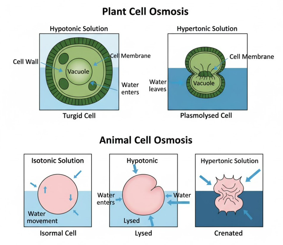
- Plant cells: Water (hypotonic) → turgid → supports stem & leaves; concentrated sugar → plasmolysis → membrane pulls from cell wall.
- Animal cells: Water → may burst (no cell wall); saline → shrinks due to water loss.
🧪 Experimental Illustration
- Beetroot / potato test: Place tissue in different sugar/alcohol solutions.
- Observe pigment leakage or tissue shrinkage/swelling.
- Higher solute concentration → more water leaves → cells shrink → more pigment in solution.
📌 Important Points to Remember
- Osmosis is passive → does not require energy.
- Only water molecules move.
- Movement always down water potential gradient.
- Partially permeable membranes are essential.
- Plant cells maintain turgor pressure → crucial for structure.
🧠 Quick Recap
Definition: Movement of free water molecules through a partially permeable membrane down a water potential gradient
Direction: High ψ → Low ψ
Energy: Passive (no ATP)
Tonicity effects: Hypotonic → swells, Hypertonic → shrinks, Isotonic → no change
Example: Beetroot/potato pigment leakage or turgidity/plasmolysis in plant cells
Membrane Transport – Passive, Active & Vesicular Transport
🌱 Introduction
Cell membranes are selectively permeable, controlling what enters and leaves the cell.
Molecules move into or out of cells by different transport mechanisms:
- Passive transport – no energy required.
- Active transport – requires energy (ATP).
- Vesicular transport – involves endocytosis and exocytosis.
Proteins in the membrane (carrier & channel proteins) help regulate transport.
🧩 (i) Types of Membrane Transport
1. Passive Transport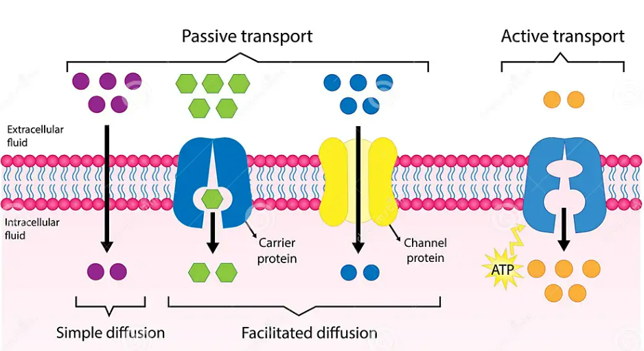
Definition: Movement of molecules down a concentration gradient without using energy.
Key Features:
- No ATP required
- High → Low concentration
Types:
- Simple diffusion: Small/nonpolar molecules (O₂, CO₂) pass directly through the lipid bilayer.
Example: Oxygen entering red blood cells. - Facilitated diffusion: Large/polar molecules (glucose, ions) use carrier or channel proteins.
Example: Glucose transport via GLUT transporters.
2. Active Transport
Definition: Movement of molecules against a concentration gradient using energy from ATP.
Key Features:
- Low → High concentration
- Requires ATP as an immediate energy source
- Involves carrier proteins (pumps)
Examples:
- Sodium-potassium pump (Na⁺/K⁺ pump): maintains ion balance in cells.
- Uptake of mineral ions in roots against concentration gradient.
3. Vesicular Transport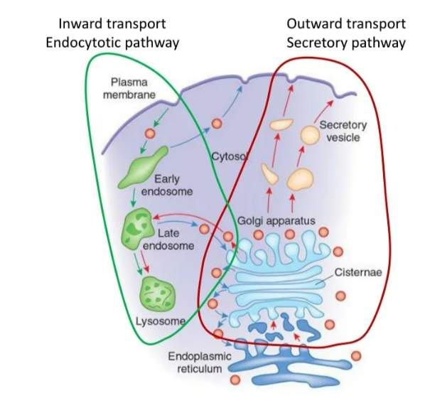
Definition: Transport of large molecules or bulk material via membrane-bound vesicles.
Types:
- Endocytosis: Cell engulfs material into vesicles.
- Phagocytosis: “cell eating” → solids (e.g., white blood cells engulf bacteria)
- Pinocytosis: “cell drinking” → liquids (e.g., uptake of extracellular fluid)
- Exocytosis: Vesicles fuse with the plasma membrane to release contents.
Example: Secretion of neurotransmitters, hormones.
🧬 (ii) Role of Carrier and Channel Proteins
| Protein Type | Function | Transport Type Example |
|---|---|---|
| Carrier proteins | Bind specific molecules, change shape, move them across the membrane | Facilitated diffusion, Active transport (Na⁺/K⁺ pump) |
| Channel proteins | Form hydrophilic pores for ions or water to pass | Facilitated diffusion (ions, water via aquaporins) |
Key Points:
- Carrier proteins are specific, may require ATP (active transport) or not (facilitated diffusion).
- Channel proteins are mostly passive, allow fast movement of ions.
- Both ensure selectivity and efficiency of membrane transport.
⚡ Summary Table – Transport Types
| Transport Type | Energy Required | Direction | Molecules | Proteins Involved | Example |
|---|---|---|---|---|---|
| Simple diffusion | No | Down gradient | O₂, CO₂ | None | Oxygen into RBCs |
| Facilitated diffusion | No | Down gradient | Glucose, ions | Carrier/Channel | Glucose via GLUT |
| Active transport | Yes (ATP) | Against gradient | Ions (Na⁺, K⁺), sugars | Carrier (pump) | Na⁺/K⁺ pump, mineral uptake |
| Endocytosis | Yes | Into cell | Solids/Liquids | Vesicles | Phagocytosis, pinocytosis |
| Exocytosis | Yes | Out of cell | Hormones, neurotransmitters | Vesicles | Secretion of insulin |
🧠 Quick Recap
Passive transport: down gradient, no energy
Active transport: against gradient, ATP required
Endocytosis: into cell (phagocytosis, pinocytosis)
Exocytosis: out of cell
Carrier proteins: bind & transport specific molecules
Channel proteins: form pores for ions/water
Mnemonic: “PANDA eats ENERGY-rich food”: Passive – ATP Not; Active – ATP Needed; Diffusion & Vesicles covered
RECOMMENDED ADDITIONAL PRACTICAL
Investigate Tissue Water Potential Using Plant Tissue & Solute Concentrations
🌱 Introduction
Water potential (Ψ) tells us how freely water molecules can move.
In this experiment, we find the water potential of plant tissues (like potato or beetroot) by placing them in different concentrations of a solute solution (usually sucrose).
The movement of water in/out of cells depends on osmosis — water moves from a region of higher water potential (less solute) to lower water potential (more solute) through a partially permeable membrane.
🧪 Aim
To determine the water potential of plant tissues by observing changes in their mass when placed in graded sucrose concentrations.
🔬 Apparatus & Materials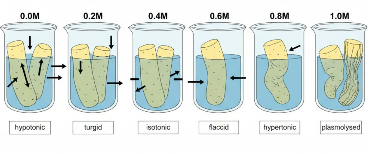
- Fresh potato cylinders/discs (or beetroot pieces)
- Sucrose solutions of different concentrations (e.g., 0.0M, 0.2M, 0.4M, 0.6M, 0.8M)
- Cork borer / knife
- Distilled water
- Beakers / test tubes
- Balance (for measuring mass)
- Paper towels
- Ruler / timer
⚗️ Procedure
- Cut equal-sized pieces of potato using a cork borer or knife.
- Blot dry with paper towel to remove surface water.
- Measure and record the initial mass of each piece.
- Place each piece in different sucrose solutions (from 0.0M → 0.8M).
- Leave for about 30–60 minutes (or as instructed).
- Remove, blot gently, and measure final mass of each piece.
- Record all readings in a table.
📊 Results & Observation
- In pure water (0.0M) → potato gains mass (water enters cells → turgid).
- In concentrated sucrose (e.g., 0.8M) → potato loses mass (water leaves cells → plasmolysed).
- At a certain concentration, no mass change occurs → the solution is isotonic with tissue → water potential of the tissue equals that solution’s potential.
📈 Data Analysis
- Plot a graph of percentage change in mass (y-axis) vs solute concentration (x-axis).
- The point where the curve crosses the x-axis (no change in mass) shows the tissue’s water potential.
💡 Key Concept
- Water moves by osmosis across the cell membrane.
- Higher solute concentration → lower water potential.
- Turgid cell = full of water
- Flaccid cell = lost some water
- Plasmolysed cell = cytoplasm shrinks away from cell wall
📘 Conclusion
The water potential of the plant tissue can be estimated from the solute concentration where no net mass change occurs.
This helps understand how plants balance water uptake and loss in different environments.
⚠️ Precautions
- Use equal-sized tissue samples.
- Dry gently to remove surface water before weighing.
- Keep time and temperature constant.
- Stir solutions gently to maintain uniform concentration.
🧠 Quick Recap
| Concept | Explanation |
|---|---|
| Aim | To find water potential of plant tissue |
| Principle | Water moves by osmosis (down water potential gradient) |
| Variable | Independent → solute concentration; Dependent → change in mass |
| Observation | Mass ↑ in hypotonic; ↓ in hypertonic |
| Isotonic Point | No change in mass → equals tissue’s water potential |
| Graph | % change in mass vs concentration |
| Key Outcome | Identifies Ψ of plant tissue |