Understanding Amino Acids, Peptides & Protein Structure
🌱 (i) Basic Structure of an Amino Acid
Amino acids are the building blocks of proteins.
🔹 General Structure: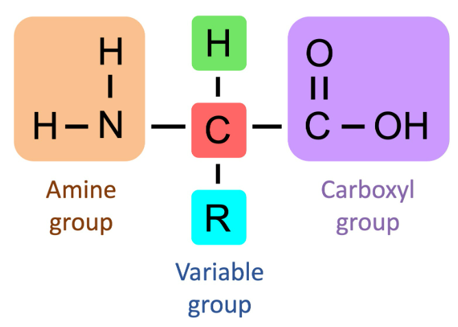
- Each amino acid has the same basic structure:
- Central carbon atom (α-carbon)
- Amino group (-NH₂)
- Carboxyl group (-COOH)
- Hydrogen atom (-H)
- R group (side chain) → differs for each amino acid
General Formula:
NH₂-CH(R)-COOH
The R group determines the type and properties of each amino acid (e.g., acidic, basic, polar, nonpolar).
Example: Glycine has R = H (simplest amino acid).
🔗 (ii) Formation of Polypeptides and Proteins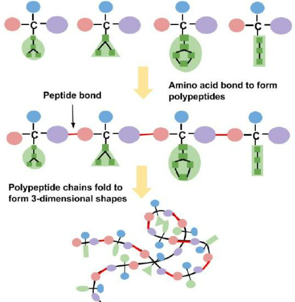
Proteins are long chains of amino acids joined together by condensation reactions.
🔹 Peptide Bond Formation:
- The carboxyl group (-COOH) of one amino acid reacts with the amino group (-NH₂) of another.
- A molecule of water (H₂O) is released.
- The resulting bond is a peptide bond (-CO-NH-).
Mnemonic: “COOH meets NH₂ → out goes H₂O → peptide bond forms.”
🔹 Chains Formed:
- 2 amino acids → dipeptide
- 3 or more → polypeptide
- Many polypeptides → protein
🧩 (iii) Protein Structure Levels & Significance
The shape (structure) of a protein determines its function. A small change in amino acid order can completely alter a protein’s function.
| Level | Description | Bonds Involved | Example / Note |
|---|---|---|---|
| Primary | Linear sequence of amino acids | Peptide bonds | Determined by DNA, a single change (e.g., in haemoglobin) can cause disease |
| Secondary | Folding of chain into α-helix or β-pleated sheet | Hydrogen bonds | Gives initial shape and strength |
| Tertiary | 3D shape (globular/fibrous) | H-bonds, ionic bonds, disulfide bridges, hydrophobic interactions | Determines overall shape & function |
| Quaternary | Multiple polypeptide chains joined | Same as tertiary + subunit interactions | e.g., haemoglobin (4 subunits) |
⚗️ (iv) Molecular Structure of Globular & Fibrous Proteins
🌐 Globular Proteins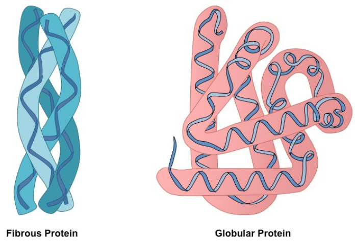
- Shape: Compact, spherical, soluble.
- Function: Metabolic (enzymes, transport, hormones).
- Bonds: Hydrogen, ionic, disulfide → maintain 3D folded shape.
- Example: Haemoglobin
- 4 polypeptide chains (2 alpha, 2 beta).
- Each contains a haem group (Fe²⁺) → binds oxygen.
- Function: Transports O₂ in blood.
- Structure-function link: Globular shape → fits through capillaries; soluble → easy transport.
🧵 Fibrous Proteins
- Shape: Long, rope-like, insoluble.
- Function: Structural (support, strength).
- Bonds: Many cross-links → tough, stable.
- Example: Collagen
- 3 polypeptide chains wound into a triple helix.
- Many hydrogen bonds between chains.
- Found in tendons, ligaments, skin.
- Structure-function link: Triple-helix + strong bonds → high tensile strength.
🧠 Summary Table
| Type | Structure | Solubility | Bonds | Function | Example |
|---|---|---|---|---|---|
| Globular | Spherical, folded | Soluble | H-bonds, ionic, disulfide | Metabolic (transport, enzyme, hormone) | Haemoglobin |
| Fibrous | Long, unbranched | Insoluble | Cross-linked, H-bonds | Structural (support, strength) | Collagen |
⚡ Quick Recap
Amino acids → building blocks of proteins
Peptide bond = CO–NH (via condensation)
Primary structure → determines higher structures
Secondary = α-helix / β-sheet (H-bonds)
Tertiary = 3D shape (H, ionic, disulfide, hydrophobic)
Quaternary = multiple chains (e.g., haemoglobin)
Globular = compact & soluble → transport/enzymes
Fibrous = long & strong → support/strength
RECOMMENDED ADDITIONAL PRACTICAL
Estimate Protein Concentration using Biuret Reagent & Colour Standards
🌱 Introduction
Proteins contain peptide bonds (-CO-NH-) that react with Biuret reagent to produce a purple/violet color.
The intensity of this color depends on how much protein is present – darker color = higher protein concentration.
This experiment uses a semi-quantitative method, meaning we compare the color of the test sample with known protein standards to estimate its concentration (not an exact numerical value, but a relative estimate).
🎯 Aim
To estimate the concentration of protein in an unknown sample using the Biuret test and color comparison with known standards.
🧰 Apparatus & Materials
- Test tubes
- Pipettes / droppers
- Test-tube rack
- Biuret reagent (alkaline copper sulfate solution)
- Protein standards (e.g., egg albumin or bovine serum albumin)
- Unknown protein solution (sample)
- Distilled water
- Color comparison chart or white background
⚗️ Reagents
Biuret Reagent Composition: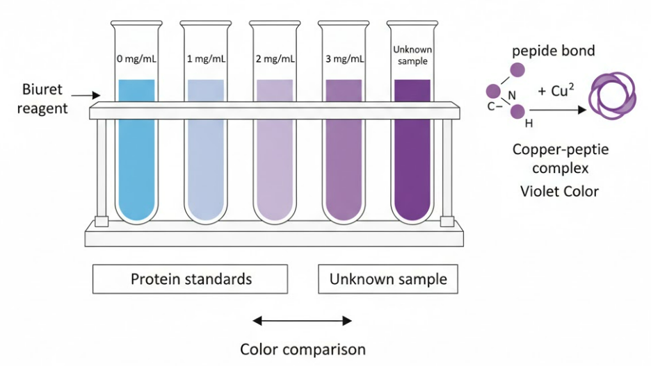
- Copper(II) sulfate (CuSO₄)
- Sodium hydroxide (NaOH)
- Potassium sodium tartrate (to stabilize Cu²⁺ ions)
🔬 Procedure
- Label test tubes for different protein concentrations, e.g., 0 mg/mL (control), 1 mg/mL, 2 mg/mL, 3 mg/mL, 4 mg/mL, and one for unknown sample.
- Pipette 2 mL of each standard solution into separate test tubes.
- Add 2 mL of Biuret reagent to each tube.
- Mix gently and leave for about 5-10 minutes at room temperature.
- Observe the color change: Light blue → violet/purple if protein present.
- Compare the color of the unknown sample with the color standards to estimate its protein concentration.
🎨 Observations
| Tube | Sample | Color After Adding Biuret | Interpretation |
|---|---|---|---|
| 1 | Distilled water (control) | Blue | No protein |
| 2 | 1 mg/mL | Light violet | Low protein |
| 3 | 2 mg/mL | Medium violet | Moderate protein |
| 4 | 3 mg/mL | Deep violet | High protein |
| 5 | Unknown sample | (Match with standard) | Estimate protein level |
🧠 Principle
Biuret reaction: peptide bonds in proteins react with Cu²⁺ ions in an alkaline medium to form a violet-colored complex.
The intensity of violet is directly proportional to the number of peptide bonds (i.e., protein concentration).
📘 Conclusion
By comparing the color intensity of the test sample with known standards, the approximate protein concentration can be estimated.
This is a semi-quantitative method – useful when exact measurement equipment (like spectrophotometers) is not available.
⚠️ Precautions
- Use equal volumes of all solutions.
- Keep time and temperature same for all tubes.
- Mix gently to avoid frothing.
- Handle Biuret reagent carefully (alkaline).
- Compare colors under same lighting conditions.
🧾 Key Notes
| Term | Meaning |
|---|---|
| Biuret test | Detects peptide bonds in proteins |
| Positive result | Violet or purple color |
| Semi-quantitative | Gives approximate, not exact, concentration |
| Color intensity | Indicates protein amount |
| Control | Distilled water → remains blue |
⚡ Quick Recap
Prepare different known protein concentrations + one unknown
Add equal volume of Biuret reagent
Wait 5–10 minutes
Observe color → light blue → violet
Compare unknown’s color with standards
💡 Darker violet = higher protein concentration