CORE PRACTICAL 5 – Observing Cells & Using a Graticule
🌱 Introduction
This practical focuses on observing animal cells under a light microscope, making labelled drawings, and measuring structures using a graticule. Understanding scale and calibration is key in microscopy.
1. Using a Light Microscope to Observe Animal Cells
- Step-by-step Procedure:
- Prepare the slide:
- Obtain a thin sample of animal tissue (e.g., cheek cells).
- Place on a clean slide, add a drop of stain (methylene blue/iodine).
- Gently place a cover slip, avoid air bubbles.
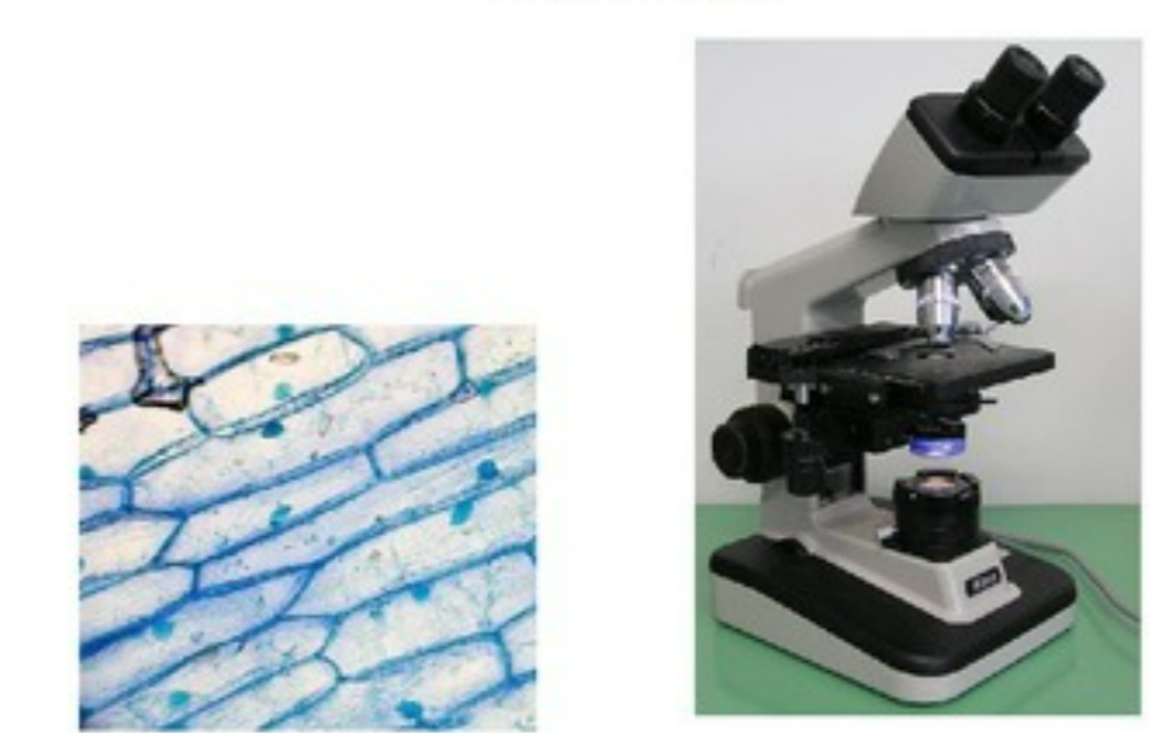
- Set up the microscope:
- Place slide on stage, secure with clips.
- Start with low power objective (×4 or ×10), use coarse focus.
- Switch to higher magnification (×40 or ×100 with immersion oil if needed).
- Adjust fine focus for clarity.
- Observation Tips:
- Look for cell shape (usually irregular).
- Identify nucleus, cytoplasm, cell membrane.
- Staining improves contrast, nucleus more visible.
- Labelled Drawings:
- Draw cells neatly with pencil.
- Include cell membrane, cytoplasm, nucleus.
- Label lines clear, include magnification, keep proportions correct.
- Prepare the slide:
2. Using a Graticule to Make Measurements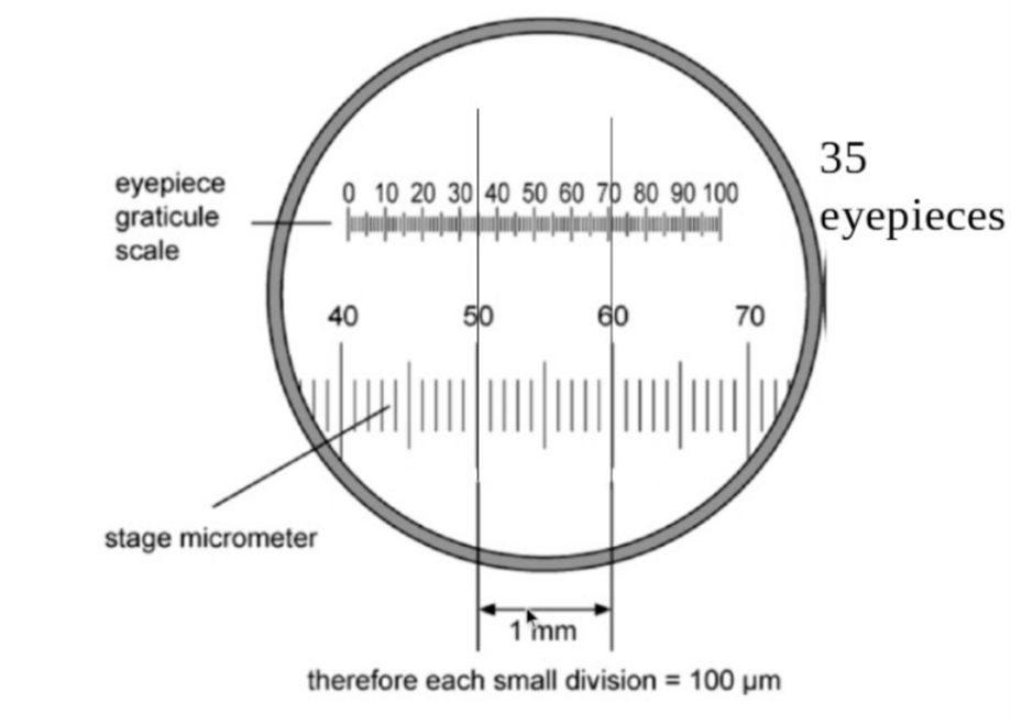
- Graticule Definition: Slide or eyepiece with a scale etched, used to measure actual sizes of cells/organelles.
- Steps for Measurement:
- Calibrate the graticule:
- Use a stage micrometer (e.g., 1 mm = 100 divisions).
- Align graticule with stage micrometer.
- Calculate value of one graticule division:
Value of 1 division = Known length on stage micrometer ÷ Number of graticule divisions
- Measure cells/organelles:
- Count graticule divisions spanned by the cell.
- Multiply by value of one division → actual size.
- Example: Cell spans 20 divisions, 1 division = 2 μm → Cell size = 40 μm.
- Calibrate the graticule:
3. Understanding Scale
- Magnification affects apparent size; graticule gives true measurement.
- Units usually in micrometers (μm).
📊 Summary Table
| Step / Concept | Key Points |
|---|---|
| Slide preparation | Thin sample, stain, cover slip, avoid air bubbles |
| Light microscope usage | Start low power → coarse/fine focus → high power |
| Observation | Look for nucleus, cytoplasm, cell membrane, irregular shape |
| Labelled drawing | Pencil, clear labels, correct magnification, proportional |
| Graticule measurement | Calibrate with stage micrometer → count divisions → calculate actual size |
| Scale understanding | Magnification changes apparent size → graticule gives real size |
⚡ Quick Recap
Staining → improves contrast, makes organelles visible.
Light microscope → observe cells at different magnifications.
Labelled drawings → neat, proportional, include magnification.
Graticule → measures cells/organelles accurately.
Calibration → converts graticule divisions to actual size.