Core Practical 7: Identifying Tissue Types Within Stems
🌱 Aim:
To use a light microscope to:
- Observe and draw plan diagrams of transverse sections (T.S.) of roots, stems, and leaves.
- Observe and draw individual cells from plant tissues.
- Identify and label sclerenchyma fibres, phloem, sieve tubes, and xylem vessels, noting their positions in each organ.
🔬 Apparatus & Materials:
- Light microscope
- Prepared T.S. slides of root, stem, and leaf
- Mounted needle, forceps, coverslip
- Microscope graticule (for measurements, if needed)
- Drawing instruments
🧩 (i) Plan Diagrams – Transverse Sections (T.S.)
1. Root (T.S.) 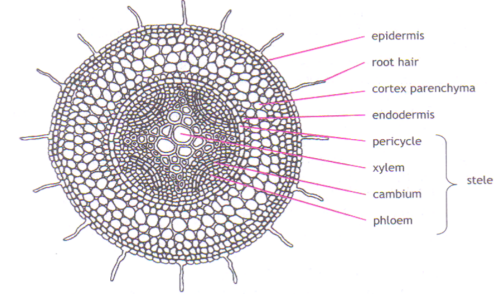
Observation:
- Circular arrangement of tissues.
- Xylem forms a central star-shaped pattern with phloem between arms.
- Endodermis forms a ring around vascular bundle.
- Cortex surrounds the endodermis.
Key Labels: Xylem (center star), Phloem (between xylem arms), Endodermis, Cortex, Epidermis
🧠 Tip: Root xylem → “X” shape (X for Xylem → Root)
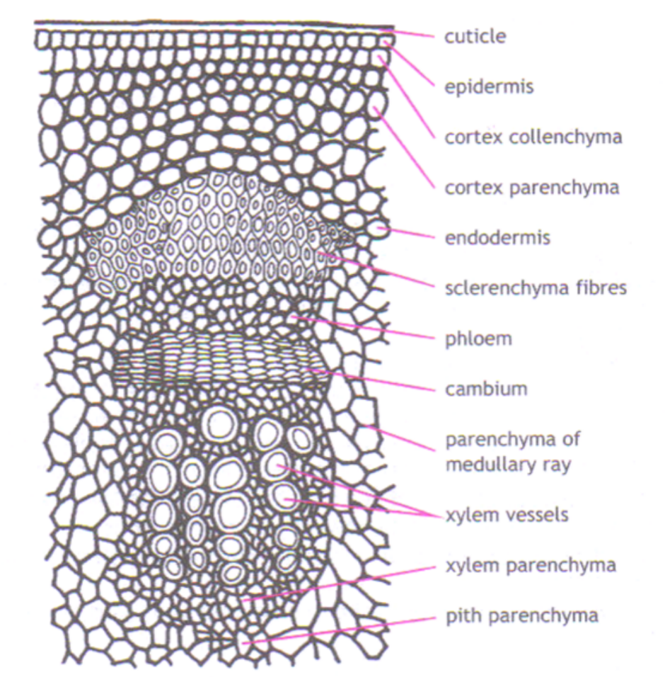
2. Stem (T.S.)
Observation:
- Epidermis forms outer boundary.
- Vascular bundles arranged in a ring around the central pith.
- Each vascular bundle has: Xylem (inner side), Phloem (outer side), and Sclerenchyma cap (outermost).
- Cortex lies between epidermis and vascular ring.
Key Labels: Xylem vessels, Phloem tissue, Sclerenchyma cap, Cortex, Epidermis, Pith
🧠 Tip: From inside → out = Xylem → Phloem → Sclerenchyma
3. Leaf (T.S.)
Observation:
- Upper & lower epidermis with a waxy cuticle.
 Mesophyll divided into:
- Palisade layer (densely packed cells → photosynthesis)
- Spongy layer (loose cells with air spaces)
- Vascular bundle (vein): Xylem on top, Phloem below.
- Stomata on lower surface.
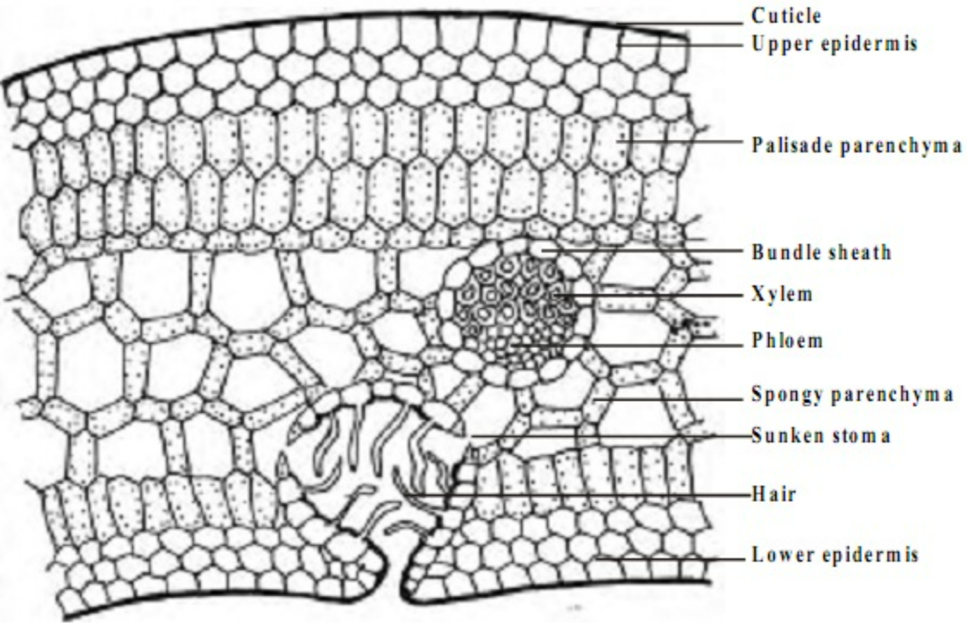 Mesophyll divided into:
Key Labels: Upper & lower epidermis, Palisade mesophyll, Spongy mesophyll, Xylem (above), Phloem (below), Stomata
🧠 Tip: “Xy up, Phlo down” in leaf veins (Xylem above Phloem)
🧫 (ii) Observation of Individual Cells in Plant Tissues
| Tissue | Cell Features Under Microscope | Notes |
|---|---|---|
| Parenchyma | Thin walls, living cells, large vacuoles | Storage & support |
| Collenchyma | Uneven thickening at corners | Flexible support (especially in young stems) |
| Sclerenchyma | Thick, lignified walls, no cytoplasm | Dead, mechanical support |
| Xylem vessels | Empty, thick walls, circular or polygonal | Water transport |
| Phloem sieve tubes | Living cells, sieve plates visible | Transport of sugars |
🧬 (iii) Identification of Major Tissues
| Tissue | Appearance Under Microscope | Position in Organ | Function |
|---|---|---|---|
| Sclerenchyma fibres | Long, narrow, thick lignified walls | Around vascular bundles (outermost) | Mechanical support |
| Phloem (sieve tubes + companion cells) | Thin-walled, living, often greenish | Just outside xylem | Translocation of sugars |
| Xylem vessels | Large, empty circles or ovals, thick lignified walls | Innermost part of vascular bundle | Water & mineral transport |
| Parenchyma | Thin walls, large vacuole | Cortex or pith | Storage, support, photosynthesis |
⚡ Quick Recap
Sclerenchyma → Dead, lignified → support
Xylem → Dead, lignified → water & mineral transport + support
Phloem → Living → sugar transport
Position (in stem): Xylem inside → Phloem → Sclerenchyma outside
In leaf vein: Xylem above, Phloem below
In root: Xylem star shape at center