Sliding Filament Theory of Skeletal Muscle Contraction
🌟 Introduction
Muscle contraction happens because myosin and actin filaments slide over each other, shortening the sarcomere. This sliding is tightly controlled by regulatory proteins and triggered by calcium ions.
🧩 Key Players and Their Roles
Actin (Thin filament)
- Long, twisted protein chain.
- Has binding sites for myosin heads.
Myosin (Thick filament)
- Has myosin heads that pull actin.
- Each head has:
- ATP binding site
- Actin binding site
Tropomyosin
- Long protein lying along actin.
- Blocks actin’s binding sites when the muscle is relaxed.
Troponin
- Attached to tropomyosin.
- Has a binding site for Ca²⁺ ions.
- Acts like a “switch” for contraction.
Calcium ions (Ca²⁺)
- Released from the sarcoplasmic reticulum (SR).
- Bind to troponin to start contraction.
ATP
- Powers myosin head movement.
- Needed for both cross bridge formation and detachment.
ATPase
- Enzyme on myosin head.
- Breaks ATP to release energy for the power stroke.
⚡ Full Process of Muscle Contraction (Sliding Filament Theory)
1. Nerve impulse arrives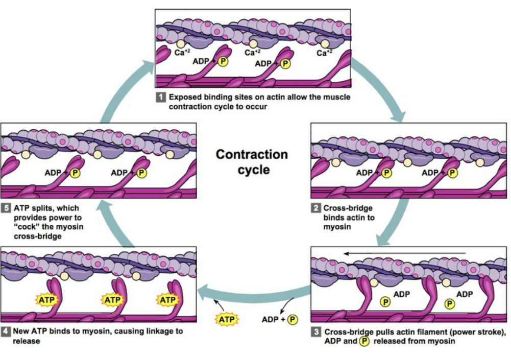
- Travels down the motor neurone.
- Causes Ca²⁺ release from the sarcoplasmic reticulum into the sarcoplasm.
2. Calcium activates the actin sites
- Ca²⁺ binds to troponin.
- Troponin changes shape.
- Pulls tropomyosin away from the actin binding sites.
- Myosin heads can now attach.
Key line: Calcium removes the “tropomyosin block”.
3. Cross-bridge formation
- Myosin head (energised by ATP breakdown) attaches to actin.
- This connection is called a cross bridge.
4. Power stroke
- Myosin head bends, pulling actin toward the centre of the sarcomere.
- This uses energy from ATP hydrolysis by ATPase.
- Result: Sarcomere shortens and the muscle contracts.
5. Detachment
- A new ATP molecule binds to the myosin head.
- This breaks the cross bridge.
- Myosin releases actin.
6. Resetting (Re-cocking)
- ATP is hydrolysed by ATPase.
- Energy resets the myosin head to its upright position ready to bind again.
7. Cycle repeats
- As long as Ca²⁺ is present and ATP is available, cross bridges keep forming and actin keeps sliding.
🧊 Muscle Relaxation
- When the nerve impulse stops, Ca²⁺ is pumped back into the SR.
- Troponin returns to its original shape.
- Tropomyosin covers actin binding sites again.
- Myosin cannot attach, so sliding stops.
- Muscle fibre relaxes.
📌 Summary Table
| Component | Role in Contraction |
|---|---|
| Actin | Thin filament pulled by myosin |
| Myosin | Thick filament forming cross bridges |
| Troponin | Binds Ca²⁺ to start contraction |
| Tropomyosin | Blocks actin sites during rest |
| Ca²⁺ ions | Remove tropomyosin block |
| ATP | Detaches myosin and resets head |
| ATPase | Breaks ATP to release energy |
🧾 Quick Recap
Ca²⁺ from SR binds troponin.
Troponin moves tropomyosin off actin.
Myosin binds actin forming cross bridges.
Power stroke uses ATP energy.
New ATP detaches myosin.
ATPase resets the myosin head.
Cycle repeats until Ca²⁺ is removed.
Ca²⁺ from SR binds troponin.
Troponin moves tropomyosin off actin.
Myosin binds actin forming cross bridges.
Power stroke uses ATP energy.
New ATP detaches myosin.
ATPase resets the myosin head.
Cycle repeats until Ca²⁺ is removed.