Detection of Stimuli by Rod Cells in Mammals
🌱 Introduction
Rod cells in the retina detect low light levels. They convert light stimuli into electrical signals (action potentials) in optic neurones.
🔍 Structure of Rod Cells
- Outer segment: contains stacked membranes with rhodopsin.
- Inner segment: contains nucleus and mitochondria for energy.
- Synaptic terminal: connects to bipolar cells, then optic neurones.
🔹 Key Molecules
| Molecule | Role |
|---|---|
| Rhodopsin | Photopigment in rods; absorbs light |
| Opsin | Protein component of rhodopsin |
| Retinal | Light-sensitive molecule (changes shape when light hits) |
| Sodium ions (Na⁺) | Maintain dark current through cation channels |
| Cation channels | Let Na⁺ flow in darkness, keeping rods depolarised |
| Hyperpolarisation | Response to light, reduces neurotransmitter release |
🧬 How Rod Cells Work
1. In the Dark
- Rhodopsin inactive, cation channels open.
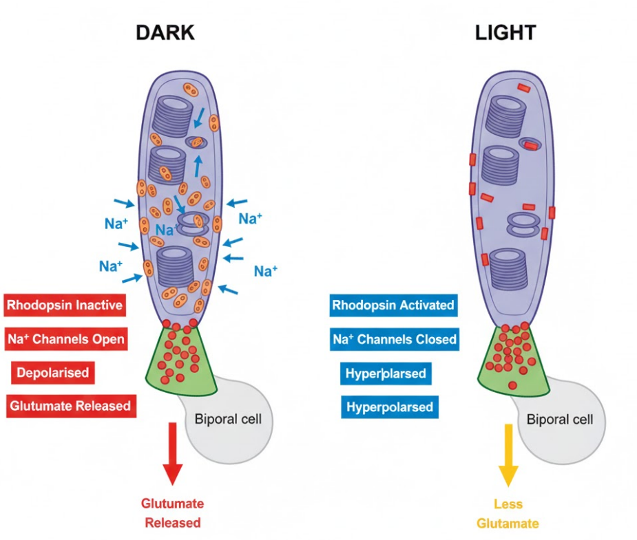 Na⁺ ions enter rod cell continuously → depolarised (~-40 mV).
- Rod releases neurotransmitter (glutamate) at synapse → inhibits bipolar cells.
- No signal sent to brain.
2. In the Light
- Light hits retinal, causing it to change shape (cis → trans).
- Activates opsin → triggers cascade → cGMP broken down.
- Cation channels close → Na⁺ entry stops.
- Rod hyperpolarises (~-70 mV).
- Glutamate release decreases → bipolar cells activated → action potentials in optic neurones.
- Signal transmitted to brain → perception of light.
🔹 Key Points
- Rods are more sensitive to low light than cones.
- Hyperpolarisation, not depolarisation, signals light detection.
- Na⁺ ion flow controls the “dark current”.
- Rods communicate with bipolar cells, which then activate optic neurones.
📊 Summary Table
| Condition | Rhodopsin | Cation Channels | Membrane Potential | Neurotransmitter |
|---|---|---|---|---|
| Dark | Inactive | Open | Depolarised (~-40 mV) | Glutamate released (inhibits bipolar cells) |
| Light | Activated | Closed | Hyperpolarised (~-70 mV) | Glutamate release decreases → bipolar cells activated |
📦 Quick Recap
Rods detect dim light, rhodopsin = opsin + retinal.
Dark: Na⁺ enters → depolarised → neurotransmitter inhibits bipolar cells.
Light: Retinal changes shape → opsin activated → cation channels close → hyperpolarisation.
Bipolar cells activated → action potential in optic neurones → brain detects light.