▶️ Answer/Explanation
Answer: D. DNA is a double helix with antiparallel sugar–phosphate backbones.
Explanation: 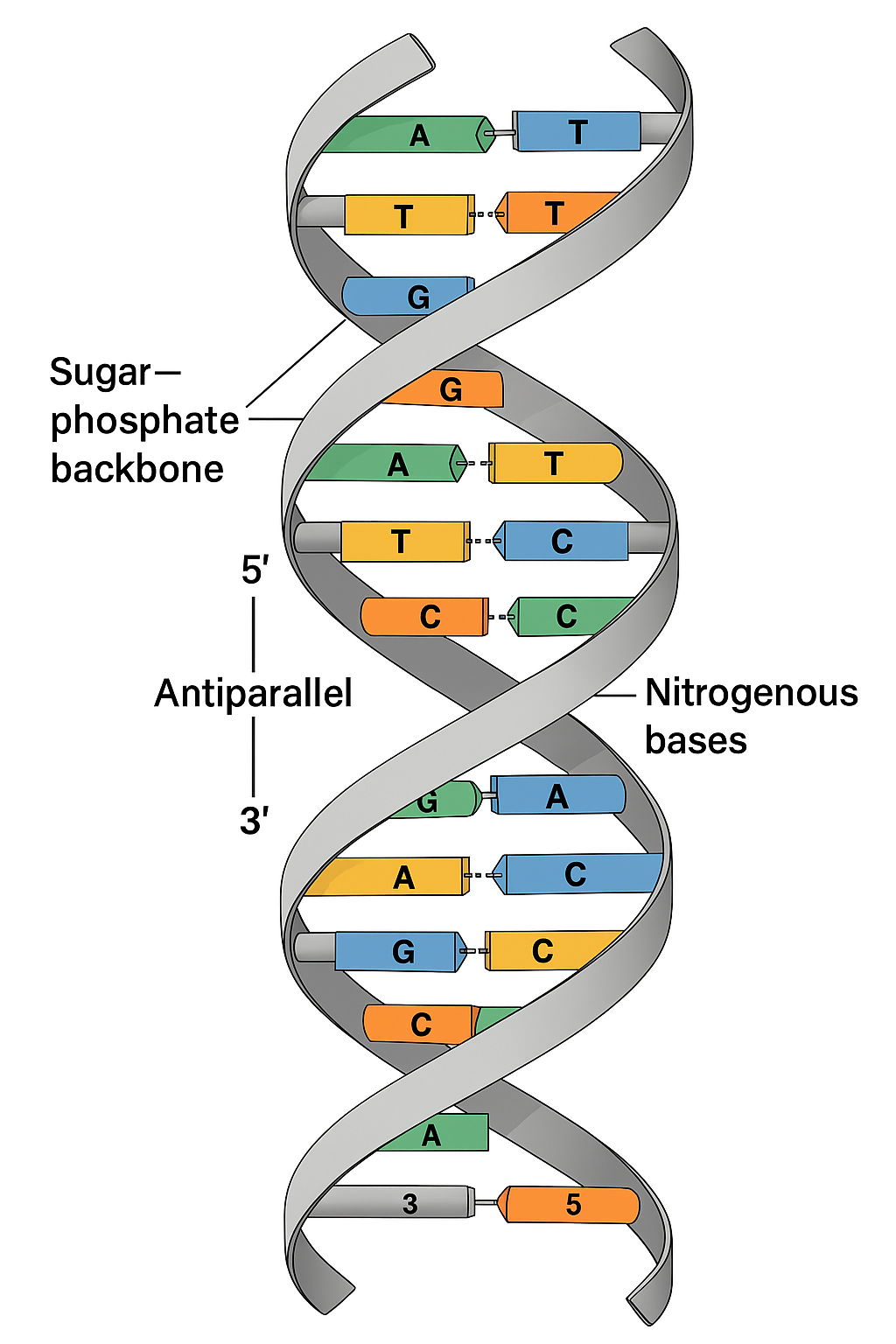
Watson and Crick proposed the first accurate three-dimensional structure of DNA. Their model revealed important details about DNA’s shape and how its strands are arranged, which explained how genetic information is stored and copied.
Evaluating the options:
A. Incorrect – It was already known that DNA is a polynucleotide (a chain of nucleotides) before their model.
B. Incorrect – The idea that adenine and thymine are present in equal amounts (Chargaff’s rules) was known before Watson and Crick’s work.
C. Incorrect – While DNA’s helical shape was suggested by earlier X-ray diffraction studies, Watson and Crick’s model went beyond just showing a helix.
D. Correct – Watson and Crick’s model was the first to show that DNA is a double helix with two antiparallel sugar–phosphate backbones, and that the bases pair specifically (A with T, G with C), explaining DNA’s structure and function.

▶️ Answer/Explanation
Answer: D. A chromosome from a cell in metaphase
Explanation:
In dividing cells, chromosomes become visible as distinct structures when they condense. During metaphase, chromosomes line up in the middle of the cell, and each one consists of two sister chromatids joined at a centromere. A micrograph from this stage often shows X-shaped chromosomes, which are duplicated chromosomes prepared for separation.
Evaluating the options:
A. Incorrect – Two chromosomes with four chromatids would suggest two duplicated chromosomes, but metaphase typically shows individual chromosomes aligned, not grouped in this way. This phrasing is not a standard description.
B. Incorrect – In telophase, chromosomes de-condense and become less visible as the nuclear envelope reforms. The micrograph would not show clearly condensed chromosomes like those seen in metaphase.
C. Incorrect – During G1 of interphase, chromosomes are not visible under a light microscope because they are in a loose, uncoiled chromatin form.
D. Correct – This image likely shows a chromosome in metaphase, where it is fully condensed and visible, typically with two sister chromatids joined at the centromere, forming an X shape.