▶️ Answer/Explanation
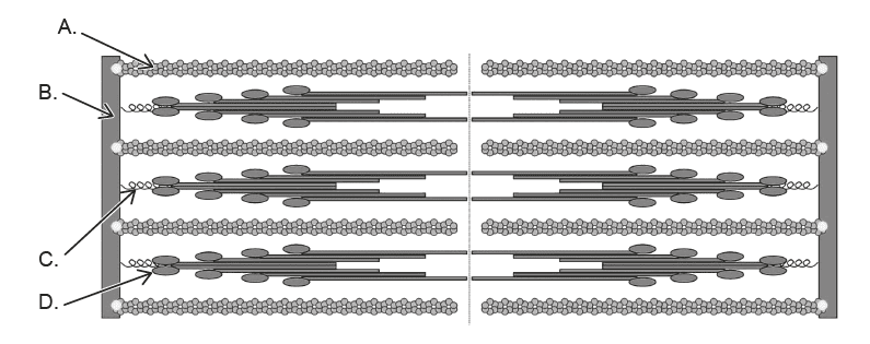
During relaxation, the H zone and I band increase in length because the thin filaments slide back outward, reducing their overlap with thick filaments. The A band remains constant. The labelled part (C) represents the region that changes length most noticeably with contraction and relaxation.
✅ Answer: (C)
✅ Answer: (C)
Question
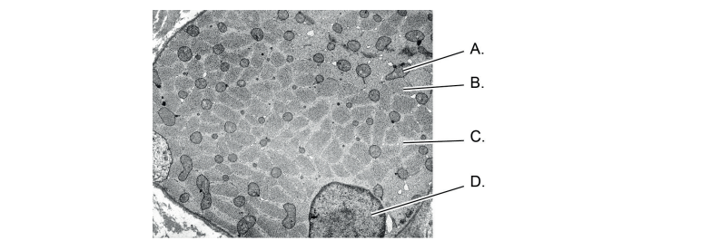
The electron micrograph displays a transverse section of a skeletal muscle fiber. Where is the specialized endoplasmic reticulum (sarcoplasmic reticulum) likely located?
▶️Answer/Explanation
Answer: C
Explanation:
Let’s analyze this skeletal muscle fiber image:
- Label A: Looks like a small vesicle or tubule near the edge could be part of SR or something else.
 Label B: The thin lines or membranes near A maybe tubules but not the main bulk.
- Label C: The large, network-like mesh surrounding the darker, denser structures inside (myofibrils).
- Label D: Large dark, dense blob definitely the nucleus.
 Label B: The thin lines or membranes near A maybe tubules but not the main bulk.
Why Label C is the sarcoplasmic reticulum?
- The sarcoplasmic reticulum (SR) forms an elaborate membranous network wrapping around the myofibrils.
- It appears as a mesh or web-like structure that surrounds the contractile elements.
- This network is visible as the light-stained areas wrapping the darker myofibrils in the image.
- So, C fits perfectly for the SR.
To clear confusion:
- A and B are likely small tubules or vesicles but not the full SR structure.
- C is the major network of tubules characteristic of the SR, which stores calcium needed for muscle contraction.
- D is the nucleus, obvious by its size and density