▶️ Answer/Explanation
Detailed solution
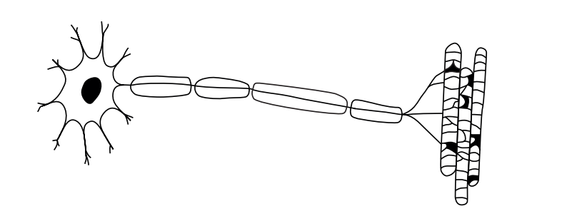
(a)
Yes. The neuron is insulated because the axon is covered by a myelin sheath, formed by Schwann cells in the diagram.
(b)
Two shared structures:
1. Ribosomes
2. Plasma membrane (phospholipid bilayer)
(Other acceptable examples: cytoplasm, DNA)
(c)
When an action potential reaches the presynaptic terminal, the membrane depolarizes and voltage-gated \(\text{Ca}^{2+}\) channels open. The influx of calcium ions triggers synaptic vesicles carrying neurotransmitters to move toward and fuse with the presynaptic membrane. The neurotransmitters are then released into the synaptic cleft via exocytosis.