C3.1.1 – System Integration
System integration is the process by which different components of a living system work together to perform an overall function. It ensures that parts do not act in isolation but contribute to the organism’s survival.
🌱 In Living Organisms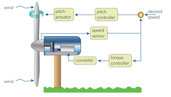
- All organisms – from unicellular to multicellular – require integration between systems.
- Communication occurs through:
- Nervous system (electrical signals)
- Endocrine system (chemical messengers)
- Local feedback loops (e.g., enzyme control)
- Example (Human): Digestion – nervous and endocrine systems coordinate muscular contractions (peristalsis) and enzyme secretion.
- Example (Plant): Phototropism – integration between light-sensing cells and growth-regulating hormones.
⚙️ Engineering Analogy: Wind Turbine
| Component | Role in Integration |
|---|---|
| Sensors | Monitor wind speed/direction |
| Controllers | Process sensor data and send commands |
| Pitch actuator | Adjusts blade angle to optimise efficiency |
| Torque controller | Maintains safe power output |
Just as a wind turbine relies on sensors, controllers, and actuators to function smoothly, organisms rely on sensing, processing, and responding systems to stay alive.
📌 Why It Matters in Biology
- Prevents conflicting actions between systems.
- Enables adaptation to changing environments.
- Allows complex behaviours and homeostasis.
– System integration = coordination of different components to achieve a shared goal.
– Communication can be electrical, chemical, or mechanical.
– Both biology and engineering use integration to optimise performance and efficiency.
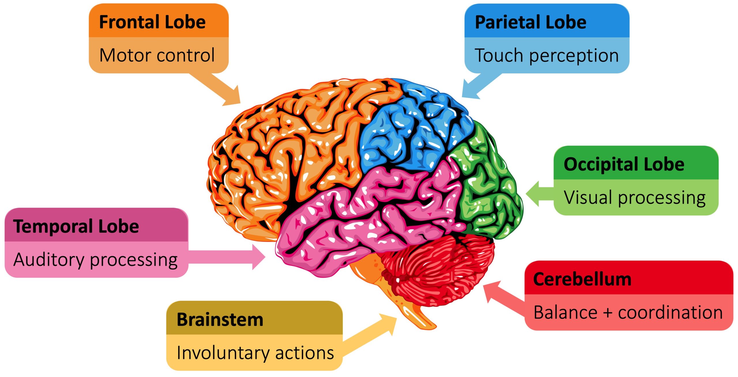
C3.1.4 – The Brain as the Central Information Integration Organ
Role of the Brain
The brain acts as the central integration hub of the body. It:
- Gathers information from multiple sensory inputs (sight, sound, touch, smell, taste, balance, and internal body signals).
- Processes and compares inputs to form a coherent understanding of the environment.
- Coordinates appropriate responses based on processed information.
Combining Multiple Inputs
Example — Catching a ball:
- Eyes judge speed and direction.
- Inner ear detects body position and balance.
- Muscles and joints send feedback on limb position.
- The brain integrates all data to guide accurate hand movement → smooth, coordinated action.
Learning and Memory
- Learning — Adapts responses through experience and practice.
- Memory — Stores and retrieves information to influence future actions.
- Example: Touching a hot pan once → stored as a painful memory → brain triggers quick withdrawal next time.
Key Features of Brain Integration
| Function | Description | Example |
|---|---|---|
| Sensory integration | Merging multiple inputs for accurate perception | Hearing footsteps + seeing shadow |
| Learning | Modifying behaviour through experience | Improving aim in basketball |
| Decision-making | Choosing the best response based on data | Stopping at a red light |
| Memory | Storing info for future use | Recognising a familiar face |
– The brain integrates information from many sources.
– It processes sensory data to guide coordinated, effective actions.
– Learning and memory allow adaptive responses over time.
– This integration is essential for survival and complex behaviour.
C3.1.5 – The Spinal Cord as an Integrating Centre for Unconscious Processes
🧠Conscious vs. Unconscious Processes
Conscious processes: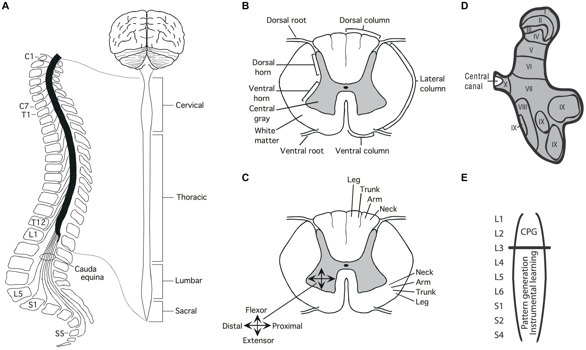
– Controlled by the brain.
– Involve awareness, thinking, and decision-making.
– Example: Deciding to wave at a friend.
Unconscious processes:
– Controlled without active thought.
– Often involve the spinal cord as an integrating centre.
– Example: Rapid withdrawal of hand from something hot.
⚡ Role of the Spinal Cord
– Acts as an integration and control centre for certain reflex actions.
– Receives sensory input → processes it → sends motor output without brain involvement.
– Advantage: Speeds up response time, increasing chances of avoiding harm.
🔄 Reflex Arc Pathway
- Stimulus detected by sensory receptor (e.g., pain receptor in skin).
- Sensory neuron sends impulse to spinal cord.
- Relay neuron in spinal cord integrates and sends signal to motor neuron.
- Motor neuron sends impulse to effector (e.g., muscle).
- Response occurs – fast, automatic, and protective.
📊 Comparison Table
| Feature | Conscious Process | Unconscious Process |
|---|---|---|
| Control centre | Brain | Spinal cord |
| Awareness | Yes | No |
| Speed | Slower | Faster |
| Example | Choosing to speak | Knee-jerk reflex |
– The spinal cord integrates unconscious processes, mainly reflexes.
– Reflex arcs allow rapid, automatic responses without brain involvement.
– This helps protect the body from injury by minimising reaction time.
C3.1.7 – Output from the Cerebral Hemispheres to Muscles Through Motor Neurons
🧾 What Are Motor Neurons?
Specialised nerve cells that carry instructions from the central nervous system (CNS) to effectors (usually muscles or glands).
They are part of the efferent pathway (signals travelling away from the CNS).
In this context: they carry messages from the cerebral hemispheres to muscles.
🔍 From Thought to Movement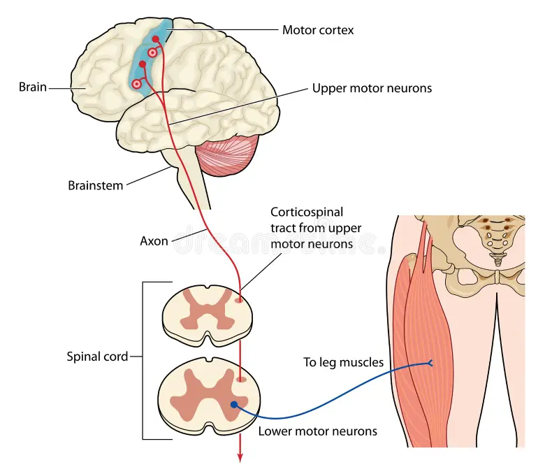
- Decision made in the cerebral hemispheres.
- Example: Deciding to wave at a friend.
- Motor commands sent down through descending pathways in the spinal cord.
- Motor neuron activation → electrical impulse travels to muscle fibres.
- Muscle contraction occurs → producing movement.
⚙️ Key Steps in the Output Pathway
- Cerebral hemispheres: Plan and initiate voluntary movement.
- Spinal cord: Acts as a transmission route for motor signals.
- Motor neurons: Connect CNS to muscles, releasing neurotransmitters (e.g., acetylcholine) at neuromuscular junctions.
- Muscles: Respond by contracting.
📊 Sensory vs Motor Neurons
| Feature | Sensory Neuron | Motor Neuron |
|---|---|---|
| Direction of impulse | Towards CNS (afferent) | Away from CNS (efferent) |
| Function | Carry sensory input from receptors | Carry motor output to effectors |
| Connected to | Receptors → CNS | CNS → Muscles/glands |
Motor neurons deliver output signals from the cerebral hemispheres to muscles, causing them to contract and produce voluntary movements.
C3.1.9 – Pain Reflex Arcs as an Example of Involuntary Responses with Skeletal Muscle as the Effector
A reflex arc is the pathway that controls an involuntary and rapid response to a stimulus.
Purpose: Protects the body from harm by bypassing conscious brain control.
🔍 Example: Pain Withdrawal Reflex in the Hand
Scenario: Touching a sharp object.
| Step | Component | Role |
|---|---|---|
| 1 | Pain receptor (free sensory nerve ending in skin) | Detects tissue damage. |
| 2 | Sensory neuron | Carries impulse towards spinal cord. |
| 3 | Interneuron (in grey matter of spinal cord) | Connects sensory neuron to motor neuron; processes signal locally. |
| 4 | Motor neuron | Sends impulse to the effector. |
| 5 | Skeletal muscle (effector) | Contracts, pulling the hand away from danger. |
📌 Key Features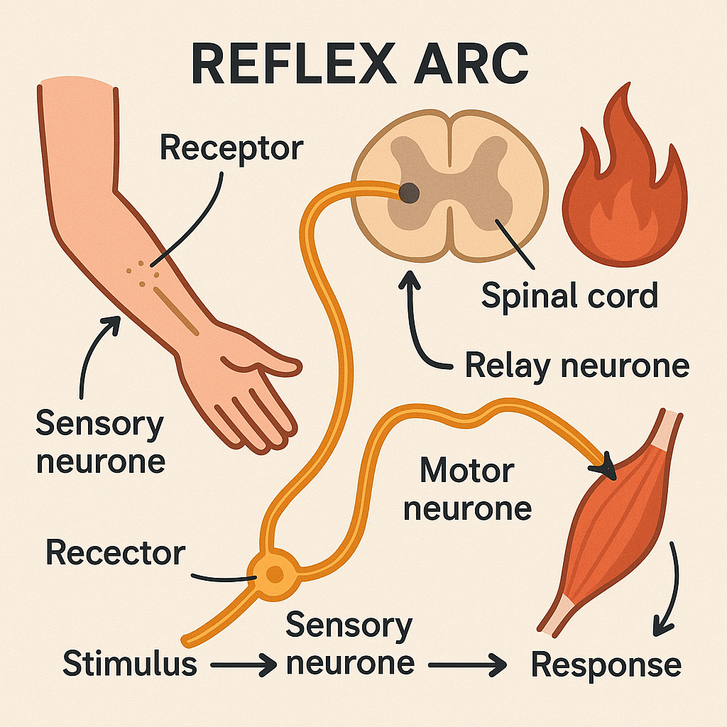
- Involuntary → no conscious control.
- Fast → avoids brain processing delays.
- Uses one interneuron → simple, rapid pathway.
- Skeletal muscle is the final effector.
🖇 Pathway in Short
Receptor → Sensory neuron → Interneuron → Motor neuron → Effector muscle
The pain withdrawal reflex protects the body by rapidly activating skeletal muscles through a spinal reflex arc. A free nerve ending detects pain, and the signal travels through a sensory neuron, is relayed by an interneuron, and triggers a motor neuron to cause muscle contraction – all without conscious thought.
C3.1.10 – Role of the Cerebellum in Coordinating Skeletal Muscle Contraction and Balance
What Is the Cerebellum?
A part of the brain located at the back of the skull, beneath the cerebrum.
Not responsible for starting movements – instead, it coordinates them.
Main Roles
| Function | Description | Example |
|---|---|---|
| Balance & posture | Adjusts muscle activity to keep the body stable, even when moving. | Standing on one foot without falling. |
| Fine-tuning movements | Ensures skeletal muscles contract in the right sequence and timing. | Throwing a ball accurately. |
| Motor learning | Helps develop and improve skills requiring precise muscle coordination. | Learning to play the piano. |
Key Points to Remember
- The cerebellum works with other brain regions to make movement smooth and coordinated.
- Without the cerebellum’s input, movements can become jerky and unsteady.
- Essential for everyday activities like walking, writing, or sports.
The cerebellum fine-tunes skeletal muscle movements, maintains balance and posture, and helps in learning new motor skills. It does not initiate movement but ensures that actions are smooth, coordinated, and well-timed.
C3.1.13 – Control of the Endocrine System by the Hypothalamus and Pituitary Gland
📍 Hypothalamus: The Master Controller
Located in the brain, just above the pituitary gland.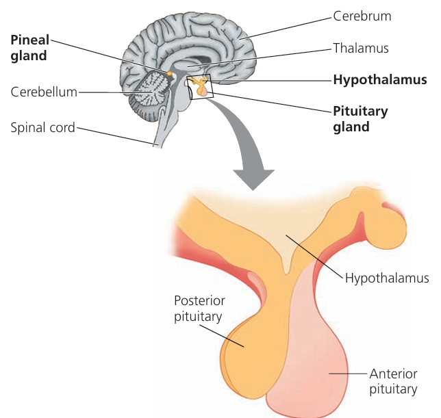
Acts as a link between the nervous system and endocrine system.
Monitors:
- Blood temperature
- Hormone levels
- Nutrient levels
Sends signals to the pituitary gland to adjust hormone secretion.
🏛 Pituitary Gland: The Master Gland
- A small gland beneath the hypothalamus.
- Called the “master gland” because it releases hormones that control other endocrine glands.
- Works under the control of the hypothalamus.
🔄 How They Work Together
| Step | Action |
|---|---|
| 1 | Hypothalamus detects change in the body (e.g., low thyroid hormone) |
| 2 | Sends releasing/inhibiting signals to pituitary |
| 3 | Pituitary releases hormones into the blood |
| 4 | These hormones target other endocrine glands (thyroid, adrenal, gonads) |
| 5 | Target glands produce their own hormones to restore balance |
🧩 Key Functions Controlled
- Growth and development (via growth hormone)
- Metabolism (via thyroid hormones)
- Stress response (via adrenal hormones)
- Reproductive functions (via gonad hormones)
The hypothalamus monitors the body’s internal environment and signals the pituitary gland to adjust hormone secretion. The pituitary, in turn, controls other endocrine glands, ensuring coordinated regulation of growth, metabolism, reproduction, and stress responses.
C3.1.14 – Feedback Control of Heart Rate via Baroreceptors and Chemoreceptors
📍 Key Sensory Receptors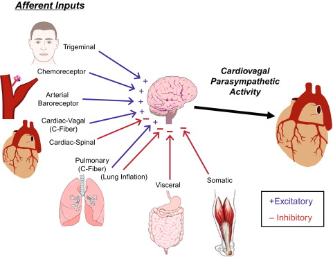
- Baroreceptors
- Location: Walls of the aorta and carotid arteries
- Function: Monitor blood pressure
- If BP rises → send signals to reduce heart rate
- If BP falls → send signals to increase heart rate
- Chemoreceptors
- Location: Same as baroreceptors (aorta & carotid arteries)
- Function: Monitor blood pH, oxygen (O₂), and carbon dioxide (CO₂) concentration
- Detect changes (e.g., low O₂, high CO₂, low pH) and signal to increase heart rate for better gas exchange
🧠 Role of the Medulla Oblongata
- Acts as the cardiovascular control centre
- Processes signals from baroreceptors and chemoreceptors
- Sends nerve impulses via:
- Sympathetic nerves → increase heart rate & stroke volume
- Vagus nerve (parasympathetic) → decrease heart rate
🔄 Feedback Loop
| Step | What Happens |
|---|---|
| 1 | Receptors detect change (BP, O₂, CO₂, pH) |
| 2 | Signals sent to medulla |
| 3 | Medulla sends impulses via appropriate nerves |
| 4 | SAN (pacemaker) adjusts heart rate |
| 5 | Blood pressure & gas levels return to normal |
⚡ Extra Factor – Epinephrine
- Released by adrenal glands during stress/exercise
- Stimulates SAN → increases heart rate
Heart rate is controlled by a feedback system involving baroreceptors, chemoreceptors, the medulla, and autonomic nerves. This ensures blood pressure, oxygen, and carbon dioxide levels remain stable, adjusting heart rate instantly to meet the body’s needs.
Additional Higher Level
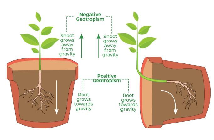
C3.1.17 – Observations of Tropic Responses in Seedlings
What Are Tropic Responses?
Tropism = growth movement in response to a stimulus.
- Phototropism → growth towards light (positive) or away from light (negative)
- Gravitropism (geotropism) → growth towards gravity (positive) or away from gravity (negative)
Making Observations
| Observation Type | What It Means | Example in Tropism Study |
|---|---|---|
| Qualitative | Descriptive, no numbers | “Seedling stem bends towards the light” |
| Quantitative | Numerical measurement | “Curvature angle = 35° towards light source” |
Practical Skills
- Qualitative data → draw diagrams showing direction and degree of bending
- Quantitative data → measure angle of curvature with a protractor
- Variables to control → light intensity, direction, duration; seedling age; soil moisture; temperature
Precision, Accuracy & Reliability
- Precision = how close repeated measurements are to each other — ↑ by using finer tools & consistent technique
- Accuracy = how close measurements are to the true value — ↑ by aligning protractor carefully & avoiding parallax errors
- Reliability = consistency of results when repeated — ↑ by repeating experiment & averaging results
In seedling tropism experiments, qualitative observations give a descriptive idea of the response, while quantitative measurements provide exact numerical data like curvature angle. Precision, accuracy, and reliability can be improved by careful measurement, consistent methods, and repetition.
C3.1.19 – Phytohormones in Plants
📖 What Are Phytohormones?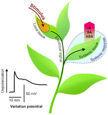
- Definition → Chemical messengers in plants that regulate growth, development, and responses to environmental stimuli.
- Similar role to hormones in animals – but produced in very small amounts and act at target tissues, often far from where they are made.
🔍 Key Characteristics
- Produced in: Specific cells/tissues in plants.
- Transported by: Diffusion, active transport, or through xylem/phloem.
- Act in low concentrations but have big effects.
- Can stimulate or inhibit processes.
🧪 Major Types and Roles
| Phytohormone | Main Functions |
|---|---|
| Auxins | Stimulate cell elongation, root initiation, phototropism, gravitropism |
| Cytokinins | Promote cell division, delay leaf senescence |
| Gibberellins | Stimulate stem elongation, seed germination, flowering |
| Abscisic Acid (ABA) | Inhibits growth, closes stomata during water stress, induces seed dormancy |
| Ethylene | Promotes fruit ripening, leaf abscission |
🌿 Why They’re Important
- Control plant architecture (height, branching)
- Coordinate timing of flowering and fruiting
- Regulate responses to light, gravity, and stress
- Help plants adapt to environmental changes
Phytohormones are plant signalling chemicals that control growth, development, and responses to the environment. Different phytohormones have specialised roles but often work together to coordinate plant life processes.
C3.1.22 – Interactions between Auxin and Cytokinin in Regulating Root and Shoot Growth
📖 Key Idea
Auxin and cytokinin work together to balance and coordinate growth of roots and shoots.
This interaction integrates whole plant growth.
🔍 How It Works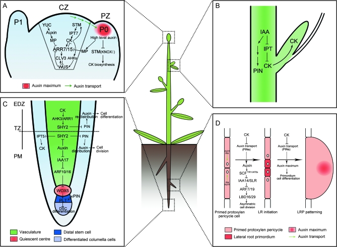
- Root tips produce cytokinin, which is transported up to shoots.
- Shoot tips produce auxin, which is transported down to roots.
- These hormones influence each other’s growth-promoting effects:
- Auxin promotes root growth and can inhibit shoot growth in some cases.
- Cytokinin promotes shoot growth and can inhibit root growth.
🌱 Integration of Growth
This cross-talk helps plants adjust growth depending on conditions:
- If the shoot grows faster, cytokinin signals encourage root growth to support it.
- If root growth is favored, auxin signals help balance shoot growth.
- Ensures roots and shoots develop proportionally to support the plant’s needs.
Auxin from shoots and cytokinin from roots travel to each other’s regions, coordinating growth so roots and shoots develop in balance.
C3.1.23 – Positive Feedback in Fruit Ripening and Ethylene Production
📖 What is Ethylene?
Ethylene, also called ethene (IUPAC name), is a simple gaseous plant hormone.
It acts as a signalling chemical that regulates many processes, including fruit ripening.
🔄 How Does Positive Feedback Work in Ripening?
When a fruit starts to ripen, it produces ethylene.
This ethylene triggers changes like:
- Softening of the fruit
- Color changes (e.g., green to red in tomatoes)
- Development of aroma and sweetness
As the fruit ripens and changes, it produces even more ethylene.
This increased ethylene stimulates even more ripening, creating a self-amplifying cycle – a positive feedback loop.
🌍 Why Is This Important?
- Rapid Ripening: The positive feedback speeds up the ripening process so the fruit doesn’t ripen too slowly or unevenly.
- Synchronization: All the fruit on a plant, or even fruits in storage, tend to ripen at the same time, which helps with:
- Seed dispersal: Animals are attracted to ripe fruit and help spread seeds effectively.
- Agriculture: Farmers can harvest crops when most fruits are ripe.
Without this feedback, ripening could be slow or happen irregularly, reducing the chance of seeds spreading or causing loss during harvesting.
🔬 Note:
Positive feedback is common in biological systems where a quick, decisive change is needed.
In fruit ripening, this feedback is an example of how plants use chemical signalling to coordinate development.
📊 Real-World Applications
- Commercial use: Ethylene gas is sometimes applied artificially to speed up ripening of fruits like bananas and tomatoes after harvest.
- Storage technology: Controlling ethylene levels helps delay ripening during shipping to extend shelf life.
Ethylene initiates fruit ripening, which in turn boosts ethylene production, creating a positive feedback loop. This ensures fruit ripening is rapid and synchronized, aiding seed dispersal and efficient harvest.