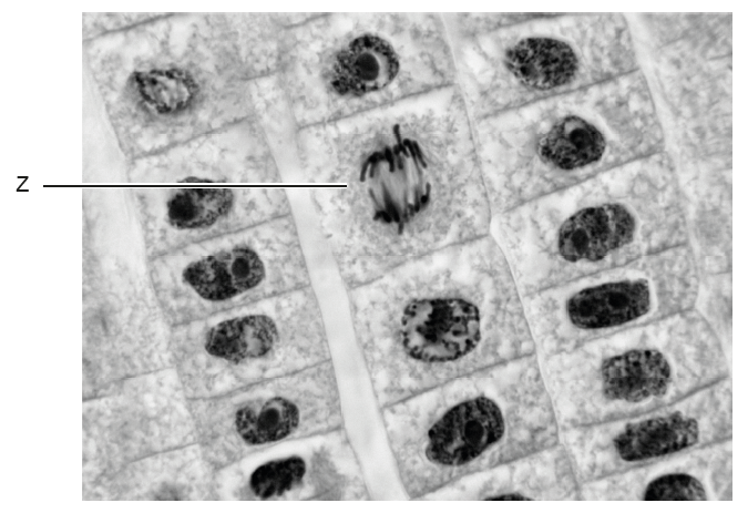
Question

▶️Answer/Explanation
During prophase I of meiosis, homologous chromosomes pair up and exchange genetic material in a process called crossing over. The points where the chromatids physically overlap are called chiasmata. These structures are only visible in prophase I, not during mitosis or other stages of meiosis.
Now let’s evaluate the options:
A. Incorrect – This refers to prophase of mitosis, but chiasmata are not formed during mitosis. Mitosis does not involve homologous pairing or crossing over.
B. Incorrect – In prophase II of meiosis, there are no chiasmata, because homologous chromosomes have already separated in meiosis I.
C. Incorrect – Eukaryotic chromosomes (like those in locusts) are linear, not circular. Circular chromosomes are found in prokaryotes like bacteria.
D. Correct – The presence of chiasmata shows that crossing over is occurring, which is specific to prophase I of meiosis.
Question
Students examined micrographs and counted cells in the different stages of mitosis as well as those cells with no visible chromosomes.
The table shows their results.
Stage | Prophase | Metaphase | Anaphase | Telophase | Interphase |
Number of cells | 10 | 3 | 2 | 5 | 30 |
What is the mitotic index?
A. 0.2
B. 0.4
C. 0.6
D. 0.7
▶️Answer/Explanation
Answer: B. 0.4
Explanation:
What is the mitotic index?
The mitotic index is the ratio of cells in mitosis (all visible stages: prophase, metaphase, anaphase, telophase) to the total number of cells observed. It gives an idea of how actively a tissue is dividing.
Formula:
\[
\text{Mitotic Index} = \frac{\text{Number of cells in mitosis}}{\text{Total number of cells}}
\]
Data from table:
- Prophase: 10
- Metaphase: 3
- Anaphase: 2
- Telophase: 5
- Interphase: 30
- Total cells = 10 + 3 + 2 + 5 + 30 = 50
- Cells in mitosis = 10 + 3 + 2 + 5 = 20
Mitotic index = 20 ÷ 50 = 0.4
A. Incorrect – 0.2 is too low
B. Correct – 0.4 is the correct calculation
C. Incorrect – 0.6 is too high
D. Incorrect – 0.7 is even higher and not supported by the data
Question
What would show that a person has developed metastatic cancer?
A. Alveolus cells forming a tumour in the lungs
B. Cancer cells producing the skin pigment melanin in the liver
C. A tumour in the prostate gland increasing levels of prostate-specific antigen
D. Cancerous lymphocytes in blood plasma
▶️Answer/Explanation
Answer: B. Cancer cells producing the skin pigment melanin in the liver
Explanation:
What is metastasis?
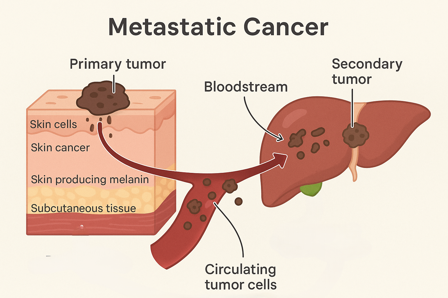
Metastasis is when cancer cells spread from their original site to other parts of the body, often via the blood or lymphatic system. When these cells grow in other tissues or organs, it is considered metastatic cancer.
Now evaluate each option:
A. Incorrect – Alveolus cells forming a tumor in the lungs could just mean lung cancer, but this does not confirm metastasis unless they originated from another tissue.
B. Correct – Melanin is produced by skin cells (melanocytes). If these pigment-producing cancer cells are found in the liver, it means skin cancer has spread, showing metastasis.
C. Incorrect – A tumor in the prostate causing higher PSA levels suggests local prostate cancer, not necessarily metastasis.
D. Incorrect – Lymphocytes are normal parts of blood plasma. Cancerous lymphocytes may indicate leukemia, but unless they originate from another tissue, this isn’t clear evidence of metastasis.