Home / IB DP Biology D2.1 Cell and nuclear division-FA 2025- IB Style Question For SL Paper 1

IB DP Biology D2.1 Cell and nuclear division-FA 2025- IB Style Question For SL Paper 1

Question

The micrograph shows some onion (Allium cepa) cells undergoing mitosis.

What is the mitotic index, taking into account all visible nuclei?
A. 0.1

B. 0.2

C. 0.4

D. 0.6

▶️Answer/Explanation

Answer: B. 0.2

Explanation:

Step 1: Count total cells with visible nuclei

By examining the micrograph:

  • There are 10 cells with clearly visible nuclei.

Step 2: Count how many are in mitosis

We are looking for cells where chromosomes are visible (condensed and distinguishable):

  • The cell on the right clearly shows condensed chromosomes = 1
  • The cell in the middle-left also has visible chromosomes, indicating it’s in a stage of mitosis (likely prophase) = 1

So: 2 cells are in mitosis

Step 3: Calculate mitotic index

\[
\text{Mitotic Index} = \frac{\text{Number of mitotic cells}}{\text{Total number of cells}} = \frac{2}{10} = 0.2
\]

Question

A tissue sample was examined under the microscope in order to determine a mitotic index. The number of cells in each stage of the cell cycle was determined and the data were entered into a table.

Stage of life cycle

Interphase

Prophase

Metaphase

Anaphase

Telophase

Total

Number of cells

120

20

10

8

2

160

What is the mitotic index?

A. 0.125

B. 0.25

C. 0.75

D. 1.00

▶️Answer/Explanation

Answer: B. 0.25

Explanation:

The mitotic index is a measure of the proportion of cells undergoing mitosis at a given time. It is calculated using the formula:

\[
\text{Mitotic Index} = \frac{\text{Number of cells in mitosis}}{\text{Total number of cells}}
\]

Step 1: Identify cells in mitosis

Cells in prophase, metaphase, anaphase, and telophase are considered to be in mitosis. Add the number of cells in these stages:

Mitotic cells=20(Prophase)+10(Metaphase)+8(Anaphase)+2(Telophase)=40

Step 2: Total number of cells

Total cells=160

Step 3: Calculate the mitotic index

\[
\text{Mitotic Index} = \frac{40}{160} = 0.25
\]

Question

Which diagram(s) represent(s) processes used in asexual reproduction?

A. I only

B. I and II only

C. II only

D. I, II and III

▶️ Answer/Explanation
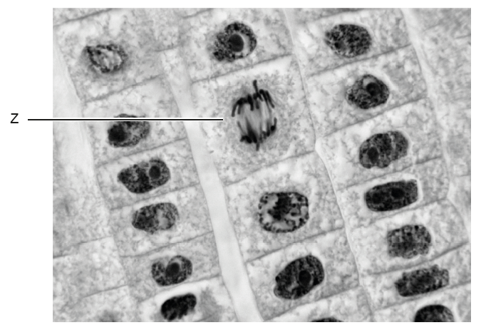
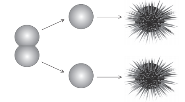
Answer: C. II only

Explanation:

Key concept: Asexual reproduction involves:

  • One parent
  • No fusion of gametes
  • Offspring genetically identical (clones)

Diagram I:

Shows a Tt parent producing T and t – this is gamete formation via meiosis, not asexual reproduction.

Not asexual reproduction.

Diagram II:

Shows a Tt parent producing two identical Tt offspring – this is consistent with mitosis, which is a form of asexual reproduction.

Diagram III:

Shows two gametes (T and t) combining to form Tt – this is fertilization, a process of sexual reproduction.

Not asexual reproduction.

Question

What occurs during meiosis but not mitosis?

A. Spindles are formed from microtubules.

B. Chromosome number is conserved.

C. Homologous chromosomes pair up.

D. Centromeres split.

▶️Answer/Explanation

Answer: C. Homologous chromosomes pair up

Explanation:

  • A key event that occurs during meiosis but not mitosis is the pairing of homologous chromosomes in a process called synapsis, which happens during prophase I of meiosis. This allows crossing over, where genetic material is exchanged between chromosomes, increasing genetic variation.
  • In contrast, mitosis does not involve pairing of homologous chromosomes. Instead, it separates sister chromatids to produce identical daughter cells.

Answer Evaluation:

A. Incorrect – Spindles form in both meiosis and mitosis to move chromosomes.
B. Incorrect – Chromosome number is halved in meiosis, but conserved in mitosis.
C. Correct – Homologous chromosomes pair up only in meiosis, not in mitosis.
D. Incorrect – Centromeres split in both processes, but at different stages: in anaphase of mitosis and anaphase II of meiosis.

Question

In which stage of mitosis is the cell labelled X?

A. Anaphase
B. Interphase
C. Metaphase
D. Prophase

▶️Answer/Explanation:

Answer: C. Metaphase

Explanation:

How to recognize Metaphase:

  • The chromosomes are lined up in the center of the cell.
  • This middle area is called the metaphase plate.
  • Spindle fibers from both sides of the cell are connected to the centromeres of the chromosomes.
  • The cell is preparing to pull the chromosomes apart in the next phase.

Why not the other options?

A. Anaphase:
In anaphase, the chromosomes would be pulling apart and moving toward opposite sides of the cell. In the image, they are still together in the middle, so it’s not anaphase.

B. Interphase:
This is the stage when the cell is not dividing. The DNA is loose, not in chromosome form, and you wouldn’t see the chromosomes clearly like in this image.

D. Prophase:
In prophase, chromosomes are just starting to become visible, and the spindle fibers are forming, but the chromosomes are not yet lined up in the middle.

Question

A two-cell sea urchin (Echinoidea) embryo was physically separated by scientists into two cells. Each cell, through further embryonic development, became an adult sea urchin.

What is the relationship between the two adult sea urchins?

A. They are equivalent to non-identical twins.

B. Half of the genes would be the same.

C. Both adults would have haploid cells.

D. They are clones.

▶️Answer/Explanation

Answer: D. They are clones.

Explanation:

  • The embryo originally came from one fertilized egg (a zygote).
  • When the embryo divides into two cells, both cells have identical genetic material (they are the result of mitotic division).
  • When scientists separated those two cells, each one still had the full set of DNA and was able to grow into a complete organism.
  • Since both sea urchins originated from the same zygote and have the same DNA, they are genetically identical.

Why the other options are incorrect:

A. Non-identical twins:
Non-identical (fraternal) twins come from two different eggs fertilized by two different sperm, so they are not genetically identical. That’s not the case here.

B. Half of the genes would be the same:
That would be true for siblings or non-identical twins, but not for clones. These sea urchins have 100% identical genes.

C. Both adults would have haploid cells:
No, fully developed organisms (like sea urchins) have diploid cells. Only gametes (sperm and eggs) are haploid.

Question

Which process occurs in meiosis but not in mitosis?

A. Attachment of spindle fibres to the centromeres of each chromosome

B. Movement of homologous chromosomes to opposite ends

C. Replication of DNA prior to the start of cell division

D. Separation of sister chromatids during anaphase

▶️Answer/Explanation

Answer: B. Movement of homologous chromosomes to opposite ends

Explanation:

  • A defining feature of meiosis—specifically in anaphase I—is the separation of homologous chromosomes, where each chromosome of a homologous pair is pulled to opposite poles of the cell.
  • This does not occur in mitosis, where sister chromatids, not homologous chromosomes, are separated.

Answer Evaluation:

A. Incorrect – Spindle fibres attach to centromeres in both mitosis and meiosis.
B. Correct – Only meiosis involves the movement of homologous chromosomes to opposite poles (anaphase I).
C. IncorrectDNA replication occurs before both mitosis and meiosis, during interphase.
D. IncorrectSister chromatids separate in both mitosis and meiosis II.

Question

When does DNA replication occur?

A. S phase of interphase

B. Early prophase

C. G phase of interphase

D. Late prophase

▶️Answer/Explanation

Answer: A. S phase of interphase

Explanation:

  • DNA replication occurs during the S phase (Synthesis phase) of interphase, which is the stage before a cell enters mitosis or meiosis. During this phase, the cell makes an exact copy of its DNA, so that each daughter cell receives a complete set of chromosomes.
  • Interphase is divided into three phases:
    • G₁ (Gap 1): Cell grows and performs normal functions.
    • S (Synthesis): DNA is replicated.
    • G₂ (Gap 2): Preparation for cell division.

Answer Evaluation:

A. Correct – DNA replication happens in the S phase of interphase.
B. Incorrect – Prophase is part of mitosis, and DNA has already been replicated before this stage.
C. Incorrect – There is no single “G phase”; G₁ and G₂ occur during interphase, but S phase is when replication happens.
D. Incorrect – Late prophase involves chromosome condensation, not DNA replication.

Question

What is a function of histones?

A.  Supercoiling of DNA during binary fission in prokaryotes

B.  Synthesis of proteins

C.  Formation of microtubules during mitosis

D.  Condensation of DNA

▶️Answer/Explanation

Answer: D. Condensation of DNA

Explanation:

  • Histones are proteins around which DNA wraps to form a structure called nucleosomes. This helps condense the long DNA molecules into a compact, organized form so they can fit inside the nucleus and be properly managed during processes like cell division.
  • This condensation is essential for protecting DNA and regulating gene expression.

Answer Evaluation:

A. Incorrect – Prokaryotes do not have histones; their DNA condensation uses different proteins.
B. Incorrect – Protein synthesis occurs in ribosomes, not histones.
C. Incorrect – Microtubules are made of tubulin, not histones.
D. Correct – Histones help condense DNA by forming nucleosomes.

Question

The image shows a micrograph of a cell.

What explains the appearance of the cell in the micrograph?

A. The cell is dying.

B. The DNA is replicating.

C. The cell is in metaphase.

D. The cell is in telophase.

▶️Answer/Explanation

Answer: C. The cell is in metaphase.

Explanation:

This micrograph shows chromosomes aligned at the center of the cell in a line-like structure, which is a hallmark of metaphase during mitosis.

In metaphase, the following occurs:

  • Chromosomes are highly condensed and visible.
  • They are aligned at the metaphase plate (the equator of the cell).
  • Spindle fibers from the centrosomes attach to the centromeres of each chromosome.

Why the other options are incorrect:

A. The cell is dying:
Dying cells usually show signs of disintegration or irregular nuclear material — this cell looks organized and structured, not dying.

B. The DNA is replicating:
DNA replication happens in interphase (S phase), during which chromosomes are not visible under the microscope like this.

D. The cell is in telophase:
In telophase, chromosomes begin to de-condense, and nuclear membranes start to re-form. The chromosomes here are still highly condensed and lined up, so this is not telophase.

Question

The diagram shows a stage in cell division.

What is the stage shown?

A. Anaphase I of mitosis

B. Anaphase II of mitosis

C. Anaphase I of meiosis

D. Anaphase II of meiosis

▶️Answer/Explanation

Answer: D. Anaphase II of meiosis

Explanation:

Let’s analyze the diagram step-by-step:

  • The diagram shows two cells, each with sister chromatids being pulled apart to opposite poles.
  • This separation of chromatids indicates a later phase, not the first part of meiosis.
  • The fact that there are two cells suggests that Meiosis I has already occurred, and we are now in Meiosis II.
  • The stage where sister chromatids are pulled apart is known as anaphase, specifically anaphase II in meiosis.

Why other options are incorrect:

A. Anaphase I of mitosis:
There is no such stage — mitosis has only one anaphase.

B. Anaphase II of mitosis:
Again, mitosis doesn’t have “Anaphase II”. That terminology belongs to meiosis.

C. Anaphase I of meiosis:
In anaphase I, homologous chromosomes (not sister chromatids) separate. But here we clearly see sister chromatids being separated.

Question

The image shows a cell from the root tip of an onion (Allium cepa) (2n = 16) during late prophase of mitosis.

How many chromatids are present in the cell?

A.  8

B.  16

C.  32

D.  64

▶️Answer/Explanation

Answer: C. 32

Explanation:

Let’s break it down:

  • The onion is diploid (2n = 16), meaning it has 16 chromosomes.
  • During late prophase (also called prophase or prophase I of mitosis), the DNA has already been replicated in interphase (S phase), but the sister chromatids are still attached at the centromere.
  • This means each chromosome consists of two sister chromatids.

Calculation:

16 chromosomes × 2 chromatids each = 32 chromatids

Why other options are incorrect:

A. 8: Too low — this would be half of the chromosome number, incorrect.

B. 16: This is the number of chromosomes, not chromatids.

D. 64: Double-counting both chromatids and chromosomes, incorrect.

Question

In the micrograph, which letter points to a cell in anaphase?

▶️Answer/Explanation

Answer: B

Explanation:

Understanding the Cell Cycle Stage – Anaphase:

Anaphase is a stage in mitosis where:

  • The sister chromatids of each chromosome separate.
  • The chromatids are pulled by spindle fibers toward opposite poles of the cell.
  • In microscope images, this looks like two groups of chromosomes moving apart from the center.

Image Analysis:

Let’s review each labeled cell:

  • A: Two nuclei visible forming → Telophase/Cytokinesis
  • B: Chromosomes are being pulled to opposite sides → Anaphase
  • C: Chromosomes are condensed but still together → Prophase
  • D: Nucleus intact, no chromosome movement → Interphase

Cell B shows the chromosomes separating and moving toward opposite ends, a key feature of anaphase. The V-shaped chromosomes indicate the chromatids being pulled apart.

Question

The micrograph shows a cross section of a lily (Lilium longiflorum) anther.

What can be found in X?
A.  Haploid nuclei produced by meiosis

B.  Diploid nuclei produced by fertilization

C.  Haploid nuclei produced by mitosis

D.  Diploid nuclei produced by pollination

▶️Answer/Explanation

Answer: A.  Haploid nuclei produced by meiosis

Explanation:

Understanding the Structure:

  • The image shows a cross section of an anther the male reproductive part of a flowering plant.
  • The central region (X) of the pollen sac (microsporangium) contains developing pollen grains.
  • Pollen grains are formed by meiosis of microsporocytes (2n), resulting in haploid (n) nuclei.

What Happens at X?

  • At X, we see cells with chromosomes aligned, some dividing → this is meiosis.
  • The result of meiosis in these pollen mother cells (microsporocytes) is four haploid microspores.
  • These haploid nuclei will develop into pollen grains (male gametophytes).

Explanation for Other Options:

B. Diploid nuclei produced by fertilization → Fertilization happens in the ovule, not in the anther.

C. Haploid nuclei produced by mitosis → Mitosis does occur later in pollen development, but the first division producing haploid cells here is meiosis.

D. Diploid nuclei produced by pollination → Pollination is the transfer of pollen, not a nuclear division process.

Question

The karyogram shown belongs to a human being.

What can be deduced from this karyogram?

A. The person is a male with Down syndrome.

B. The person is a female with Down syndrome.

C. The person is a male with a genetic disorder.

D. The person is a female with a missing chromosome.

▶️Answer/Explanation

Answer: A. The person is a male with Down syndrome.

Explanation:

Step-by-step Analysis:

Chromosome Count:

  • Humans normally have 46 chromosomes arranged in 23 pairs.
  • This karyogram has:
  • 22 autosome pairs (1–22)
  • An extra chromosome at pair 21 → Trisomy 21
  • Indicates Down syndrome

Sex Chromosomes:

  • One X chromosome and one Y chromosome are present.
  • Indicates male

Correct Interpretation:

  • This individual has Down syndrome (extra chromosome 21)
  • The presence of XY confirms the person is male

Why Other Options Are Incorrect:

B. Female with Down syndrome → Incorrect: XY sex chromosomes = male, not female

C. Male with a genetic disorder → Partially correct, but Down syndrome is the specific disorder — too vague

D. Female with a missing chromosome → Incorrect: No missing chromosomes and not female (XY present)

Scroll to Top