IB DP Biology Topic 11: Animal physiology : 11.2 Movement HL Paper 1

Question

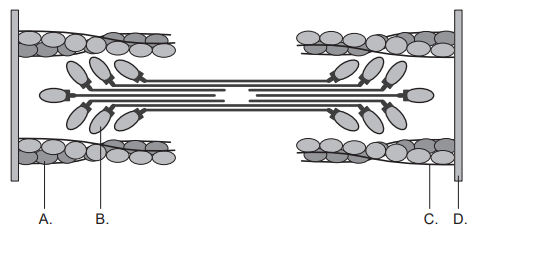
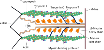
The diagram represents a sarcomere. Which structure is myosin?

▶️Answer/Explanation

Ans: B

Myosin consists of an elongated tail region attached to a globular head via a flexible neck structure.

Therefore, the correct answer is B

Question

What is the role of calcium in muscle contraction?

A. To release tropomyosin from myosin

B. To bind to troponin so myosin-binding sites on actin are exposed

C. To bind to tropomyosin so ATP can bind to actin

D. To release ATP from actin so myosin can bind to troponin

▶️Answer/Explanation

Markscheme

B

The role of calcium in muscle contraction is to bind to troponin so myosin-binding sites on actin are exposed.

When a muscle is stimulated to contract, calcium ions are released from the sarcoplasmic reticulum into the cytoplasm of the muscle fiber. The calcium ions then bind to troponin, which causes a conformational change in the troponin-tropomyosin complex, exposing the myosin-binding sites on the actin filament.

This allows the myosin heads to bind to the actin filament, forming cross-bridges. The energy from ATP hydrolysis is used to power the movement of the myosin heads, which slide along the actin filament, causing the muscle to contract.

Therefore, the correct answer  is B.

Question

Which process is part of the mechanism that controls muscle contraction?

A. Troponin enables actin heads to attach to ATP and slide along myosin.
B. Myosin heads attach to troponin and tropomyosin pulls on actin filaments.
C. Tropomyosin attaches to calcium and breaks the bond between actin and myosin.
D. Calcium frees actin filaments for myosin heads to attach

▶️Answer/Explanation

Markscheme

D

The process that is part of the mechanism that controls muscle contraction is calcium freeing actin filaments for myosin heads to attach.

When a muscle is stimulated to contract, calcium ions are released from the sarcoplasmic reticulum into the cytoplasm of the muscle fiber. The calcium ions then bind to troponin, which causes a conformational change in the troponin-tropomyosin complex, exposing the myosin-binding sites on the actin filament.

Once the myosin-binding sites are exposed, myosin heads bind to the actin filament, forming cross-bridges. The energy from ATP hydrolysis is used to power the movement of the myosin heads, which slide along the actin filament, causing the muscle to contract.

Therefore, the correct answer is D.

Question

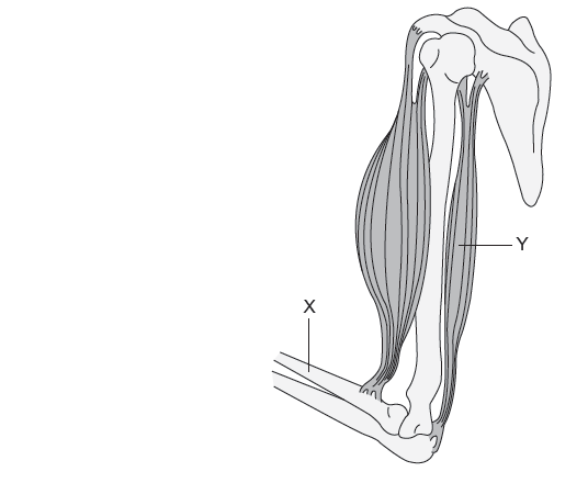
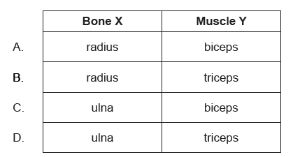
What is bone X and muscle Y in the diagram of the elbow joint?

▶️Answer/Explanation

Markscheme

B

X – radius and Y – triceps
The triceps brachii is located in the dorsal compartment of the arm. 
The radius and ulna are long bones that make up the forearm, extending from the elbow to the wrist. In the anatomical position, the radius is found in the lateral forearm, while the ulna is found in the medial forearm.

Question

What is the main role of nerves in human movement?

A. To cause muscles to stretch
B. To move joints
C. To transport pain signals that indicate muscle injuries
D. To stimulate muscle contraction

▶️Answer/Explanation

Markscheme

D

The main role of nerves in human movement is to stimulate muscle contraction.

Nerves are responsible for transmitting signals from the brain and spinal cord to the muscles, which causes the muscles to contract. When a nerve impulse reaches the end of a motor neuron, it triggers the release of a neurotransmitter called acetylcholine.

Acetylcholine then binds to receptors on the muscle fiber, causing a depolarization of the muscle cell membrane, which leads to the release of calcium ions from the sarcoplasmic reticulum. The calcium ions then bind to troponin, which exposes the myosin-binding sites on the actin filaments, allowing the myosin heads to bind to the actin filament and form cross-bridges.

The energy from ATP hydrolysis is used to power the movement of the myosin heads, which slide along the actin filament, causing the muscle to contract.

Therefore, the correct answer is D.

Question

What is a function of synovial fluid in the elbow joint?

A. Joins the humerus to the radius and ulna

B. Grows red blood cells

C. Protects the biceps

D. Allows easy movement

▶️Answer/Explanation

Markscheme

D

The function of synovial fluid in the elbow joint is to allow easy movement.

Synovial fluid is a clear, viscous fluid that is secreted by the synovial membrane that lines the joint capsule. It acts as a lubricant, reducing friction between the articular cartilage surfaces of the bones in the joint.

In the elbow joint, synovial fluid helps to reduce friction between the humerus, radius, and ulna bones, allowing for easy movement of the joint.

Therefore, the correct answer is D.

Question

Movement of insects requires muscles in antagonistic pairs. The diagram shows an insect leg with muscles labelled $\mathrm{X}$ and $\mathrm{Y}$.

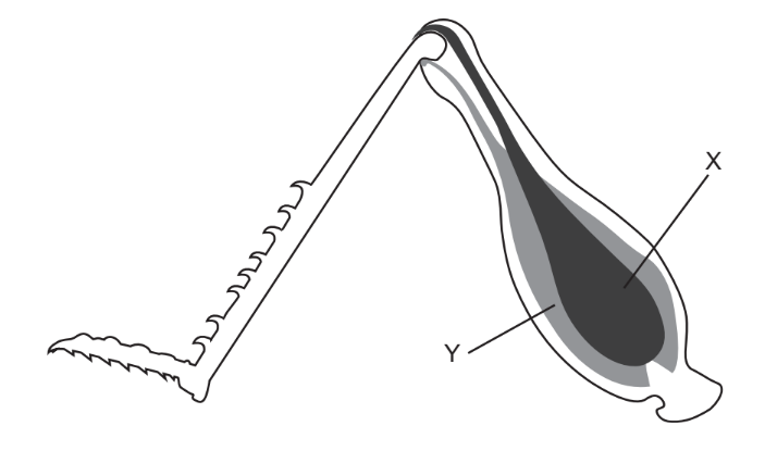
What actions in the human arm are equivalent to muscle X contracting and muscle Y relaxing?
A. triceps contracts, biceps relaxes, arm extends

B. biceps contracts, triceps relaxes, arm flexes

C. triceps contracts, biceps relaxes, arm flexes

D. biceps contracts, triceps relaxes, arm extends

▶️Answer/Explanation

Ans:A

Muscle $\mathrm{X}$ contracting and muscle $\mathrm{Y}$ relaxing in an insect leg is equivalent to the biceps contracting and the triceps relaxing in the human arm. This action causes the arm to flex.

Therefore, the correct answer is B. biceps contracts, triceps relaxes, arm flexes.

Scroll to Top