IB DP Biology Topic 6: Human physiology : 6.5 Neurons and synapses Question Bank HL Paper 2

Question

The image shows a transverse section of an intestinal wall at 100 x magnification.

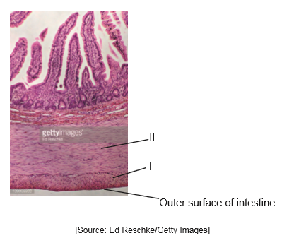
Identify the tissues labelled I and II on the image.

I:  ……………………………………………………………
II:  …………………………………………………………… 

[2]
a.

All motor neurons use acetylcholine to activate skeletal muscle. Explain the effect of neonicotinoid pesticides in insect synapses in the central nervous system.

[3]
b.

Resistance to neonicotinoid pesticides has been observed in some insects. Describe briefly how this resistance could have arisen in populations of insects.

[2]
c.
▶️Answer/Explanation

Markscheme

I and II are both muscle
circular and longitudinal

a.

Neonicotinoid pesticides are similar to nicotine «chemically»

Bind to nicotinic/acetylcholine receptors

Not broken down by «acetyl» cholinesterase
OR
binding is irreversible

Prevents/blocks acetylcholine binding

Blocks transmission from CNS  Reject slows transmission.
OR
blocks signals going to muscle
OR
muscle contraction blocked
OR
causes paralysis

b.

Mutations «for resistance in some insects»  Do not award mark if the answer implies directed mutations or that the pesticide causes the mutation.

«Mutation causes» breakdown of pesticide/detoxification of pesticide/changes to receptor site

Natural selection for resistance Do not accept natural selection if not in context.
OR
resistant insects survive and reproduce
OR
non-resistant killed leaving only resistant insects

Do not accept answers that use the term immunity instead of resistance.

c.

Question

Draw a labelled diagram to show the structure of a sarcomere.

[5]
a.

Explain how an impulse passes along the axon of a neuron.

[8]
b.

Describe the process of endocytosis.

[5]
c.
▶️Answer/Explanation

Markscheme

Award [1] for each structure clearly drawn and correctly labelled.

a. sarcomere – clearly indicated between Z lines (whether Z lines named or not);

b. Z lines – shown at the ends of a sarcomere;

c. actin (filaments) – drawn as thin lines attached to Z lines;

d. myosin (filaments) – drawn as thick lines interdigitating with thin/actin filaments;

e. myosin heads – on both sides of at least one myosin filament;

f. light band and dark band – indicating regions of actin only and myosin plus actin;

a.

a. resting potential is –70mV / relatively negative inside in comparison to the outside;

b. Na+/Kpumps maintain/re-establish (the resting potential);

c. more sodium ions outside than inside (when at the resting potential);

d. more potassium ions inside than outside (when at the resting potential);

e. nerve impulse is an action potential that stimulates a (wave of) depolarization along the membrane/axon;

f. if neuron is stimulated/threshold potential/–50mV is reached sodium ion channels open;

g. sodium ions diffuse/move in;

h. (Namove in) causing depolarization;

i. potassium ion channels open / potassium ions diffuse/move out;

j. (Kmove out) causing repolarization;

k. local currents / description of Naion diffusion between depolarized region and next region of axon to depolarize;

Accept any of the above points clearly explained in an annotated diagram.

b.

a. (plasma) membrane encloses/engulfs solid particles/droplets of fluid/molecules;

b. fluidity of the membrane allows endocytosis;

c. plasma membrane forms pit/forms indentation/pulled inwards/invaginates;

d. membrane pinches off/seals back on itself/edges fuse;

e. vesicle/vacuole formed;

f. inside of plasma membrane becomes outside of vesicle membrane / converse;

g. vesicle breaks away from plasma membrane/moves into cytoplasm;

h. active process / endocytosis/vesicle formation requires energy;

Accept any of the above points clearly described in an annotated diagram.

c.

Question

Draw a labelled diagram to show the molecular structure of a membrane.

[4]
a.

Some proteins in membranes act as enzymes. Describe a model that accounts for the ability of enzymes to catalyse reactions.

[6]
b.

Membranes of pre-synaptic and post-synaptic neurons play an important role in transmission of nerve impulses. Explain the principles of synaptic transmission.

[8]
c.
▶️Answer/Explanation

Markscheme

Award [1] for each of the following clearly drawn and correctly labelled.

phospholipid bilayer; (double row of opposing phospholipids, tails to inside)

hydrophilic/phosphate/polar (heads) and hydrophobic/hydrocarbon/fatty acid/nonpolar (tails) labeled;

integral protein; (embedded in the phospholipid bilayer)

protein channel/channel protein; (integral protein showing clear channel/pore)

peripheral protein; (shown on surface or slightly embedded on either side)

glycoprotein; (with carbohydrate attached on outer side)

cholesterol; (shown embedded in bilayer and smaller than the hydrophobic tail)

a.

induced fit model; (do not accept lock and key hypothesis)
accounts for ability of some enzymes to bind to several substrates;
enzyme with active site to which substrate(s) binds;
enzyme active site and substrate do not match up exactly;
enzyme-substrate complex forms;
enzyme changes shape once bound / enzyme moulds to substrate/ hand in glove;
change in shape strains bonds/facilitates bonds breaking/product formation;
reduces activation energy;
once reaction is complete, products leave and enzyme can work again;
Award any of the above points for a clearly drawn correctly annotated diagram.

b.

synapse is gap between adjacent neurons;
(arriving) action potential depolarizes pre-synaptic membrane;
opens (voltage-gated) calcium channels in membrane;
causes influx of calcium ions;
causes synaptic vesicles to fuse with pre-synaptic membrane;
vesicles release/exocytose neurotransmitter into the synaptic cleft;
neurotransmitter diffuses/moves across synaptic cleft;
neurotransmitter binds to receptors on post-synaptic membrane;
opens channels allowing sodium ions/potassium ions to diffuse;
initiation of action potential/depolarization in post-synaptic membrane;
removal/breakdown of neurotransmitter stops effect on post-synaptic membrane;
Award any of the above points for a clearly drawn correctly annotated diagram.
(Plus up to [2] for quality)

c.

Question

Multicellular organisms benefit from cell specialization and division of labour.

(a) Outline the processes occurring during interphase in the cell cycle.[4]

(b) Describe what occurs in a neuron when an action potential is propagated along the axon. [4]

(c) Explain how cells in the bloodstream cause a specific immune response.[7]

▶️Answer/Explanation

a a. growth/increase in cell size;

b. division of mitochondria/chloroplasts/production of more organelles/number of organelles doubled;
c. replication of DNA/amount of DNA is doubled;
d. transcription of genes/production of mRNA;
e. protein synthesis;
f. cell respiration/production of ATP;

b a. sodium ions/Na+ enter/diffuse in;

b. depolarization/membrane potential/voltage changes from negative to positive;
c. potassium channels open AND potassium ions/K+ exit/diffuse out;
d. repolarization/membrane potential/voltage changes back from positive to negative;
e. local current due to diffusion of sodium ions along the neuron;
f. (local currents) cause next sodium channels to open/next part of axon to depolarize;
g. opening of sodium channels triggered when threshold potential/-50mV reached;

c a. (specific immune response is) production of antibodies in response to a particular pathogen;
b. antibody is specific to/binds to a specific antigen;
c. macrophages/phagocytes engulf/present antigens from pathogens/viruses/bacteria;
d. T lymphocytes activated by antigens/antigen presentation/antigens presented by macrophage;
e. (activated) T lymphocytes activate B lymphocytes;
f. only B lymphocytes that produce antibodies against the antigen/pathogen are activated;
g. (activated) B lymphocytes clone/divide by mitosis to form plasma cells;
h. plasma cells then secrete (large quantity) of an antibody/secrete antibodies of same type;
i. some B lymphocytes/plasma cells form memory cells;
j. memory cells give long lasting immunity/faster response to a disease/pathogen;

Scroll to Top