Question (21 marks)
Developments in the understanding of atomic physics have led to many useful applications in industry, medicine and technology.
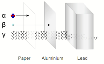
Through understanding patterns in the properties of alpha ( $\alpha)$, beta $(\beta)$ and gamma $(\gamma)$ radiation, scientists have developed ways in which the specific properties may be used.
One useful property is the difference in the penetration of alpha, beta and gamma radiation. The diagram compares the penetration of the three types of radiation.
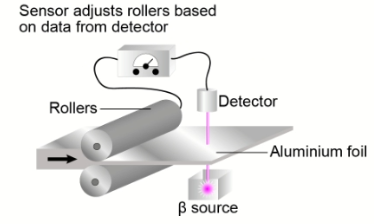
The amount of beta radiation absorbed depends on the thickness of the materials. This property of beta radiation is used to monitor the thickness of aluminium foil produced in an aluminium factory.
The following video shows how the absorption of radiation can be measured with a Geiger-Muller tube.
The Geiger-Muller tube is linked to a digital rate meter. The rate meter gives readings in counts per second, which is a measure of the number of ionization’s detected by the Geiger-Muller tube each second.
You are provided with the following equipment:
- A source of beta radiation. In this case strontium-90 will be used. The radioactive source is in a sealed lead-lined container and produces a beam of beta radiation.
- A number of sheets of aluminium foil, with a thickness of $0.04 \mathrm{~mm}$
- Standard laboratory equipment (metre rules, clamps and clamp stands, etc.).

Question g (4 mark)
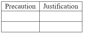
State and justify two precautions you would take to ensure that the method was carried out safely.
▶️Answer/Explanation
Ans:
Precaution 1: Use appropriate shielding for radioactive sources.
Justification: Radioactive sources, such as the strontium-90 used in this experiment, emit ionizing radiation that can be harmful to living organisms if not properly shielded. It is essential to enclose the radioactive source in a sealed lead-lined container to prevent the direct exposure of individuals to the radiation. The lead lining effectively absorbs the radiation and minimizes the risk of exposure, ensuring the safety of the experimenters and others in the vicinity.
Precaution 2: Wear appropriate personal protective equipment (PPE).
Justification: Personal protective equipment should be worn to minimize the potential exposure to radiation during the experiment. This includes wearing lab coats, gloves, and safety goggles. Lab coats provide an additional barrier between the radioactive source and the experimenter’s skin, reducing the risk of direct contact. Gloves protect the hands from potential contamination and reduce the risk of radioactive material transferring to other surfaces. Safety goggles protect the eyes from any potential splashes or airborne particles. Wearing appropriate PPE ensures the safety of the experimenters and prevents unnecessary exposure to radiation.
These precautions help to ensure that the experiment is carried out safely by minimizing the risks associated with working with radioactive sources and reducing the potential exposure of individuals to ionizing radiation.
Question (12 marks)
We have seen that strontium- 90 has useful applications in industry but uncontrolled release of strontium-90 into the environment has negative consequences.
In 2011, there was a disaster at the Fukushima Daiichi nuclear power plant in Japan. This disaster resulted in the release of large amounts of radioactive water into the local environment; scientists think that this process is still occurring. The water contains many different radioisotopes including strontium-90. Some of this material made its way into the sea and has been spread globally as a result.
The map below shows a model of how the radioactive material released from Fukushima might spread across the world in seawater. The darker colour shows where the readings of radiation are predicted to be the highest.

The negative environmental effects of strontium-90 are clear, but to understand how long-lasting these effects will be, scientists must know the half-life of the radioisotope. The half-life is the time taken for the number of radioactive nuclei to decrease by half.
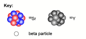
Radioactive decay is a random process. Strontium- 90 decays to yttrium- 90 by beta decay.
$$
{ }_{38}^{90} \mathrm{Sr} \rightarrow{ }_{39}^{90} \mathrm{Y}+{ }_{-1}^0 \beta
$$
Two years after the Fukushima Daiichi disaster, scientists wanted to investigate what was happening inside the reactor. Radiation levels inside the reactor were too high for humans to investigate directly so the scientists used a probe specially designed to work in radioactive conditions. The scientists found evidence of highly radioactive water leaking from the reactor.
Question a (6 mark)
The simulation below shows what could happen to a sample of 100 strontium- 90 nuclei over a period of 100 years.
You are going to collect data to enable you to plot a decay curve. You need to know how the number of strontium-90 nuclei varies with time. Time is your independent variable and number of strontium-90 nuclei is your dependent variable.
The table on the left below shows the data that you will record during the simulation.
Identify six times at which you will record data. Run and pause the simulation to collect this data.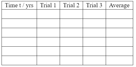
Calculate mean averages and enter them in the column headed “Average”. You should give your values to an appropriate number of significant figures.
▶️Answer/Explanation
Ans:
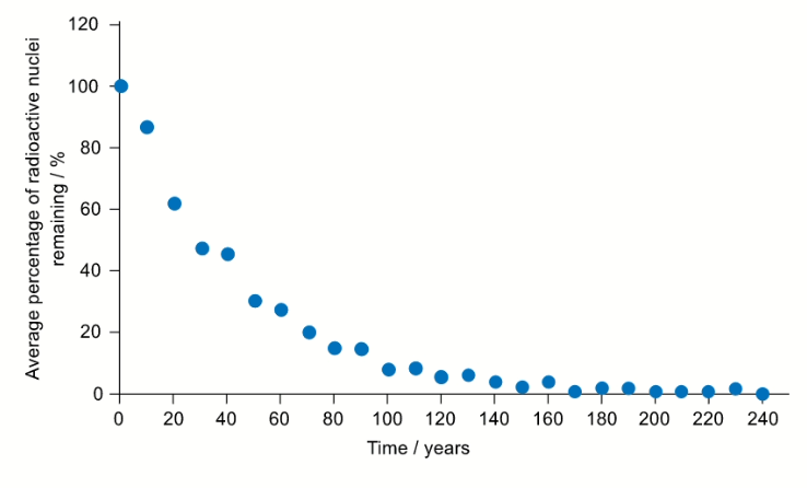
Another radioisotope that was released in the Fukushima Daiichi nuclear disaster was caesium-137. Another student used a simulation to collect decay data for caesium-137. Their data is presented in the graph below where time is on the $x$-axis and average percentage of caesium-137 remaining is on the y-axis.
Question b (1 mark)
Some of the data is also presented in the graphs below. Click on the tabs to view the different graphs.
Select the most appropriate graph to present the data.
▶️Answer/Explanation
Ans:
Question (4 marks)
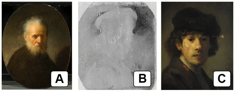
Compare image C to images A and B.
Some art historians suggest that Rembrandt reused the canvas shown in image B.
Question c (1 mark)
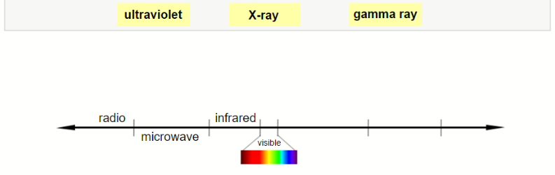
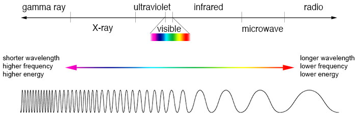
X-rays, gamma rays and ultraviolet light are all forms of electromagnetic radiation.
Label the diagram of the electromagnetic spectrum.
▶️Answer/Explanation
Ans:
Question d (3 mark)
X-rays and gamma rays can both be used by doctors to produce images of the internal structure of the human body. The different properties of X-rays and gamma rays produce different types of image.
An X-ray image is formed by projecting X-rays, and then capturing the “shadow” on a surface that reacts to X-ray radiation.
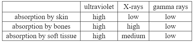
Using information from the table, discuss why X-rays are used, rather than ultraviolet or gamma rays, when doctors wish to make images of a person’s bones.
▶️Answer/Explanation
Ans:
When doctors wish to make images of a person’s bones, X-rays are used rather than ultraviolet or gamma rays. This choice is based on the differential absorption properties of these types of radiation as indicated in the table.
X-rays have a relatively low absorption by skin, allowing them to penetrate through the body’s outer layers and reach the underlying bones. This characteristic is advantageous for imaging purposes since it enables X-rays to pass through the soft tissues and reach the bones, which have a relatively high absorption of X-rays. As a result, the bones appear as contrasting structures on the X-ray image, while the surrounding soft tissues and skin contribute less to the overall image formation.
On the other hand, ultraviolet rays have high absorption by both the skin and bones. Their high absorption by the skin limits their penetration depth, making it difficult for them to reach the bones effectively. This characteristic makes ultraviolet rays less suitable for bone imaging purposes.
Similarly, gamma rays have low absorption by both bones and soft tissues. While this property allows gamma rays to penetrate through the body, it also means that they are less likely to be absorbed or attenuated by the bones. As a result, gamma rays do not produce distinct contrast between bones and surrounding tissues, making them less effective for bone imaging compared to X-rays.
In summary, X-rays are used for imaging bones because they have relatively low absorption by skin, allowing them to penetrate through the body and reach the bones, which have a higher absorption of X-rays. This differential absorption property of X-rays enables the production of clear and contrasting images of the bones while minimizing the contribution of surrounding soft tissues and skin.
Question e (14 mark)
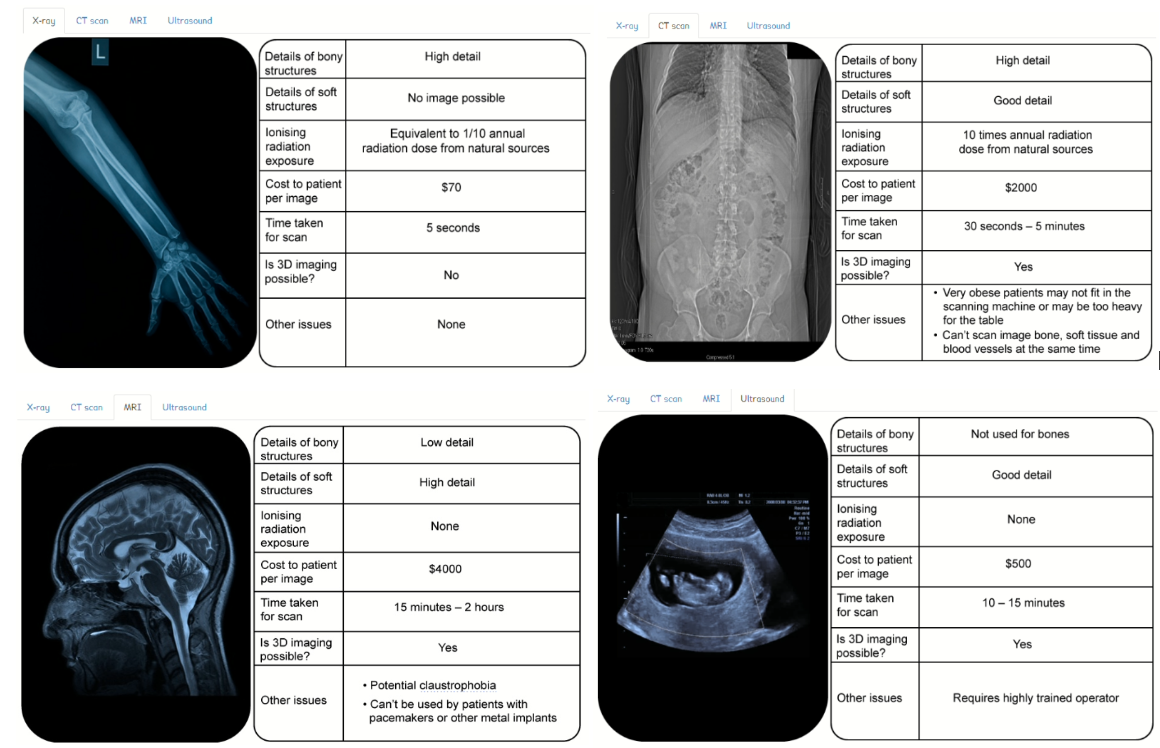
X-rays are not the only means of producing medical images. There are different options for producing medical images. Information about some of the options is presented in the tables below.
All hospitals have a limited amount of money to spend on medical equipment. Hospital managers have to balance the advantages and disadvantages of different types of equipment when they decide how to spend their money.
Using the information in the tables, discuss and evaluate the medical imaging equipment you would recommend to the hospital manager, clearly justifying your recommendation. In this extended piece of writing, you should consider the social and economic factors and include:
- the advantages of your chosen equipment
- the disadvantages of your chosen equipment
- the perspective of the hospital
- the perspective of the patients.
▶️Answer/Explanation
Ans:
To provide a comprehensive evaluation and recommendation for medical imaging equipment, let’s consider the information from the tables and the perspectives of both the hospital and the patients. The tables provide information on four different types of medical imaging options: X-ray, computed tomography (CT), magnetic resonance imaging (MRI), and ultrasound.
1. X-ray Imaging:
Advantages:
- Cost-effective: X-ray equipment is generally more affordable compared to other imaging modalities, making it a suitable choice for hospitals with limited budgets.
- Quick imaging: X-rays provide rapid results, allowing for efficient diagnosis and treatment decisions.
- Wide availability: X-ray technology is widely accessible and commonly used in hospitals, ensuring familiarity and ease of use for medical professionals.
Disadvantages:
- Limited tissue differentiation: X-rays primarily highlight differences in tissue density, which means they may not provide detailed information on soft tissues or subtle abnormalities.
- Ionizing radiation exposure: X-rays involve ionizing radiation, which carries a potential risk of cumulative radiation exposure over time. Steps must be taken to minimize patient radiation dose.
Hospital Perspective:
X-ray imaging is a fundamental and cost-effective imaging modality that can provide valuable information for a wide range of medical conditions. It is especially useful for bone imaging and initial assessments. Considering the limited budget, the hospital can allocate funds to maintain and upgrade X-ray equipment while ensuring proper radiation safety protocols.
Patient Perspective:
From the patient’s viewpoint, X-ray imaging offers advantages such as quick results, relatively low cost, and non-invasive nature (as it does not involve injections or invasive procedures). However, patients may have concerns about radiation exposure. It is essential for the hospital to prioritize patient safety, implement appropriate dose optimization techniques, and educate patients about the benefits and risks of X-ray imaging.
2. Computed Tomography (CT):
Advantages:
- Rapid and detailed imaging: CT scans provide detailed cross-sectional images, allowing for accurate diagnosis and treatment planning.
- Wide range of applications: CT is useful for imaging various body regions, including the chest, abdomen, and pelvis.
- Advanced imaging capabilities: Modern CT scanners can perform advanced techniques such as angiography and 3D reconstruction.
Disadvantages:
- Ionizing radiation exposure: CT uses ionizing radiation, which carries a higher radiation dose compared to other modalities, posing potential health risks.
- Cost and space requirements: CT equipment and maintenance are costly, and a dedicated space with radiation shielding is necessary.
Hospital Perspective:
CT imaging plays a crucial role in emergency medicine, trauma assessment, and detailed anatomical evaluation. However, due to the high initial and operational costs, the hospital must carefully consider the patient population’s needs and allocate resources accordingly.
Patient Perspective:
CT scans provide rapid and detailed imaging, enabling timely diagnosis and treatment. However, patients may have concerns about radiation exposure. Hospitals should prioritize radiation dose optimization, follow appropriate imaging protocols, and communicate the benefits and risks to patients.
3. Magnetic Resonance Imaging (MRI):
Advantages:
- Excellent soft tissue contrast: MRI provides superior soft tissue visualization, making it ideal for evaluating the brain, spinal cord, joints, and soft organs.
- Multi-planar imaging: MRI can acquire images in multiple planes, facilitating comprehensive anatomical assessment.
- No ionizing radiation: MRI does not involve ionizing radiation, eliminating radiation exposure concerns.
Disadvantages:
- High cost: MRI equipment and maintenance are expensive, making it a significant investment for hospitals.
- Patient limitations: Certain patients with claustrophobia, pacemakers, or metal implants may face challenges in undergoing an MRI examination.
Hospital Perspective:
MRI is a valuable imaging modality for detailed anatomical assessment and characterization of various pathologies. However, the high cost of MRI equipment and the need for dedicated space and specialized technical expertise should be considered in the budget allocation.
Patient Perspective:
Patients appreciate the superior image quality and the absence of radiation exposure in MRI. However, the lengthy scanning time and potential claustrophobia can be sources of anxiety for some patients. Hospitals should prioritize patient comfort by offering open MRI options, ensuring clear communication, and providing necessary support during the examination.
4. Ultrasound Imaging:
Advantages:
- Non-ionizing radiation: Ultrasound uses sound waves instead of ionizing radiation, making it a safe option for repeated imaging and monitoring during pregnancy or pediatric cases.
- Real-time imaging: Ultrasound provides real-time imaging, allowing dynamic visualization of structures and functions.
- Portable and versatile: Ultrasound machines are typically portable and can be used at the bedside or in various clinical settings, enhancing accessibility and flexibility.
Disadvantages:
- Operator-dependent: Obtaining high-quality ultrasound images requires skilled operators, and the interpretation of images can be subjective.
- Limited penetration: Ultrasound may have limitations in imaging deep-seated structures or patients with obesity or gas-filled intestines.
Hospital Perspective:
Ultrasound imaging offers versatility and flexibility for various clinical scenarios, including obstetrics, cardiology, and point-of-care assessments. The hospital can consider investing in advanced ultrasound systems with additional features and training opportunities for operators to maximize the benefits of this modality.
Patient Perspective:
Patients often appreciate the non-invasive nature of ultrasound imaging, absence of radiation exposure, and the ability to visualize real-time images. Ultrasound is commonly used in prenatal care and can provide reassurance for patients during the imaging process.
In conclusion, considering the social and economic factors, the hospital manager should prioritize allocating funds to maintain and upgrade the existing X-ray equipment due to its cost-effectiveness, wide availability, and usefulness in bone imaging. Simultaneously, investments in advanced ultrasound systems and operator training can enhance imaging capabilities and expand its utility in various clinical scenarios. While MRI and CT offer superior imaging capabilities, their high costs and specific patient requirements warrant careful consideration and budget allocation based on the hospital’s specific needs and patient population. Ultimately, a balanced approach that considers both the hospital’s financial constraints and the patients’ needs for safe and accurate diagnosis should guide the equipment recommendation.
Question (3 marks)
Look at the two very different images of feet.
The second image is by the artist Hugh Turvey who uses X-rays in his work. This piece of art is called Femme Fatale and shows the foot of a woman wearing a high-heeled shoe. The artist has used an X-ray, normally used in science, medicine or industry to create this artistic image.
Outline what the use of science can reveal that a photograph does not. Refer to the image and apply the ideas that you have been introduced to in this task in your answer.
▶️Answer/Explanation
Ans:
The use of science, specifically X-ray imaging, in creating the artistic image by Hugh Turvey can reveal certain aspects that a traditional photograph may not capture. Here are a few points to consider:
1. Internal Structure: X-ray imaging allows us to see beyond the external appearance of an object or subject. In the case of the X-ray image of the foot in Femme Fatale, it reveals the internal structure of the foot, showcasing the skeletal framework and the positioning of bones. This provides a unique perspective and adds a layer of depth to the image that a photograph cannot convey.
2. Transparency: X-rays have the ability to penetrate certain materials, such as soft tissues, and showcase their level of transparency. In the image, the X-ray reveals the transparency of the shoe, allowing us to see through the material and observe the underlying foot structure. This transparency aspect adds an intriguing and somewhat surreal quality to the artwork, providing a different aesthetic experience compared to a traditional photograph.
3. Hidden Details: X-ray imaging can unveil hidden or obscured details that are not visible in a photograph. It can highlight subtle nuances or features that are not easily perceivable through conventional photography. In the case of Femme Fatale, the X-ray image exposes the intricate details of the foot’s bone structure and the alignment of joints, presenting an anatomical perspective that is typically hidden from plain sight.
4. Scientific Context: The use of X-rays, a scientific technology primarily employed in fields such as medicine and industry, brings a scientific context to the artwork. It bridges the gap between art and science, creating an intriguing juxtaposition that stimulates thought and reflection on the intersection of these disciplines.
Overall, by utilizing X-ray imaging in the creation of the artistic image, the artist, Hugh Turvey, offers a unique perspective that goes beyond what a photograph can convey. The scientific application of X-rays reveals the internal structure, transparency, hidden details, and scientific context, enhancing the aesthetic and conceptual dimensions of the artwork.
Studying how soft X-rays pass through air
A student knows that soft X-rays are known to be blocked by air easily and that hard X-rays can travel long distances through air. She forms a hypothesis that the distance X-rays can travel is directly proportional to the frequency of the X-rays. In order to test her hypothesis, she tries to find some data. She discovers this graph in a scientific paper. It shows the percentage of X-rays which can travel a certain distance in air. The graph shows results for different wavelengths of X-rays.

The student uses this data from this experiment to find the amount of air required to block half of the X-rays at different wavelengths.
Question:
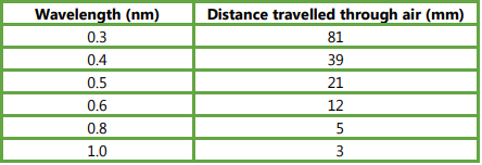
Read off values from the graph to find the distance the different wavelengths of X-rays travel before half are absorbed. Record your data in a suitable table.
▶️Answer/Explanation
Ans: Table of six values; correct values (see below, allow ±1 mm); column headings; with units.
Question:
Plot a graph of your data.
▶️Answer/Explanation
Ans: 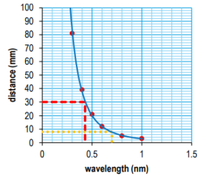
Question:
Add a line of best fit to your graph.
▶️Answer/Explanation
Ans: