IB DP Biology Membranes and membrane transport Study Notes
IB DP Biology Membranes and membrane transport Study Notes at IITian Academy focus on specific topic and type of questions asked in actual exam. Study Notes focus on IB Biology syllabus with guiding questions of
- How do molecules of lipid and protein assemble into biological membranes?
- What determines whether a substance can pass through a biological membrane?
Standard level and higher level: 4 hours
Additional higher level: 1 hour
B2.1.1 – Lipid Bilayers as the Basis of Cell Membranes
🌿 What Are Cell Membranes?
Cell membranes surround all living cells and many organelles inside them. They act as selective barriers controlling what enters and leaves the cell. In eukaryotic cells, internal membranes also form compartments (organelles) for specialized functions.
🧪 Structure of the Membrane: The Lipid Bilayer
The core of all biological membranes is a phospholipid bilayer. These bilayers form spontaneously in water because of the special structure of phospholipids.
🧬 Phospholipids – Amphipathic Molecules
Amphipathic = having both a hydrophilic (water-loving) head and a hydrophobic (water-fearing) tail.
- The hydrophilic heads face the water (inside and outside of the cell).
- The hydrophobic tails face each other, avoiding water.
This arrangement creates a double layer (bilayer) that is stable and self-sealing.
📌 Why Do Lipid Bilayers Form Naturally?
Water is a polar solvent, and amphipathic molecules interact with it in predictable ways. This causes phospholipids to spontaneously organize into a bilayer, minimizing energy and creating a stable boundary. No energy or enzymes are required it’s a self-assembly process driven by the laws of chemistry.
🧬 Fluid Mosaic Model of the Membrane
The membrane isn’t rigid it’s fluid, allowing movement of molecules within it. Described as a “mosaic” of:
- Phospholipids (forming the bilayer)
- Proteins (embedded or attached)
- Carbohydrates (on the outer surface)
This structure allows for flexibility, repair, and cell interactions.
📊 Key Features of the Lipid Bilayer
Feature | Description |
---|---|
Selective Permeability | Only certain substances can cross (e.g. small, non-polar molecules) |
Fluidity | Lipids and proteins can move laterally within the layer |
Self-healing | Minor tears in the membrane can reseal automatically |
Dynamic Nature | Constantly changing and responsive to cellular needs |
Barrier Function | Keeps internal environment separate from the external one |
🔍 Real-World Example
Soap micelles in water form spontaneously due to amphipathic molecules similar to how phospholipids form bilayers. Liposomes (artificial vesicles made of lipid bilayers) are used in drug delivery because they mimic natural membranes.
🧠 Key Takeaways
- All cell membranes are based on a phospholipid bilayer.
- Phospholipids are amphipathic, leading to spontaneous bilayer formation in water.
- The bilayer is fluid and dynamic, not static.
- The fluid mosaic model describes the membrane’s mixed composition and movement.
- The membrane enables selective transport, compartmentalization, and homeostasis.
B2.1.2 – Lipid Bilayers as Barriers
🧬 What Makes a Lipid Bilayer a Barrier?
The lipid bilayer is not just a structure it also functions as a selective barrier. It separates the aqueous (water-based) environments inside and outside the cell. This barrier function is mainly due to its hydrophobic core formed by the fatty acid tails of phospholipids.
🌿 Amphipathic Phospholipids – The Key to Selectivity
Phospholipids have:
- A hydrophilic (polar) head that faces water
- A hydrophobic (non-polar) tail that avoids water
When they form a bilayer:
- The hydrophobic tails face inward, creating a non-polar core
- This core resists the passage of polar or charged (hydrophilic) substances
🚫 What Can’t Easily Pass Through the Bilayer?
The hydrophobic core is a major obstacle for:
- Ions (e.g., Na⁺, Cl⁻, K⁺) → strongly attracted to water, not the membrane interior
- Polar molecules (e.g., glucose, amino acids)
- Large molecules (e.g., proteins, nucleotides)
These substances cannot diffuse freely across the bilayer—they need help (e.g., protein channels or transporters).
🧪 Who Gets Through Easily?
- Small non-polar molecules (like oxygen, carbon dioxide) can pass through easily
- Small uncharged polar molecules (like water, ethanol) may pass slowly
- Large or charged molecules = blocked without help
📊 Membrane Permeability Overview
Molecule Type | Examples | Permeability |
---|---|---|
Small non-polar | O₂, CO₂ | High |
Small uncharged polar | H₂O, ethanol | Moderate |
Large polar molecules | Glucose, sucrose | Very low |
Ions | Na⁺, Cl⁻, K⁺ | Very low |
Large molecules | Proteins, DNA | Blocked |
🔬 Why Is This Important for Cells?
The barrier allows the cell to:
- Control what enters and exits
- Maintain ion gradients (used in nerve impulses, muscle contractions, etc.)
- Protect its internal chemistry
Without this barrier, the cell would lose control of its internal environment, and vital processes would fail.
🧠 Key Takeaways
- The hydrophobic interior of the bilayer blocks water-soluble substances.
- Only small, non-polar molecules cross easily.
- This selective permeability helps maintain a stable internal environment.
- Membrane proteins help transport the substances that can’t pass through on their own.
B2.1.3 – Simple Diffusion Across Membranes
🧬 What is Simple Diffusion?
Simple diffusion is the passive movement of particles from a region of high concentration to low concentration. It happens due to the random motion of molecules and does not require any energy (no ATP). The process continues until particles are evenly distributed (equilibrium).
🌿 Where Does Simple Diffusion Occur in Cells?
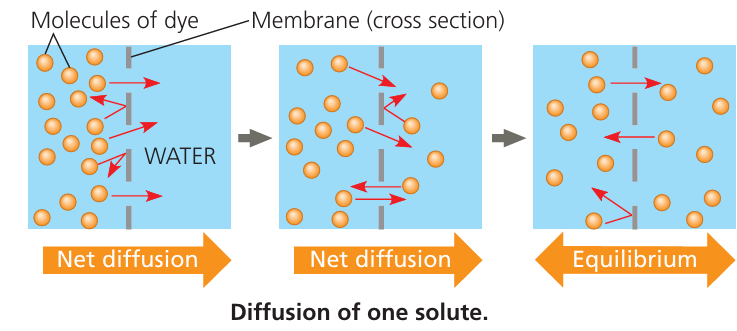
It occurs across the lipid bilayer of the plasma membrane, mainly for small, non-polar molecules.
- Oxygen (O₂) and carbon dioxide (CO₂) are classic examples:
- Both are non-polar and small, so they can move freely between the phospholipids of the membrane.
🧪 How Does It Work Through the Membrane?
The hydrophobic core of the bilayer:
- Blocks polar and charged substances
- Allows small non-polar molecules to pass directly through the phospholipids
No transport proteins or energy are involved
📌 Example – Oxygen and Carbon Dioxide Movement
Oxygen (O₂): Moves into cells where it’s needed for respiration
Carbon dioxide (CO₂): Moves out of cells as a waste product
Both diffuse across cell membranes in opposite directions, depending on their concentration gradients.
🔍 Real-Life Application – Oxygen Diffusion in the Eye
The cornea (transparent front part of the eye) has no blood vessels. It gets oxygen from the air through:
Simple diffusion across the tear film → cornea → inner corneal cells
This passive process is essential for keeping corneal cells alive.
📊 Key Features of Simple Diffusion
Feature | Description |
---|---|
Passive process | No energy required |
Concentration gradient | Particles move from high to low concentration |
No proteins needed | Molecules pass directly between phospholipids |
Selective | Only works for small, non-polar molecules |
Examples | Oxygen (O₂), Carbon dioxide (CO₂), some small lipids |
🧠 Key Takeaways
- Simple diffusion is a passive and energy-free process.
- It allows small, non-polar molecules like O₂ and CO₂ to move directly through the membrane.
- This is essential for gas exchange in cells and tissues, such as the cornea of the eye.
- Larger or polar substances cannot use this method—they require facilitated transport.
B2.1.4 – Integral and Peripheral Proteins in Membranes
🧬 What Are Membrane Proteins?
Cell membranes aren’t just made of phospholipids—they also contain proteins that carry out essential cellular functions. These proteins vary in structure, position, and role, depending on the needs of the membrane.
Membrane proteins can be grouped into two main types based on how they interact with the lipid bilayer:
- Integral proteins
- Peripheral proteins
🌿 Integral Proteins (Transmembrane or Embedded Proteins)
Located inside the lipid bilayer.
- Can either span the entire membrane (transmembrane proteins)
- Or be partially embedded in one lipid layer
Often have both hydrophobic and hydrophilic regions.
Key roles:
- Transport (e.g. channels, carriers for ions or glucose)
- Cell signaling (e.g. hormone receptors)
- Cell recognition (e.g. antigens, glycoproteins)
🧪 Peripheral Proteins
Not embedded in the lipid bilayer.
Instead, they are loosely attached to either:
- The inner or outer surface of the membrane
- Or to integral proteins
Usually hydrophilic in nature.
Key roles:
- Support and stabilization of the membrane
- Signal relay inside the cell
- Cell shape and movement (attached to cytoskeleton)
📊 Comparison: Integral vs Peripheral Proteins
Feature | Integral Proteins | Peripheral Proteins |
---|---|---|
Location | Embedded in one or both lipid layers | Attached to surface of bilayer |
Span membrane? | Often span the full membrane | Do not span the membrane |
Hydrophobic region? | Yes | No (mostly hydrophilic) |
Functions | Transport, signaling, recognition | Support, signal relay, structural roles |
Stability | Strongly attached | Loosely attached |
🔍 Real-World Examples
Transport proteins in root cell membranes:
Positioned to take up potassium ions (K⁺) from the soil into the plant root.
Mitochondrial membranes:
Have many integral proteins for electron transport and ATP synthesis.
Myelin sheath membranes:
Mostly lipid, with fewer proteins due to lower activity.
📌 Protein Content Varies by Function
- Active membranes (e.g. mitochondria, chloroplasts): → High protein content (up to 75% of membrane)
- Passive membranes (e.g. myelin sheath): → Lower protein content (around 18%)
🧠 Key Takeaways
- Integral proteins are embedded in the membrane and carry out major functions like transport and signaling.
- Peripheral proteins sit on the membrane surface and help with support and communication.
- The type and amount of membrane proteins depend on the function of the membrane.
- Together, these proteins make membranes dynamic and functional, not just barriers.
B2.1.5 – Movement of Water Molecules by Osmosis & the Role of Aquaporins
🌿 What is Osmosis?
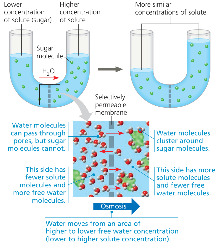
Osmosis is the passive movement of water molecules across a semi-permeable membrane. Water moves from a region of high water concentration (low solute concentration) to a region of low water concentration (high solute concentration). Driven by random movement of particles—no energy is required.
🧬 Why Does Osmosis Happen?
Most biological membranes are impermeable to solutes (like salts, sugars, ions) but partially permeable to water.
This creates an imbalance:
- Solutes can’t move, but water can.
- Water flows across the membrane to balance solute concentration on both sides.
Osmosis continues until equilibrium is reached or physical resistance (like pressure) stops it.
📌 Key Features of Osmosis
Feature | Description |
---|---|
Type of transport | Passive (no energy required) |
Direction of flow | From low solute to high solute (i.e., high water to low water) |
Driven by | Random movement of water molecules |
Blocked solutes | Solutes can’t cross the membrane freely |
Effect | Causes cells to swell or shrink based on water gain/loss |
🧪 What Are Aquaporins?
Aquaporins are protein channels that allow water to pass through membranes more efficiently. They are selective for water only—prevent passage of ions or protons (H⁺). Water moves through aquaporins in single file, maintaining control and speed.
🔍 Why Are Aquaporins Important?
Although some water can pass directly through the lipid bilayer, it’s very slow.
Aquaporins make water movement rapid and regulated, especially in cells with high water demands:
- Kidney cells: Reabsorb water to prevent dehydration
- Root hair cells: Absorb water from soil into plants
📊 Osmosis vs. Aquaporin-Facilitated Water Movement
Aspect | Osmosis Through Bilayer | Osmosis via Aquaporins |
---|---|---|
Speed | Slow | Fast |
Selective? | Somewhat | Highly selective for water |
Direction | Down water potential gradient | Same |
Examples | All cells | Kidney, root hair, brain cells |
🧠 Key Takeaways
- Osmosis is water movement from low solute to high solute concentration, driven by random motion.
- It is passive, does not use energy, and balances water levels across membranes.
- Aquaporins are special water channels that make osmosis faster and more efficient in key cells.
- Without osmosis and aquaporins, cells couldn’t maintain proper hydration, turgor pressure, or reabsorb water effectively.
B2.1.6 – Channel Proteins for Facilitated Diffusion
🌿 What is Facilitated Diffusion?
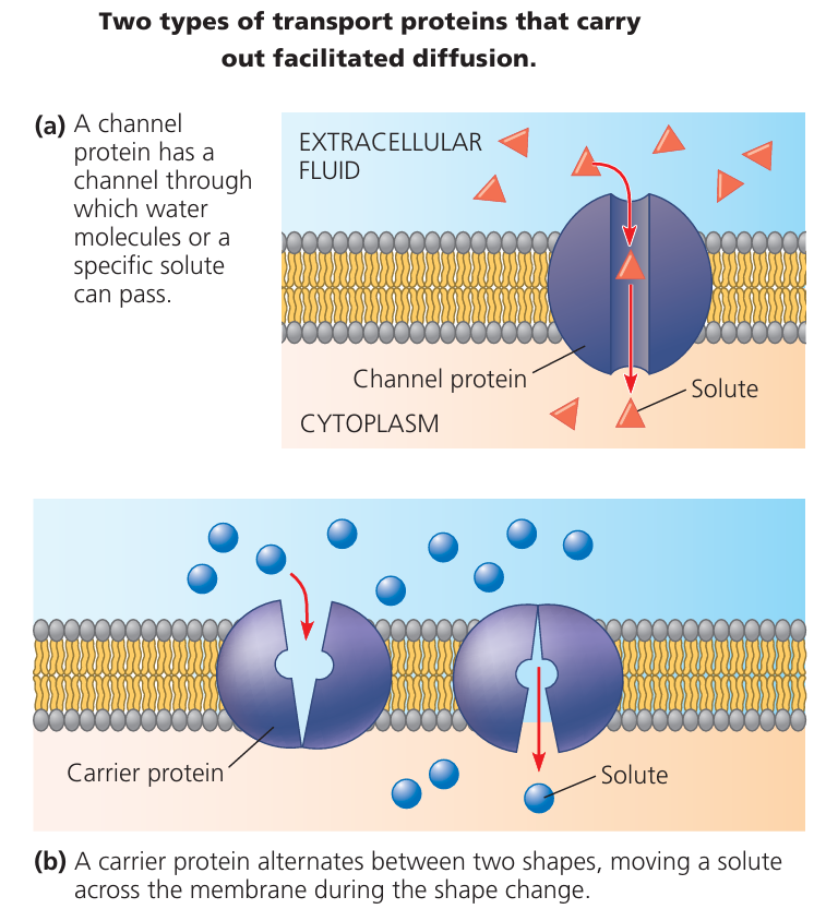
Facilitated diffusion is a type of passive transport where certain molecules move across membranes with help. It requires no energy (ATP) and moves substances down their concentration gradient (from high to low).
However, large polar molecules or charged ions cannot pass through the lipid bilayer on their own—they need help from channel proteins.
🧪 What Are Channel Proteins?
Channel proteins are integral membrane proteins that form pores in the lipid bilayer. They allow specific ions or small polar molecules to cross the membrane. These channels are highly selective each type is designed to allow only one kind of ion or molecule through.
🔐 How Do Channel Proteins Work?
Channels may be:
- Always open (leak channels), or
- Gated—open or close in response to signals like:
- Voltage changes (voltage-gated)
- Binding of a molecule (ligand-gated)
- Mechanical forces (mechanically-gated)
When open, particles diffuse through quickly, just like in simple diffusion—but only for specific substances.
📌 Examples of Channel Protein Action
- Sodium (Na⁺) channels: Only allow Na⁺ ions through.
- Potassium (K⁺) channels: Selectively allow K⁺ ions to exit or enter.
- Chloride (Cl⁻) channels: Maintain electrical balance and water movement.
Example: In nerve cells, voltage-gated Na⁺ and K⁺ channels enable the rapid transmission of impulses.
📊 Simple vs Facilitated Diffusion
Feature | Simple Diffusion | Facilitated Diffusion (via channels) |
---|---|---|
Needs proteins? | No | Yes (channel proteins) |
Type of molecules | Small, non-polar | Ions or polar molecules |
Energy used? | No | No |
Selectivity | Low | High (specific to one type of ion) |
Speed | Depends on gradient only | Faster due to specialized pathways |
🧬 Selective Permeability in Action
Channel proteins help make membranes selectively permeable:
- Only certain ions can cross, depending on:
- Which channel proteins are present
- Whether those channels are open or closed
This allows cells to:
- Control internal ion concentrations
- Respond to stimuli
- Maintain homeostasis
🧠 Key Takeaways
- Facilitated diffusion uses channel proteins to transport specific ions or polar molecules.
- The process is passive—no energy needed.
- Channel proteins provide selective, controlled entry into the cell.
- Gated channels respond to signals and play key roles in nerve impulses, muscle contraction, and more.
B2.1.7 – Pump Proteins for Active Transport
🌿 What is Active Transport?
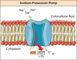
Active transport is the movement of substances against their concentration gradient: from low concentration → high concentration. This process requires energy, usually from ATP (adenosine triphosphate). Active transport allows cells to take in essential particles, even when they’re scarce outside the cell.
🔋 What Are Pump Proteins?
Pump proteins are integral membrane proteins that perform active transport. They use energy from ATP to force molecules or ions across membranes. Each pump is specific for certain substances (e.g. sodium, potassium, calcium).
🔄 How Do Pump Proteins Work?
The pump binds to a specific particle on one side of the membrane. ATP is used, causing a conformational (shape) change in the protein. This change moves the particle to the other side and releases it. The pump then resets to repeat the cycle.
📌 Key Features of Pump Proteins
Feature | Description |
---|---|
Energy required? | Yes, typically from ATP |
Direction of movement | Against concentration gradient (low → high) |
Specificity | Each pump is selective for certain particles |
One-way transport | Pump action is usually unidirectional |
Conformational changes | Change in shape helps move substances across membrane |
🧪 Real-World Examples
- Sodium-Potassium Pump (Na⁺/K⁺ pump): Moves 3 Na⁺ out of the cell and 2 K⁺ in. Essential for nerve impulses, muscle contraction, and osmotic balance.
- Calcium pumps: Maintain low Ca²⁺ inside the cell and high outside. Important for cell signaling and muscle function.
- Proton pumps: Move H⁺ ions across membranes (e.g., in stomach lining or mitochondria).
📊 Passive vs. Active Transport
Feature | Passive Transport | Active Transport |
---|---|---|
Energy required? | No | Yes (ATP) |
Direction | High → Low | Low → High |
Uses protein? | Sometimes (channels/carriers) | Yes (pumps) |
Examples | Diffusion, Osmosis | Na⁺/K⁺ pump, Ca²⁺ pump |
🧠 Key Takeaways
- Pump proteins carry out active transport by moving substances against the gradient.
- They use ATP and undergo shape changes to move specific particles.
- These proteins are essential for life, helping maintain ion balance, absorb nutrients, and support nerve/muscle function.
- Unlike channels, pumps work in one direction only and allow cells to control their internal environment precisely.
B2.1.8 – Selectivity in Membrane Permeability
🌿 What Does Selectively Permeable Mean?
A selectively permeable membrane allows some substances to pass through but blocks others. This is essential for maintaining internal balance (homeostasis) and supporting specialized cellular functions. Cell membranes are not just passive barriers—they use proteins to control what goes in and out.
🧪 Three Types of Permeability Mechanisms
1. Simple Diffusion – Not Selective
No energy or proteins needed.
Only depends on:
- Size of the molecule
- Polarity or hydrophobic/hydrophilic nature
Small, non-polar molecules like O₂ and CO₂ can pass freely. But large or polar molecules cannot cross without help.
2. Facilitated Diffusion – Selective
Uses channel or carrier proteins to allow specific substances through (e.g., Na⁺, glucose).
Still passive (no ATP used).
Selectivity is based on:
- Shape
- Charge
- Size
Each channel allows only one or a few types of particles.
3. Active Transport – Highly Selective
Uses pump proteins to move particles against their concentration gradient.
Requires ATP energy.
Pumps are extremely specific (e.g., sodium-potassium pump moves only Na⁺ and K⁺).
Enables cells to accumulate substances even when they’re scarce outside.
📊 Comparison of Permeability Mechanisms
Transport Type | Protein Involved | Energy Used? | Selective? | Example |
---|---|---|---|---|
Simple Diffusion | No | No | Low selectivity | O₂, CO₂ |
Facilitated Diffusion | Yes (channel/carrier) | No | Yes | Glucose, Na⁺ |
Active Transport | Yes (pumps) | Yes (ATP) | Highly selective | Na⁺/K⁺ pump, Ca²⁺ pump |
🧬 Why Is Selectivity Important?
Cells must import nutrients (e.g., glucose, amino acids) and exclude toxins. They must also maintain specific ion balances for processes like:
- Nerve impulses
- Muscle contractions
- Water balance
Without selectivity, cells would lose control over their internal environment and could not function properly.
🧠 Key Takeaways
- Simple diffusion is based only on size and polarity—not selective.
- Facilitated diffusion and active transport involve proteins that make membranes highly selective.
- This selective permeability allows cells to:
- Maintain homeostasis
- Control internal composition
- Respond to external signals precisely
B2.1.9 – Structure and Function of Glycoproteins & Glycolipids
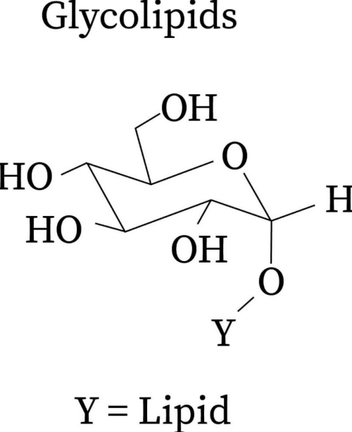
🧬 What Are Glycoproteins and Glycolipids?
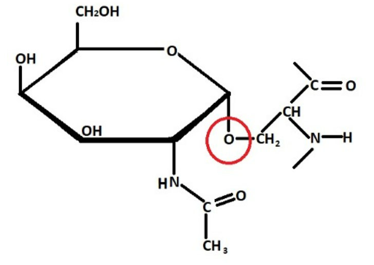
These are molecules with carbohydrate chains attached to:
Proteins → called glycoproteins
Lipids → called glycolipids
They are found in the cell membrane, with their carbohydrate portions always facing outward into the extracellular space.
🌿 Basic Structure
Component | Structure | Location |
---|---|---|
Glycoprotein | Protein + carbohydrate chain | Protein spans membrane; sugar sticks out |
Glycolipid | Lipid + carbohydrate chain | Lipid embedded in membrane; sugar sticks out |
Carbohydrate | Usually short chains (oligosaccharides) | Always on the outer surface of membrane |
These carbohydrate chains together form a sticky outer coat called the glycocalyx.
📌 Key Functions of Glycoproteins & Glycolipids
- Cell Recognition: Cells use glycoproteins/glycolipids to identify each other.
- Immune system → detecting self vs non-self
- Blood groups → e.g., ABO blood types are determined by glycolipids
- Cell Adhesion: Carbohydrate chains from neighboring cells interact and bind, helping cells stick together. Essential for tissue structure and wound healing.
- Protection and Signaling:
- The glycocalyx acts as a protective layer
- Shields the membrane from mechanical damage
- Participates in cell signaling and receptor interactions
🧪 Real-Life Example – Blood-Brain Barrier
In brain capillaries, the glycocalyx is dense and forms part of the blood-brain barrier. It prevents large or harmful substances from entering brain tissue and helps maintain a protected brain environment.
📊 Overview Table
Function | Role of Glycoproteins/Glycolipids |
---|---|
Cell recognition | Identify other cells (e.g., immune cells, pathogens) |
Cell adhesion | Help cells stick together in tissues |
Signaling | Involved in hormone or molecule binding |
Protection | Form glycocalyx to cushion and shield membrane |
Membrane location | Carbohydrate part always faces the extracellular space |
🧠 Key Takeaways
- Glycoproteins = proteins with sugars; Glycolipids = lipids with sugars.
- Carbohydrates always project outward from the membrane, forming the glycocalyx.
- These structures are vital for cell-cell communication, recognition, protection, and tissue organization.
B2.1.10 – Fluid Mosaic Model of Membrane Structure
🧬 What Is the Fluid Mosaic Model?
The fluid mosaic model is the widely accepted model that describes the structure of the plasma membrane. It shows that the membrane is:
- Fluid: The components (like lipids and proteins) move laterally, making the membrane flexible.
- Mosaic: It’s a patchwork of many molecules—phospholipids, proteins, cholesterol, and carbohydrates—like tiles in a mosaic.
🌿 Main Components of the Membrane
Component | Description |
---|---|
Phospholipids | Form the basic bilayer; have hydrophilic heads (face water) and hydrophobic tails (face inward) |
Integral Proteins | Span across the bilayer; help in transport, reception, and cell communication |
Peripheral Proteins | Attached to the inner or outer surface; support and assist in signaling |
Glycoproteins | Proteins with carbohydrate chains; involved in cell recognition and adhesion |
Glycolipids | Lipids with carbohydrate chains; also help in cell recognition |
Cholesterol | Scattered within phospholipids; stabilizes membrane fluidity and reduces permeability |
🌊 Why “Fluid”?
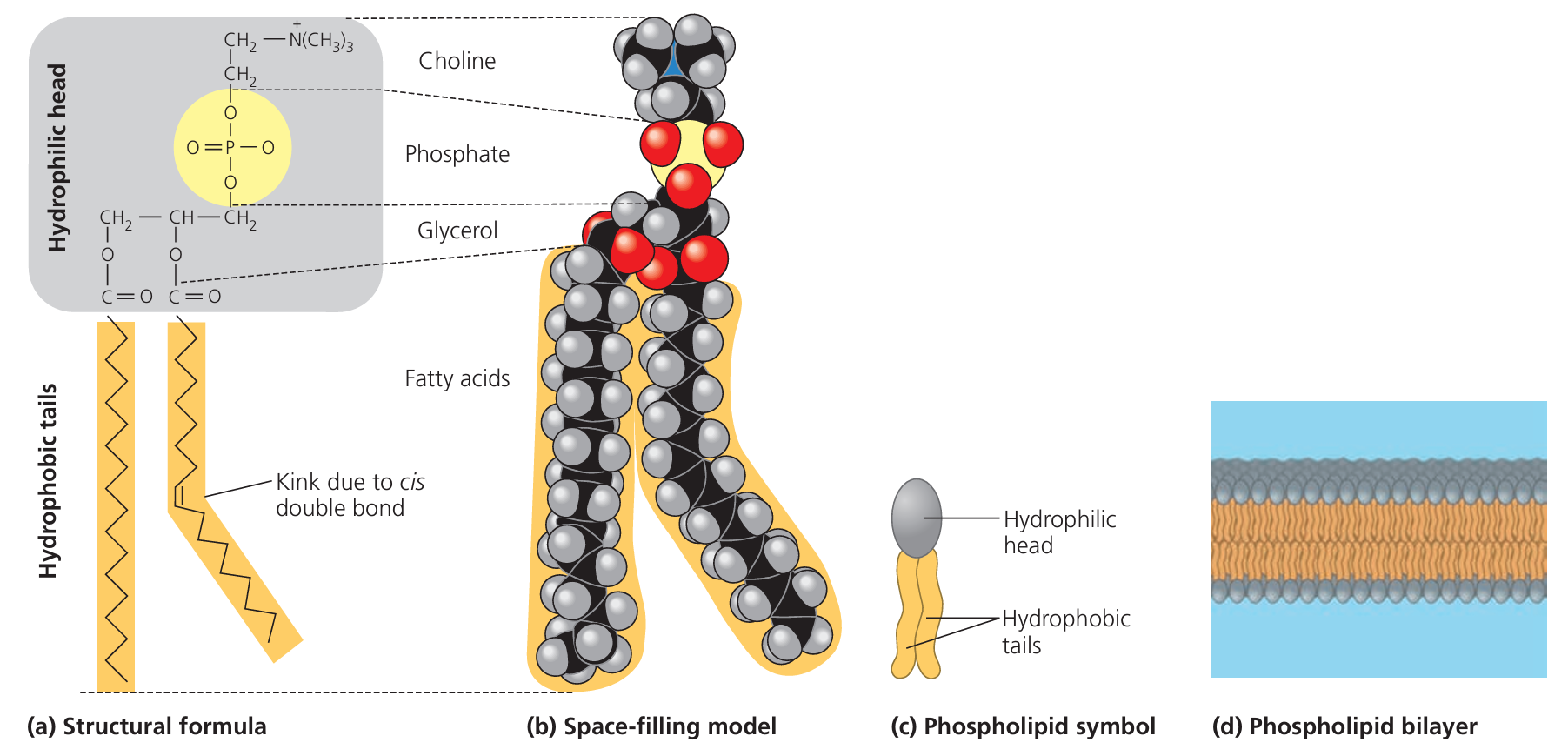
Phospholipids and proteins can move side to side within the layer.
- Membrane flexibility
- Self-repair
- Movement of embedded proteins
🧲 Hydrophilic vs Hydrophobic Regions
- Hydrophilic regions: “Water-loving” — Found on the outside of the membrane (phosphate heads of phospholipids)
- Hydrophobic regions: “Water-fearing” — Found in the core of the membrane (fatty acid tails)
This dual nature (amphipathic property) is essential to form bilayers in water.
🧠 Key Takeaways
- The fluid mosaic model describes a flexible, selectively permeable cell membrane made of lipids, proteins, and carbohydrates.
- Its fluidity allows for protein movement and cell interaction.
- The phospholipid bilayer is arranged with hydrophilic heads outside and hydrophobic tails inside.
- Proteins and carbs give the membrane its function—transport, communication, and recognition.
Additional Higher Level
B2.1.11 – Fatty Acid Composition & Membrane Fluidity
🧬 Why Does Fatty Acid Composition Matter?
The type of fatty acids in the phospholipid bilayer directly affects how fluid or rigid a cell membrane is.
- Transport of substances
- Protein mobility
- Cell flexibility
- Proper functioning at different temperatures
🌿 Saturated vs Unsaturated Fatty Acids
Fatty Acid Type | Structure | Effect on Membrane |
---|---|---|
Saturated | Straight chains, no double bonds | Tightly packed → Less fluid |
Unsaturated | Kinks in chains due to double bonds | Loosely packed → More fluid |
Saturated fats: Have a higher melting point, so membranes become more rigid.
Unsaturated fats: Have a lower melting point, so membranes stay fluid and flexible at lower temperatures.
🌡️ Role of Temperature in Membrane Fluidity
Cell membranes must remain fluid across temperature changes.
Organisms adjust the ratio of saturated and unsaturated fatty acids to cope with their environment:
- Cold environments: More unsaturated fatty acids to prevent stiffness
- Hot environments: More saturated fatty acids to maintain structure
🐟 Adaptation Example: Antarctic Fish
- Live in cold waters where low temperatures could freeze or stiffen membranes.
- Their membranes have a high proportion of unsaturated fatty acids.
- This keeps membranes fluid enough for normal cell function, even in freezing conditions.
📌 Key Points on Membrane Fluidity
Factor | Increases Fluidity | Decreases Fluidity |
---|---|---|
Fatty acid type | Unsaturated | Saturated |
Temperature | Higher temps | Lower temps |
Packing of fatty acids | Loose (kinks) | Tight (straight chains) |
Organismal adaptations | Cold: more unsaturated | Hot: more saturated |
🧠 Key Takeaways
- Membrane fluidity is crucial for cell survival and function.
- Unsaturated fatty acids increase fluidity, helping cells remain flexible in cold temperatures.
- Saturated fatty acids decrease fluidity, helping membranes stay intact at high temperatures.
- Organisms adjust their membrane composition based on habitat temperature for optimal performance.
B2.1.12 – Cholesterol and Membrane Fluidity in Animal Cells
🧬 What Is Cholesterol Doing in the Membrane?
Cholesterol is a lipid found only in animal cell membranes.
It is amphipathic:
- Has a hydrophilic (polar) OH group that aligns near the phospholipid heads
- Has a hydrophobic steroid ring that sits among the fatty acid tails
This position allows cholesterol to interact with both parts of the bilayer and influence its behavior.
🌡️ How Cholesterol Regulates Fluidity
Temperature Condition | Cholesterol’s Role | Effect on Membrane |
---|---|---|
High temperatures | Restricts phospholipid movement | Prevents membrane from becoming too fluid or leaky |
Low temperatures | Prevents tight packing of fatty acid tails | Keeps membrane from becoming too rigid |
So, cholesterol acts as a buffer it stabilizes membrane structure across different temperatures.
🧲 Other Key Roles of Cholesterol
- Maintains membrane integrity: Keeps it from breaking down under stress
- Regulates permeability: Helps reduce unwanted leakage of ions (like Na⁺, H⁺)
- Supports membrane protein function: Creates the right environment for embedded proteins to work
🐾 Why Only in Animal Cells?
Animal cells lack a cell wall, so cholesterol helps:
- Reinforce the membrane
- Protect against temperature changes
Plant cells don’t need cholesterol they have stiff cell walls and use other lipids like phytosterols for stability.
📌 Summary Table
Feature | Cholesterol’s Effect |
---|---|
At high temp | Reduces fluidity, stabilizes membrane |
At low temp | Prevents stiffening, keeps membrane flexible |
Membrane permeability | Decreases leakiness of hydrophilic substances |
Found in | Animal cell membranes only |
🧠 Key Takeaways
- Cholesterol acts as a fluidity regulator in animal membranes.
- It helps maintain the right balance—not too rigid, not too fluid.
- This ensures membranes stay functional and protective in changing conditions.
- Without cholesterol, animal cells would be more vulnerable to temperature-related damage.
B2.1.13 – Membrane Fluidity & Vesicle Fusion/Forming
🌿 Why Is Membrane Fluidity Important for Transport?
Cell membranes are fluid, meaning phospholipids can move sideways within the bilayer.
This fluidity allows the membrane to:
- Bend
- Curve
- Fuse with other membranes
- Form vesicles (small membrane-bound sacs)
These actions are essential for moving substances in and out of cells.
📥 Endocytosis – Bringing Substances Into the Cell
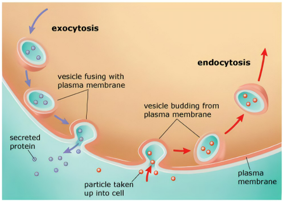
Definition: The plasma membrane folds inward, forming a vesicle that encloses external materials and brings them into the cell.
- Requires energy (ATP)
- Phagocytosis (“cell eating”): Engulfing large particles like bacteria
- Pinocytosis (“cell drinking”): Engulfing fluids or small molecules
- Receptor-mediated endocytosis: Targeted uptake using surface receptors
📤 Exocytosis – Releasing Substances From the Cell
Definition: Vesicles formed inside the cell fuse with the plasma membrane and release their contents outside.
- Also requires ATP
- Secretion of hormones (e.g., insulin from pancreatic cells)
- Release of neurotransmitters at nerve endings
- Excretion of waste materials
🧬 How Membrane Fluidity Enables These Processes
Process | Role of Fluidity |
---|---|
Vesicle formation | Fluid membrane bends and pinches off to form vesicles |
Vesicle fusion | Vesicle merges with the membrane to deliver or export contents |
Endocytosis | Membrane wraps around material and buds inward |
Exocytosis | Vesicle fuses outward to release contents |
Without membrane fluidity, vesicles couldn’t form or fuse, and the cell would lose its ability to transport materials efficiently.
🧠 Key Takeaways
- Fluid membranes enable vesicle formation and fusion, which are key to cellular transport.
- Endocytosis brings substances into the cell using vesicles.
- Exocytosis releases substances out of the cell using vesicles.
- Both processes are active transport and require ATP energy.
- These mechanisms help cells absorb nutrients, communicate, and get rid of waste efficiently.
B2.1.14 – Gated Ion Channels in Neurons
🧬 What Are Gated Ion Channels?
Gated ion channels are protein channels in the membrane that open or close in response to specific stimuli.
They allow charged ions (like Na⁺, K⁺, Cl⁻) to move in or out of the neuron, helping transmit nerve impulses.
There are two main types:
1. Voltage-Gated Ion Channels
Open or close in response to changes in membrane potential (electrical charge difference across the membrane).
Ion Channel | Opens in Response to… | Function in Neurons |
---|---|---|
Sodium (Na⁺) Channel | Membrane becoming more positive (depolarization) | Allows Na⁺ to rush in → initiates action potential |
Potassium (K⁺) Channel | Membrane becoming more positive (later in action potential) | K⁺ flows out → helps repolarize membrane |
2. Ligand-Gated (Neurotransmitter-Gated) Ion Channels
Open when a specific chemical (ligand) binds to them often a neurotransmitter.
📌 Example: Nicotinic Acetylcholine Receptor
Found in neuromuscular junctions.
Opens when acetylcholine (ACh) binds to it.
Allows Na⁺ to enter the muscle cell, triggering muscle contraction.
This is an example of chemical signaling leading to electrical activity.
🔁 How Ion Channels Help in Nerve Impulse Transmission
Stage of Impulse | Channel Involved | What Happens? |
---|---|---|
Resting potential | Most gated channels closed | Neuron is ready to fire |
Depolarization | Na⁺ channels open | Na⁺ rushes in, inside becomes more positive |
Repolarization | K⁺ channels open | K⁺ flows out, restoring negative charge |
Return to rest | All channels reset | Na⁺/K⁺ pump restores ion balance |
🧠 Key Takeaways
- Gated ion channels are essential for nerve signal transmission.
- Voltage-gated channels open due to changes in electrical charge.
- Ligand-gated channels open when a chemical (like acetylcholine) binds.
- Examples: Voltage-gated Na⁺ and K⁺ channels → action potentials; Nicotinic acetylcholine receptors → muscle activation
B2.1.15 – Sodium-Potassium Pumps as Exchange Transporters
🔬 What Is the Sodium-Potassium Pump?
- The sodium–potassium pump is a type of active transport protein found in the membranes of most animal cells.
- It uses energy from ATP to move ions against their concentration gradients.
- This pump is a classic example of an exchange transporter: it swaps Na⁺ and K⁺ ions across the membrane.
⚙️ How the Pump Works (Step-by-Step)
Step | What Happens |
---|---|
1 | 3 Na⁺ ions from inside the cell bind to the pump. |
2 | ATP is hydrolyzed → phosphate group attaches to the pump. |
3 | Pump changes shape and releases Na⁺ outside the cell. |
4 | 2 K⁺ ions from outside bind to the pump. |
5 | Phosphate is released → pump returns to original shape. |
6 | K⁺ is released into the cell. |
Cycle repeats, using ATP each time.
Why Is the Sodium-Potassium Pump Important?
1. Maintains Membrane Potential
The pump helps create a net negative charge inside the cell because:
- 3 positive ions (Na⁺) leave
- 2 positive ions (K⁺) enter
This difference in charge across the membrane is called the membrane potential.
Crucial for:
- Nerve impulses
- Muscle contractions
- Maintaining ion balance
2. Prevents Cell Swelling
If Na⁺ builds up inside, water would follow by osmosis → the pump prevents this by removing excess Na⁺.
🔁 Summary Table: Sodium-Potassium Pump
Feature | Description |
---|---|
Type of transport | Active (uses ATP) |
Ions moved | 3 Na⁺ out, 2 K⁺ in |
Energy source | ATP hydrolysis |
Direction of movement | Against concentration gradient |
Function | Maintains membrane potential, cell volume, and ion balance |
🧠 Key Takeaways
- The sodium–potassium pump actively exchanges Na⁺ and K⁺ ions, powered by ATP.
- It is essential for generating membrane potential and keeping the inside of the cell negative.
- Supports nerve and muscle function and prevents swelling by removing excess Na⁺.
- It’s a continuous, vital process for maintaining homeostasis in animal cells.
B2.1.16 – Sodium-Dependent Glucose Cotransporters (SGLTs)
🧬 What Are Sodium-Dependent Glucose Cotransporters?
These are membrane proteins that transport glucose into cells along with sodium ions (Na⁺).
Also known as SGLTs (Sodium-Glucose Linked Transporters).
They use a process called indirect active transport:
- ATP is not directly used by the SGLT itself
- Instead, energy comes from a sodium ion gradient created by the sodium–potassium pump
⚙️ How Indirect Active Transport Works
Step | Process |
---|---|
1 | Na⁺/K⁺ pump uses ATP to pump Na⁺ out of the cell (creates low Na⁺ inside) |
2 | Na⁺ wants to re-enter the cell (down its gradient) |
3 | SGLT uses the energy from Na⁺ re-entry to transport glucose into the cell (against its gradient) |
4 | Glucose builds up in the cell and is then moved into the blood by facilitated diffusion |
🌿 Where Are SGLTs Used in the Body?
Location | Function |
---|---|
Small intestine | Absorb glucose from digested food into epithelial cells |
Nephron (kidney) | Reabsorb glucose from the filtrate back into the blood |
Both sites use SGLTs to ensure glucose is efficiently captured, even when external concentrations are low.
📌 Why Is This Important?
- Prevents loss of glucose in urine (in kidneys)
- Allows maximum glucose absorption from food (in intestines)
- Maintains blood glucose levels for energy supply
🧠 Key Takeaways
- SGLTs use sodium gradients (set up by the sodium–potassium pump) to pull glucose into cells.
- This is indirect active transport-ATP is used indirectly.
- Essential in the small intestine (glucose absorption) and nephron (glucose reabsorption).
- Supports efficient nutrient uptake and prevents glucose loss in urine.
B2.1.17 – Cell Adhesion and Tissue Formation
🧬 What Is Cell Adhesion?
Tissues are formed when individual cells stick together using special proteins called Cell-Adhesion Molecules (CAMs).
CAMs help cells:
- Recognize and bind to one another
- Organize themselves into layers or structures
- Form stable tissues like skin, muscle, or nerve networks
🔗 What Are CAMs? (Cell-Adhesion Molecules)
Feature | Description |
---|---|
Location | Found on the cell surface (embedded in membrane) |
Structure | Part inside the cell, part sticking out into extracellular space |
Function | Allow cells to connect to each other or to surroundings |
Specificity | Different CAMs are used for different types of junctions between cells |
🧬 Types of CAM Interactions
Interaction Type | Description |
---|---|
Homophilic | CAMs of the same type bind with each other (e.g., same cell type) |
Heterophilic | CAMs of different types interact (e.g., between different cell types) |
This flexibility allows tissues to be organized in complex ways, depending on the function and type of tissue.
How CAMs Help Build Tissues
CAMs allow cells to link up and communicate, forming:
- Tight junctions – e.g., in the intestine to prevent leakage
- Adherens junctions – for structural support
- Desmosomes – resist mechanical stress (e.g., in skin)
- Gap junctions – for exchanging signals and ions
🧠 Key Takeaways
- CAMs are proteins that allow cells to stick together and form functional tissues.
- They are crucial in tissue and organ development, and maintaining structural integrity.
- They also help cells communicate with their neighbors.
- Different CAMs create different types of junctions, depending on tissue function.