A2.2.2 – Microscopy Skills
🧪 Microscopes in Cell Biology
Microscopy is essential for studying cells and tissues. It goes beyond observation – enabling measurement, analysis, and visualization of microscopic structures.
🧷 Using a Light Microscope
| Part | Function |
|---|---|
| Eyepiece | Lens you look through (usually 10× magnification). |
| Objective lenses | Rotating lenses of 4×, 10×, 40×, or 100× magnification. |
| Stage | Holds the slide in position. |
| Coarse focus knob | Quickly adjusts focus to locate specimen. |
| Fine focus knob | Sharpens the image after coarse focusing. |
| Light source | Provides illumination for viewing. |
| Diaphragm | Controls light intensity through the specimen. |
| Condenser | Focuses light onto the specimen. |
Preparing a Temporary Slide
Place a thin sample on a clean slide → Add a drop of water o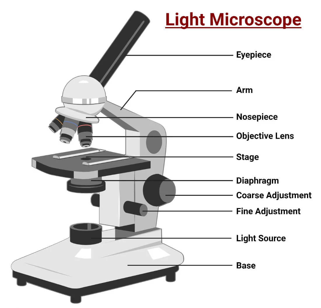 r stain → Lower coverslip at an angle → Dab excess fluid with tissue.
Using Stains
| Stain | Used for |
|---|---|
| Iodine | Highlights starch granules and nuclei. |
| Methylene blue | Stains nuclei blue for contrast. |
| Eosin | Colors cytoplasm pink. |
🔎 Focusing Your Image
- Begin with the lowest magnification (4× or 10×).
- Center specimen in the field of view.
- Use the coarse focus, then fine focus for sharpness.
- Switch to higher magnification if needed.
Measuring and Magnification
Magnification = Image Size ÷ Actual Size
Actual Size = Image Size ÷ Magnification
📐 Eyepiece Graticule & Calibration
An eyepiece graticule is a transparent scale used for measuring microscopic objects. It must be calibrated using a stage micrometer (a slide with a precise scale).
Capturing and Annotating Images
- Use a smartphone or digital camera with the microscope.
- Add a scale bar based on known measurements.
- Label key features like nuclei, cytoplasm, or chloroplasts.
🧠 Nature of Science (NOS): Microscopy turns visual observation into measurable, testable data – transforming biology into a more evidence-based science.
✅ Summary Table
| Skill | Description |
|---|---|
| Mounting | Place specimen on slide, add liquid, lower coverslip. |
| Focusing | Start with coarse focus, refine with fine focus. |
| Staining | Increases visibility and contrast of cell structures. |
| Measuring | Use graticule and magnification formula for accuracy. |
| Scale bar | Calculate real dimensions and add to image. |
| Photos | Capture labeled, clear microscopy images for reports. |
A2.2.3 – Developments in Microscopy
🧠 Why is this important?
Microscopy has revolutionized cell biology. As technologies advanced, newer microscopes and techniques allowed scientists to visualize structures beyond the reach of traditional light microscopes.
Key Modern Microscopy Techniques
🔬 Electron Microscopy (EM)
- How it works: Uses electron beams instead of light; image forms from electron scattering or absorption.
- Resolution: Extremely high (up to 0.1 nm).
- Reveals: Fine ultrastructure – mitochondria, membranes, ribosomes, viruses.
Types:
- TEM: Electrons pass through thin sections → reveals internal details.
- SEM: Electrons bounce off surface → 3D surface views.
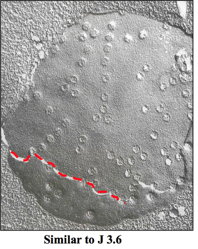
❄️ Freeze Fracture Microscopy
- How it works: Sample is frozen and fractured → coated with metal → viewed under EM.
- Reveals: Internal membrane details, embedded proteins, and bilayer separation.
🧊 Cryogenic Electron Microscopy (Cryo-EM)
- How it works: Rapid freezing preserves sample in near-native state.
- Advantages: Reduces damage, no staining or fixation needed.
- Used for: Imaging proteins, viruses, and molecular complexes.
💡 Fluorescent Stains in Light Microscopy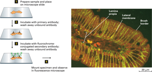
- How it works: Dyes absorb light and emit fluorescence to highlight structures.
- Examples:
- DAPI: Binds DNA → emits blue.
- Rhodamine: Stains actin (red).
- FITC (fluorescein): Green → often paired with antibodies.
- Benefits: High contrast, allows multi-labeling of structures.
 🧬 Immunofluorescence Microscopy
- How it works: Fluorescent antibodies bind to specific proteins or antigens.
- Types: Direct (tagged antibody) and Indirect (uses a fluorescent secondary antibody).
- Uses: Localizing proteins, diagnosing diseases, studying expression patterns.
🧪 Summary Table
| Technique | Key Benefit | Used For |
|---|---|---|
| Electron Microscopy (TEM/SEM) | Very high resolution | Cell ultrastructure, viruses |
| Freeze Fracture EM | Views membrane interiors | Bilayers, membrane proteins |
| Cryo-EM | Preserves native structure | Protein complexes, viruses |
| Fluorescent Stains | Color-specific labeling | DNA, cytoskeleton, organelles |
| Immunofluorescence | Protein targeting | Protein expression/localization |
A2.2.5 – Prokaryote Cell Structure
🔍 What are prokaryotes?
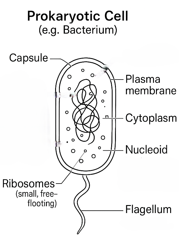
Prokaryotes are unicellular organisms that lack a membrane-bound nucleus. Their genetic material floats freely in the cytoplasm. Common examples include Gram-positive bacteria such as Bacillus and Staphylococcus.
Basic Prokaryotic Cell Components (with Functions)
| Structure | Function |
|---|---|
| Cell Wall | Rigid layer of peptidoglycan that provides shape and protection |
| Plasma Membrane | Selectively permeable; controls entry and exit of materials |
| Cytoplasm | Watery fluid where cellular processes and reactions occur |
| Naked DNA | Circular loop of double-stranded DNA located in the nucleoid |
| 70S Ribosomes | Sites of protein synthesis; smaller than eukaryotic ribosomes |
🧬 Optional Structures (may be present)
| Structure | Function |
|---|---|
| Plasmid | Small circular DNA carrying extra genes (e.g., antibiotic resistance) |
| Pili | Aid attachment to surfaces, involved in conjugation (DNA exchange) |
| Flagella | Provide motility in liquid environments |
| Capsule | Sticky outer coating for protection and surface adhesion |
🌟 Special Features of Prokaryotic Cells
- Small size: typically 1–5 µm in diameter
- No membrane-bound organelles
- Reproduce by binary fission (asexual)
- All metabolic processes occur in the cytoplasm or plasma membrane

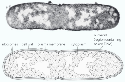
🧪 Example: E. coli Cell
A typical E. coli cell includes a peptidoglycan cell wall, plasma membrane, cytoplasm, naked circular DNA (nucleoid), 70S ribosomes, and sometimes plasmids, flagella, and pili.
✅ Summary Table
| Feature | Prokaryotes |
|---|---|
| Nucleus | No true nucleus |
| DNA form | Circular, naked (not wrapped in histones) |
| Ribosome type | 70S (smaller) |
| Organelles | No membrane-bound organelles |
| Reproduction | Asexual (binary fission) |
| Examples | Bacteria (E. coli, Staphylococcus) |
A2.2.6 – Eukaryote Cell Structure
– Plant cells also have a cell wall (for support) and chloroplasts (for photosynthesis).
– Animal cells have centrioles and sometimes cilia/flagella for movement.
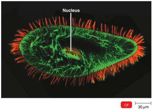
🧬 What is a Eukaryotic Cell?
Eukaryotic cells have a true nucleus and membrane-bound organelles. These organelles carry out specialized functions that allow the cell to work efficiently.
Examples of Eukaryotes:
Unicellular: Protists
Multicellular: Fungi, Plants, Animals
🔍 Key Features Found in All Eukaryotic Cells:
| Structure | Description |
|---|---|
| Plasma Membrane | Phospholipid bilayer that controls what enters and exits the cell |
| Cytoplasm | Watery environment for biochemical reactions |
| 80S Ribosomes | Site of protein synthesis (larger than 70S ribosomes in prokaryotes) |
| Nucleus | Contains DNA; surrounded by a double membrane with pores |
| Membrane-bound Organelles | Includes mitochondria, ER, Golgi apparatus, lysosomes, etc. |
| Cytoskeleton | Microtubules and microfilaments that support cell shape and transport |
🧠 Why Is Compartmentalization Important?

- Allows different environments inside organelles (e.g., acidic lysosomes)
- Concentrates enzymes and substrates (e.g., in mitochondria)
- Isolates harmful reactions (e.g., digestion inside lysosomes)
🧪 Important Organelles & Functions:
- Nucleus: Stores DNA, controls gene expression
- Mitochondria: Site of aerobic respiration, ATP production
- Rough ER: Has ribosomes, synthesizes and transports proteins
- Smooth ER: Lipid synthesis, detoxification, glucose release
- Golgi Apparatus: Modifies and packages proteins, forms vesicles
- Lysosomes: Contain enzymes to break down waste (animal cells only)
- Vacuoles: Large in plants (stores water), small in animals
- Vesicles: Transport and storage sacs inside cells
- Cytoskeleton: Provides structure, movement, organelle anchoring
- Centrioles: (Animals only) Organize spindle fibers during cell division
- Chloroplasts: (Plants only) Site of photosynthesis, contain thylakoids
A2.2.9 – Atypical Cell Structure in Eukaryotes
Some eukaryotic cells show atypical features, particularly in the number of nuclei they possess. This variation is closely tied to their structure and specialized function.
1. Aseptate Fungal Hyphae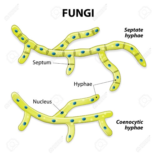
Nuclei: Multinucleate (many nuclei per cell)
Structure: Long filamentous cells without cross-walls (septa)
Explanation: Without septa, cytoplasm and nuclei flow freely, supporting rapid growth and nutrient sharing.
Example: Fungi like Rhizopus (bread mold)
2. Skeletal Muscle Cells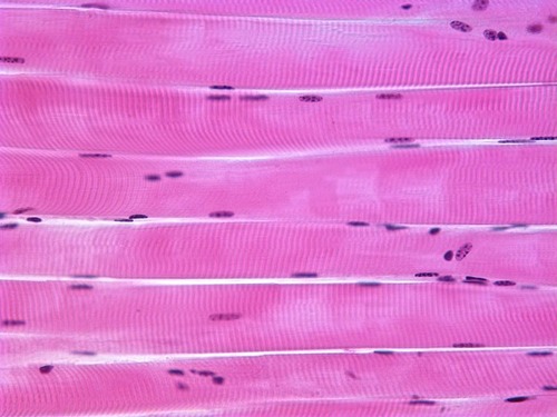
Nuclei: Multinucleate
Structure: Long, cylindrical fibers formed by fusion of myoblasts
Explanation: Multiple nuclei support high protein synthesis and contraction over the entire cell length.
Example: Human skeletal muscle fibers
3. Red Blood Cells (RBCs)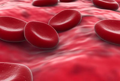
Nuclei: Anucleate (no nucleus)
Structure: Biconcave disc with no organelles
Explanation: Ejecting the nucleus allows more room for hemoglobin, boosting oxygen transport.
Example: Mammalian red blood cells
4. Phloem Sieve Tube Elements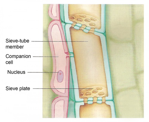
Nuclei: Absent or highly reduced
Structure: Elongated tube-like cells stacked end-to-end
Explanation: The lack of nuclei allows free flow of sap. Companion cells nearby provide metabolic support.
Example: Phloem tissue in vascular plants
✅ Summary Table
| Cell Type | Nucleus Type | Unique Feature |
|---|---|---|
| Aseptate Fungal Hyphae | Multinucleate | No septa – nuclei share one cytoplasm |
| Skeletal Muscle Cell | Multinucleate | Formed by fusion of myoblasts |
| Red Blood Cell (RBC) | Anucleate | Ejects nucleus to hold more hemoglobin |
| Phloem Sieve Tube Element | Absent / Reduced | Relies on companion cells for support |
These examples challenge the standard idea of “one nucleus per eukaryotic cell” and reveal how structure relates to function and adaptation in specialized cells.
A2.2.11 – Drawing and Annotation Based on Electron Micrographs
✏️ Why Drawing from Micrographs Is Important
- Simplifies complex visuals → Helps identify structures more easily
- Focuses on key features → Omits distracting artifacts
- Develops understanding → You learn by translating images into labeled forms
🧠 Tips Before Drawing
- Only include what’s visible in the micrograph.
- Keep drawings clear and uncluttered.
- Use straight lines for labels.
- Label with functions, not just names.
📋 Structures You Should Be Able to Draw and Annotate
| Structure | Description in EM | Function (for annotation) |
|---|---|---|
| Nucleus | Double membrane with pores, contains chromatin | Stores genetic material and controls cell activities |
| Mitochondrion | Oval, double membrane, folded inner (cristae) | Site of ATP production via aerobic respiration |
| Chloroplast | Double membrane, stacks of thylakoids (grana) | Carries out photosynthesis to produce glucose |
| Sap Vacuole (plant) | Large central, membrane-bound | Maintains turgor pressure; stores ions, waste, nutrients |
| Golgi Apparatus | Series of flattened sacs (cisternae), no ribosomes | Modifies, sorts, and packages proteins for secretion |
| Rough ER | Flattened sacs with attached ribosomes | Synthesizes proteins and sends them to the Golgi |
| Smooth ER | Flattened sacs without ribosomes | Synthesizes lipids; detoxifies harmful substances |
| Chromosomes | Condensed chromatin strands during division | Carries genetic information (visible during division) |
| Cell Wall (plant) | Thick rigid layer outside membrane | Provides support and protection |
| Plasma Membrane | Thin boundary around cell | Controls movement of substances in/out |
| Secretory Vesicles | Small membrane sacs | Transport proteins/lipids for secretion |
| Microvilli (animal) | Finger-like membrane extensions | Increase surface area for absorption |
Additional Higher Level
A2.2.12 – Origin of Eukaryotic Cells by Endosymbiosis
🌱 What Is Endosymbiosis?
Endosymbiosis is a widely accepted theory that explains how complex eukaryotic cells originated from simpler prokaryotic ancestors.
🧬 Step-by-Step Evolutionary Process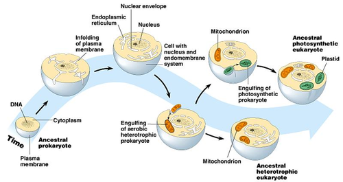
- Formation of a primitive eukaryotic cell: A unicellular ancestor developed a nucleus (membrane-bound DNA) and began reproducing sexually.
- Endosymbiosis of aerobic prokaryote: An ancestral eukaryotic cell engulfed an aerobic prokaryote (e.g. a proteobacterium). Instead of digesting it, the prokaryote survived inside and produced ATP. This evolved into the mitochondrion.
- Secondary endosymbiosis in some lineages: A second engulfing event occurred where a photosynthetic prokaryote (likely a cyanobacterium) was engulfed. This led to the formation of chloroplasts in plant and algal cells.
🧪 Evidence Supporting the Endosymbiotic Theory
| Feature | Explanation |
|---|---|
| Double Membranes | Inner membrane from engulfed cell; outer from host vesicle |
| Circular Naked DNA | Like prokaryotes, both mitochondria and chloroplasts have circular DNA |
| 70S Ribosomes | Same size as those found in prokaryotes (vs 80S in cytoplasm) |
| Self-replication | Mitochondria and chloroplasts divide independently by binary fission |
| Antibiotic Sensitivity | Some antibiotics that affect bacteria also affect mitochondria and chloroplasts |
🧠 Nature of Science (NOS) Insight
Theory strength = Explanatory Power + Predictive Ability
The endosymbiotic theory is supported by a wide range of independent observations from genetics, microscopy, and biochemistry. It provides a unifying explanation for the origin of key eukaryotic features and predicts similarities between prokaryotes and organelles.
Mitochondria & chloroplasts = “cells within cells”
Look for: 70S ribosomes, circular DNA, binary fission, double membranes
A2.2.13 – Cell Differentiation as the Process for Developing Specialized Tissues in Multicellular Organisms
🧬 What is Cell Differentiation?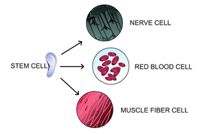
Cell differentiation is the biological process where unspecialized cells (e.g. stem cells) become specialized to carry out distinct functions.
🧪 How Does Differentiation Happen?
- Gene Expression Changes: Only some genes are activated, while others are switched off, leading to the production of proteins needed for a specific function.
- Environmental Triggers: Internal/external cues (e.g. growth factors, chemical signals) influence gene expression. Triggers include embryo position, signaling, and hormones.
🌱 Examples of Differentiation in Humans
| Stem Cell | Differentiates Into |
|---|---|
| Hematopoietic stem cell | Red blood cells, white blood cells, platelets |
| Neural stem cell | Neurons, glial cells |
| Embryonic stem cell | Any cell type (pluripotent) |
🧠 Why Is Differentiation Important?
- Tissue Specialization: Enables cells to perform unique roles (e.g., muscle contraction, oxygen transport).
- Organ Development: Specialized cells combine to form functional organs and systems.
- Efficient Functioning: Specialization improves cellular performance and energy use.
🔁 Key Steps in Differentiation
- Undifferentiated cell (e.g. embryonic stem cell)
- Receives signal (environmental or internal)
- Activates specific genes
- Produces specialized proteins
- Change’s structure and function → Becomes a specialized cell
Differentiation = Selective gene expression
Stem cells → Specialized cells
Triggered by signals like chemicals, hormones, or environment
A2.2.14 – Evolution of Multicellularity
🌍 What Is Multicellularity?
Multicellularity refers to organisms made up of more than one cell that work together to form tissues, organs, and complex systems.
🧬 How Did Multicellularity Evolve?

Repeated Evolution: Multicellularity evolved independently in several groups, including:
- Fungi
- Eukaryotic algae
- Plants
- Animals
Key Process: Cell Aggregation
Individual single-celled organisms clustered together into groups.
These clusters allowed better:
- Nutrient sharing
- Protection from predators
- Cooperation
🔄 From Cluster to Complexity
- Loose Grouping: Cells lived together but acted independently
- Permanent Association: Cells began sticking together permanently (e.g. via adhesion molecules)
- Specialization: Some cells took on specific roles (e.g., movement, feeding)
- Communication & Coordination: Cells developed ways to cooperate, leading to true multicellularity
🧪 Why Is Multicellularity an Advantage?
| Advantage | Explanation |
|---|---|
| Larger Body Size | Helps escape predators and access new environments |
| Cell Specialization | Increases efficiency — cells perform specific functions |
| Damage Resistance | Organism survives injury to individual cells |
| Division of Labour | Improves survival, growth, and reproduction |
Multicellularity evolved multiple times in different groups.
It began with cell aggregation, then developed specialization.
Key benefits: larger body size, division of labour, and adaptability.