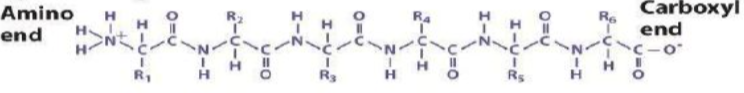
B1.2.1 – Generalized Structure of an Amino Acid
🧬 What Are Amino Acids?
Amino acids are the basic building blocks of proteins. They all have a standard core structure and a variable R-group that determines their properties. There are 20 common amino acids used in protein synthesis.
🔬 General Structure of an Amino Acid
Each amino acid contains four groups attached to the central α-carbon:
| Group | Symbol | Function |
|---|---|---|
| Amino group | –NH₂ | Basic → Can accept a proton (acts as a base) |
| Carboxyl group | –COOH | Acidic → Can donate a proton (acts as an acid) |
| Hydrogen atom | –H | Simple hydrogen bonded to central carbon |
| R-group (side chain) | –R | Variable → Gives each amino acid its unique traits |
🌊 Amphiprotic Nature of Amino Acids
Amino acids are amphiprotic, meaning they can act as both acids and bases because:
- –COOH group donates H⁺ (acidic)
- –NH₂ group accepts H⁺ (basic)
🔁 Importance of the R-Group
The R-group determines the chemical nature and function of each amino acid:
- Polarity: Hydrophilic or hydrophobic
- Charge: Acidic, basic, or neutral
- Size and shape


Examples:
Glycine: R = –H (smallest)
Alanine: R = –CH₃
Cysteine: R = –CH₂–SH (forms disulfide bonds)
🧠 Summary Box – General Structure of Amino Acids
| Component | Symbol | Description |
|---|---|---|
| Central carbon | Cα | Backbone of the amino acid |
| Amino group | –NH₂ | Acts as a base (accepts H⁺) |
| Carboxyl group | –COOH | Acts as an acid (donates H⁺) |
| Hydrogen atom | –H | Present in all amino acids |
| R-group | –R | Variable → determines identity and behavior |
B1.2.2 – Condensation Reactions Forming Dipeptides and Polypeptides
🧬 What Is a Condensation Reaction?
A condensation reaction joins two molecules together by removing a molecule of water (H₂O). In protein formation, amino acids are linked via condensation to form dipeptides and longer polypeptide chains.
🔗 Formation of a Peptide Bond
When two amino acids join:
- The carboxyl group (–COOH) of one reacts with the amino group (–NH₂) of another.
- This forms a peptide bond (–CO–NH–) and releases one water molecule.
Word Equation:
Amino acid + Amino acid → Dipeptide + Water (H₂O)
🔬 Generalized Dipeptide Structure
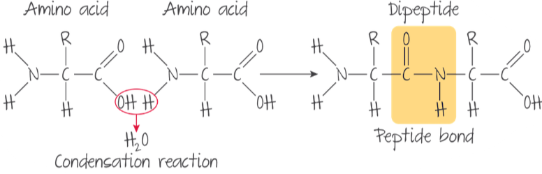
The peptide bond forms between the carbon of the carboxyl group and the nitrogen of the amino group. The resulting dipeptide retains two R-groups – one from each amino acid and a stable backbone.
Polypeptide Formation
Multiple condensation reactions add amino acids to a growing chain, forming a polypeptide. These chains fold into functional proteins, and their order of amino acids determines the protein’s function (its primary structure).
⚙️ Biological Context
| Step | Details |
|---|---|
| Site of reaction | Inside ribosomes during protein synthesis |
| Enzyme involvement | Catalyzed by ribosomal enzymes |
| Directionality | Chain grows by adding amino acids to the C-terminal end |
| Consistency of bond | Peptide bond is structurally the same, regardless of R-groups |
🧠 Summary Box – Peptide Bond Formation
| Component | Description |
|---|---|
| Reactants | 2 amino acids |
| Reaction type | Condensation (removal of water) |
| Bond formed | Peptide bond (–CO–NH–) |
| Product | Dipeptide (or longer polypeptide) |
| Location in cell | Ribosome (site of protein synthesis) |
🧩 The peptide bond is the molecular glue that strings amino acids into functional proteins — forming enzymes, hormones, and the structural core of all life.
B1.2.3 – Dietary Requirements for Amino Acids
🧬 What Are Amino Acids in the Diet?
Amino acids are the building blocks of proteins, essential for growth, repair, and metabolism. While the body can make some amino acids, others must be obtained from food sources.
🍽️ Types of Amino Acids
| Type | Description |
|---|---|
| Essential amino acids | Cannot be synthesized by the human body → must be obtained from the diet |
| Non-essential amino acids | Can be synthesized from other amino acids in the body |
| Conditionally essential | Usually non-essential, but needed in the diet under specific conditions (e.g., infancy, illness) |
Note: Students are not required to memorize specific amino acids by name.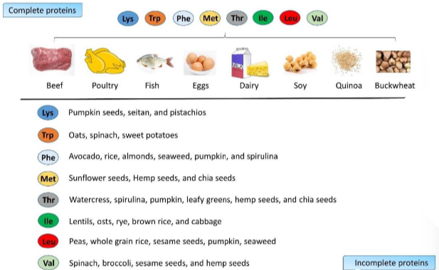
🌱 Why It Matters in Diets
Omnivorous diets (including meat, eggs, dairy) usually provide all essential amino acids. In contrast, vegan or plant-based diets may lack one or more essential amino acids unless foods are carefully combined.
🔁 Protein Complementation
This is the practice of combining different plant protein sources to provide all essential amino acids.
| Example Combinations | Why It Works |
|---|---|
| Rice + lentils | Complements limiting amino acids |
| Corn + beans | Each compensates for the other’s gaps |
| Peanut butter + whole wheat | Together provide a complete amino acid profile |
🧠 Summary Box – Amino Acids in Nutrition
| Concept | Explanation |
|---|---|
| Essential amino acids | Must come from food |
| Non-essential amino acids | Made by the body |
| Vegan diet consideration | Requires food combinations to get all essential amino acids |
| Protein complementation | Strategy to ensure complete protein intake in plant-based diets |
🧩 Balanced protein intake is vital for maintaining muscles, enzymes, immune function, and overall health — especially on restricted or plant-based diets.
B1.2.4 – Infinite Variety of Possible Peptide Chains
🧬 Why Are There So Many Different Polypeptides?
Polypeptides are chains of amino acids joined by peptide bonds. These chains can vary endlessly in:
- Length (from a few to thousands of amino acids)
- Sequence (order of amino acids)
- Composition (which amino acids are included)
 🔤 The Genetic Basis of Variety
| Source of Diversity | Explanation |
|---|---|
| 20 amino acids | Every position in a chain can be any of the 20 |
| Any length of polypeptide | Some proteins have 5 amino acids, others have 5,000+ |
| Amino acids in any order | Different orders = different structures and functions |
🧠 Mathematical Insight
For a peptide with 7 amino acids: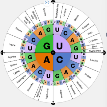
Possible combinations = 20⁷ = 1,280,000,000+ unique sequences
🧪 Real-Life Examples of Polypeptides
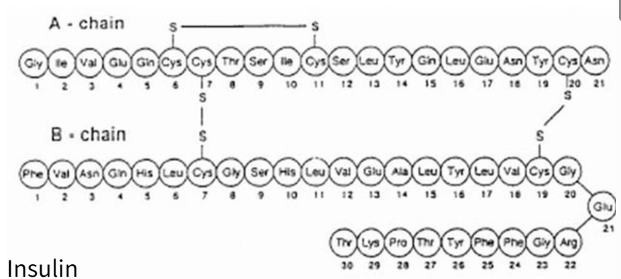
| Polypeptide | Length | Function |
|---|---|---|
| Insulin | 51 amino acids | Regulates blood glucose levels |
| Hemoglobin | ~574 amino acids | Transports oxygen in the blood |
| Keratin | Hundreds–thousands | Forms hair, nails, feathers |
| Actin | 375 amino acids | Involved in muscle contraction & cell shape |
Each polypeptide folds into a specific 3D shape based on its amino acid sequence – this determines its biological function.
🧠 Summary Box – Infinite Polypeptide Diversity
| Key Idea | Why It Matters |
|---|---|
| 20 different amino acids | Provide huge combinatorial diversity |
| Sequences can be any length/order | Allows billions of possible chains |
| Each sequence = unique polypeptide | Structure determines specific function in cells and organisms |
| Examples include insulin, keratin | Real proteins illustrate functional diversity of polypeptides |
🧩 This incredible variation is why proteins can serve so many roles – from enzymes and hormones to structural components and transporters.
B1.2.5 – Effect of pH and Temperature on Protein Structure
🧬 Proteins Depend on Structure to Function
Proteins must maintain a specific 3D shape (tertiary structure) to perform biological roles such as enzyme catalysis, transport, and hormonal signaling.
- Hydrogen bonds
- Ionic bonds
- Disulfide bridges
- Hydrophobic interactions
🔥 Effect of Temperature
- Mild heat can increase activity up to an optimum point.
- High heat breaks hydrogen and ionic bonds.
- Protein unfolds, loses functional shape – usually irreversible.
⚗️ Effect of pH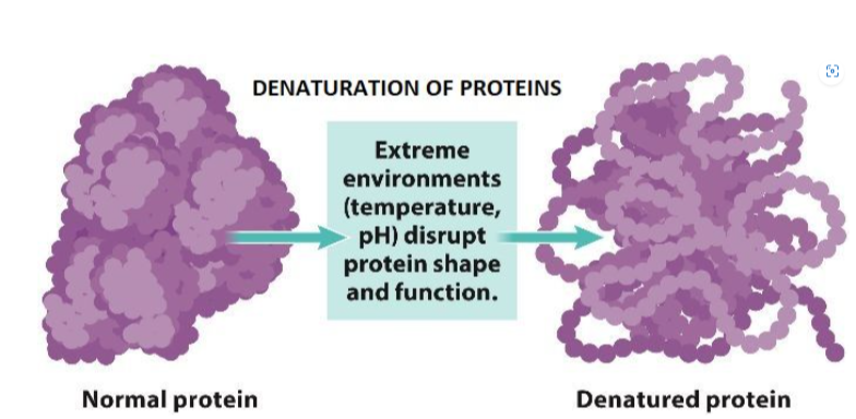
- pH affects charged R-groups of amino acids.
- Disrupts ionic interactions and hydrogen bonds.
- Extremely high or low pH causes denaturation.
🌡️ Example: Heat-Resistant Enzymes
Taq DNA polymerase from Thermus aquaticus is used in PCR because it works efficiently even at 80–90°C, unlike most enzymes which denature at high temperatures.
💧 What Happens During Denaturation?
| Before Denaturation | After Denaturation |
|---|---|
| Stable, folded 3D structure | Unfolded and irregular |
| Hydrophobic regions hidden inside | Hydrophobic groups exposed |
| Active site has specific shape | Active site lost → enzyme inactivated |
🧠 Summary Box – Protein Structure and Denaturation
| Factor | Effect on Protein |
|---|---|
| High temperature | Breaks bonds → irreversible denaturation |
| Extreme pH | Alters charge on amino acids → disrupts shape |
| Denatured protein | Loses function → may precipitate |
| Structure = Function | Shape change = function loss (especially for enzymes) |
🔍 Protein function entirely depends on maintaining correct shape. Heat and pH changes can disrupt life-sustaining reactions by denaturing key proteins.
Additional Higher Level
B1.2.6 – Chemical Diversity in the R-Groups of Amino Acids and Its Role in Protein Form and Function
🧬 What Are R-Groups?
Each of the 20 amino acids shares a basic structure:
- Amine group (–NH₂)
- Carboxyl group (–COOH)
- Hydrogen atom
- Unique side chain – the R-group
🧪 Types of R-Groups and Their Properties
| R-Group Type | Description | Effect on Protein |
|---|---|---|
| Hydrophobic | Non-polar, water-repelling | Fold into interior of proteins |
| Hydrophilic | Polar or charged, water-attracting | Found on outer surfaces in water-rich environments |
| Polar (uncharged) | Partial charges (–OH, –SH, etc.) | Form hydrogen bonds with water or other residues |
| Charged (acidic) | Negative charge (–COO⁻) | Repel other negatives, attract positives |
| Charged (basic) | Positive charge (–NH₃⁺) | Attract acidic groups, help bond formation |
🧠 Why R-Groups Matter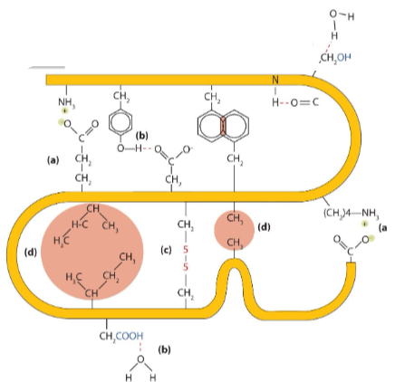
- Protein Folding: Interactions between R-groups (attraction, repulsion, bonding) drive 3D structure formation.
- Active Sites: Enzyme function depends on specific R-group arrangements to bind substrates.
- Solubility: Hydrophilic R-groups face outward; hydrophobic ones face inward – determines location and function.
- Flexibility & Strength: Cysteine R-groups form disulfide bridges, stabilizing protein structure.
🧩 R-Groups Enable Protein Diversity
With 20 amino acids, endless combinations of R-groups across long chains enable millions of different proteins – each tailored for a specific function:
- Enzymes: Catalyze biochemical reactions
- Structural Proteins: Provide support (e.g., keratin, collagen)
- Transport Proteins: e.g., hemoglobin
- Hormones & Receptors: Signal and regulate
- Antibodies: Immune defense
📌 Protein Form = Function: The sequence and chemistry of R-groups determines folding → shape → biological role.
🧠 Summary Box – Why R-Groups Are Essential
| Feature | Importance |
|---|---|
| R-group variation | Basis of protein diversity and function |
| Hydrophobic vs. hydrophilic | Determines folding, solubility, and location in cells |
| Polar and charged R-groups | Enable bonding, attraction/repulsion, and active sites |
| Disulfide bridges (Cys) | Add structural stability and strength |
B1.2.7 – Impact of Primary Structure on the Conformation of Proteins
🔹 What is Protein Conformation?
Conformation refers to the precise 3D shape of a protein, which is essential for its biological function (e.g., enzymes, transporters, receptors).
🧪 What Determines Protein Shape?
A protein’s shape is dictated by its primary structure – the specific sequence of amino acids.
The folding process is influenced by:
- Chemical nature of R-groups
- Hydrogen and ionic bonding
- Hydrophobic interactions
- Disulfide bridges (S–S bonds)
Proteins fold automatically into stable structures guided by these interactions – no external templates are required.
📌 Four Levels of Protein Structure
(a) Primary structure
(b) Secondary structure
(c) Tertiary structure
(d) Quaternary structure
| Level | Description |
|---|---|
| Primary | Linear sequence of amino acids joined by peptide bonds |
| Secondary | Local folding into alpha-helices or beta-pleated sheets via hydrogen bonding |
| Tertiary | 3D folding due to R-group interactions (e.g., hydrophobic, ionic, disulfide) |
| Quaternary | Multiple polypeptide chains joined to form one functional protein (e.g., haemoglobin) |
🧠 Why Does This Matter?
- Protein structure directly affects function — enzymes must fold correctly to bind substrates.
- Primary sequence determines the folding pattern → defines the functional shape.
- AI tools like AlphaFold can now accurately predict protein 3D structure from sequence alone.
- The Protein Data Bank (PDB) holds over 180,000 confirmed protein structures for research and medical use.
Structure = Function: A protein’s job depends on its folding – misfolding can cause disease, while correct folding enables everything from digestion to immunity.
✅ Summary Box
| Concept | Key Point |
|---|---|
| Primary structure | Determines all levels of folding and function |
| Folding depends on | R-group chemistry, hydrogen/ionic bonding, hydrophobic interactions |
| Mutation or misfolding | Disrupts shape → loss of function or disease |
| Prediction tools | AlphaFold and PDB help visualize protein structures from amino acid sequences |
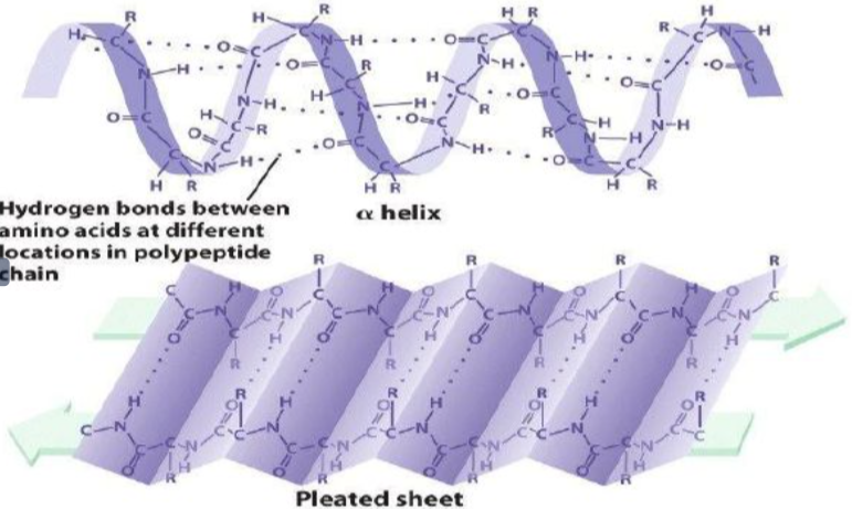
B1.2.8 – Pleating and Coiling of Secondary Structure of Proteins
🔹 What Is the Secondary Structure?
The secondary structure of a protein refers to regular, repetitive folding patterns within a polypeptide chain caused by hydrogen bonding between backbone atoms.
🔬 How Is It Stabilized?
Hydrogen bonds form between the carbonyl group (C=O) and the amino group (N–H) of the peptide backbone.
These bonds occur at regular intervals and stabilize specific folding patterns:
- Intrachain: Hydrogen bonding within the same chain
- Interchain: Hydrogen bonding between separate regions or strands
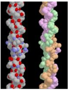
🌀 Alpha Helix (α-Helix)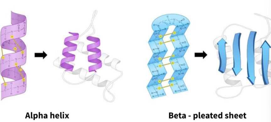
- The polypeptide chain coils into a right-handed spiral
- Hydrogen bonds form between every 4th amino acid
- Stabilizes the helical structure
- Common in structural proteins like keratin (hair, nails)
📄 Beta-Pleated Sheet (β-Sheet)
- Polypeptide chains lie side by side in a zig-zag arrangement
- Can be parallel or antiparallel in orientation
- Hydrogen bonds form between backbone atoms in adjacent strands
- Seen in fibrous proteins like silk (e.g., fibroin)
✅ Summary Box
| Feature | Alpha Helix | Beta-Pleated Sheet |
|---|---|---|
| Shape | Coiled spiral | Zig-zag sheet |
| H-bonding | Within the same chain (intrachain) | Between adjacent segments (inter/intrachain) |
| Stabilization role | Maintains coiling | Aligns and stabilizes strands |
| Common location | Hair, nails (keratin), membrane proteins | Silk, enzymes, transport proteins |
B1.2.9 – Dependence of Tertiary Structure on Hydrogen Bonds, Ionic Bonds, Disulfide Bonds, and Hydrophobic Interactions
What Is Tertiary Structure?
- The tertiary structure is the complete three-dimensional shape of a single polypeptide chain, formed by interactions between R-groups that are far apart in the sequence.
- This structure determines the protein’s biological function (e.g., enzyme activity, receptor binding, transport).
🔑 Types of Bonds and Interactions Stabilizing Tertiary Structure:
1. Ionic Bonds (Salt Bridges)
- Form between oppositely charged R-groups (e.g., –NH₃⁺ and –COO⁻).
- Result from ionization of amine and carboxyl side chains.
- Sensitive to pH – changes in pH can disrupt these bonds.
2. Hydrogen Bonds
- Occur between polar R-groups such as –OH, –NH, and =O.
- Weak individually but collectively important for structural stability.
- Form when partially positive hydrogen bonds with electronegative atoms (O or N).
3. Disulfide Bonds (Disulfide Bridges)
- Strong covalent bonds formed between sulfur atoms in two cysteine residues.
- Result from oxidation of thiol groups: –SH + –SH → –S–S– + 2H⁺ + 2e⁻
- Stabilize the folded shape — e.g., in hormones like insulin.
4. Hydrophobic Interactions
- Nonpolar R-groups cluster away from water in aqueous environments.
- Help form the inner core of globular proteins.
- Drive folding and maintain compact protein shape.
🎯 Summary: Why These Interactions Matter
| Interaction Type | Bond Strength | Involves | Role in Structure |
|---|---|---|---|
| Hydrogen Bond | Weak (but many) | Polar R-groups | Maintains internal folds and loops |
| Ionic Bond | Medium | Charged R-groups | Stabilizes tertiary structure; pH-dependent |
| Disulfide Bond | Strong covalent | Cysteine (–SH) groups | Locks protein conformation |
| Hydrophobic Interaction | Weak individually | Nonpolar R-groups | Drives folding in watery environments |
B1.2.10 – Effect of Polar and Non-Polar Amino Acids on Tertiary Structure of Proteins
🔹 Hydrophilic vs. Hydrophobic Amino Acids
| Property | Polar (Hydrophilic) Amino Acids | Non-Polar (Hydrophobic) Amino Acids |
|---|---|---|
| Nature | Charged or polar side chains | Uncharged, non-polar side chains |
| Water Affinity | Attracted to water | Repelled by water |
| Location in Protein | Surface of water-soluble proteins | Core of water-soluble proteins |
🌀 Effect on Protein Folding and Tertiary Structure
1. In Soluble (Globular) Proteins
- Hydrophilic amino acids are located on the outer surface, exposed to the watery environment.
- Hydrophobic amino acids cluster within the interior of the protein.
- This folding pattern maintains solubility and functional conformation in aqueous environments.
2. In Membrane (Integral) Proteins
- Hydrophobic regions interact with the fatty acid tails of the lipid bilayer.
- Hydrophilic regions extend into the cytoplasm or extracellular fluid.

🌐 Special Case: Channel Proteins
- Form hydrophilic pores across cell membranes.
- Allow polar or charged molecules to pass through the otherwise hydrophobic membrane interior.
🎯 Summary
| Protein Type | Hydrophobic Amino Acids Role | Hydrophilic Amino Acids Role |
|---|---|---|
| Globular (e.g. enzymes) | Clustered inside to avoid water | On the surface to interact with water |
| Integral Membrane | Interact with membrane’s hydrophobic core | Face aqueous interior/exterior |
| Channel Proteins | Line membrane-embedded region | Line water-filled channel for transport |
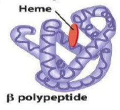
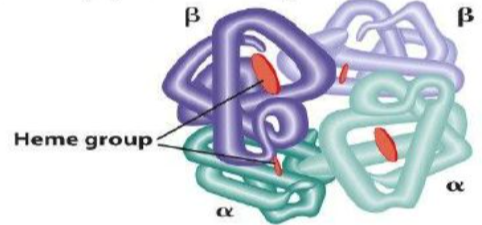
B1.2.11 – Quaternary Structure of Non-Conjugated and Conjugated Proteins
🔹 What Is Quaternary Structure?
Quaternary structure refers to the 3D arrangement formed when two or more polypeptide chains (subunits) come together to make a functional protein. These subunits are held together by hydrogen bonds, ionic interactions, disulfide bridges, and hydrophobic interactions.
🧩 Non-Conjugated Proteins
INSULIN COLLAGEN
These consist only of polypeptide chains, without any non-protein (prosthetic) groups.
- Insulin: Two polypeptide chains (A and B) linked by disulfide bridges. A globular hormone that regulates blood glucose.
- Collagen: Three polypeptide chains forming a triple helix. A fibrous protein providing strength to skin, tendons, and bones.
Conjugated Proteins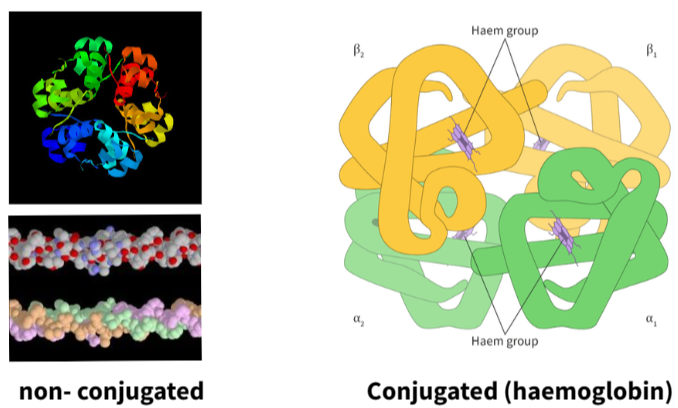
These contain one or more non-polypeptide components (called prosthetic groups) attached to the protein.
- Haemoglobin: Made of four subunits (2 alpha and 2 beta). Each contains a haem group with iron. It transports oxygen in red blood cells.
🔍 NOS Insight: Seeing the Invisible
Cryogenic Electron Microscopy (Cryo-EM)
• Visualizes proteins at near-atomic resolution
• Reveals shape, folding, and interaction
• Does not require crystallization
• Useful for studying complex protein structures
Cryo-EM is a revolutionary technique that allows scientists to image single protein molecules directly – even those that do not form crystals.
📦 Summary Table
| Feature | Non-Conjugated Proteins | Conjugated Proteins |
|---|---|---|
| Components | Only polypeptide chains | Polypeptides + prosthetic groups |
| Examples | Insulin, Collagen | Haemoglobin |
| Function | Hormonal signaling, structural support | Oxygen transport |
| Bonds/Interactions | Disulfide bridges, hydrogen bonds | Same + binding to prosthetic group |