IB DP Biology Organelles and compartmentalization Study Notes
IB DP Biology Organelles and compartmentalization transport Study Notes at IITian Academy focus on specific topic and type of questions asked in actual exam. Study Notes focus on IB Biology syllabus with guiding questions of
- How are organelles in cells adapted to their functions?
- What are the advantages of compartmentalization in cells?
Standard level and higher level: 4 hours
Additional higher level: 1 hour
B2.2.1 – Organelles as Specialized Cell Subunits
🧫 What Are Organelles?
Organelles are distinct functional structures found inside cells.
Each organelle performs specific roles that are vital for cell survival- just like organs in a body.
Most organelles are membrane-bound and are mainly found in eukaryotic cells.
🔬 Examples of Recognized Organelles
Organelle | Function Summary |
---|---|
Nucleus | Contains DNA; controls gene expression and cell activity |
Vesicles | Small membrane sacs used for storage and transport |
Ribosomes | Make proteins from mRNA (protein synthesis) |
Plasma membrane | Controls entry/exit of substances; maintains cell shape |
🌿 Double-Membrane Organelles (in Eukaryotes)
Structures like the cell wall, cytoskeleton, and cytoplasm are important, but they are not classified as organelles.
These organelles have two membranes for compartmentalization:
- Nucleus – Stores DNA and protects genetic material
- Mitochondria – Site of aerobic respiration (energy production)
- Chloroplasts – Photosynthesis (in plant cells)
- Amyloplasts – Store starch
- Chromoplasts – Contain pigments (in plant cells)
📚 Eukaryotic vs Prokaryotic Cells
Feature | Eukaryotic Cells | Prokaryotic Cells |
---|---|---|
Nucleus | Present – stores DNA | Absent – DNA floats freely in cytoplasm |
Ribosomes | Present | Present |
Post-transcriptional modification | Happens in the nucleus before translation | Does not occur – translation begins immediately |
🔍 Nature of Science (NOS) Insight
Scientific Progress via New Technology:
The study of organelles became possible only after the invention of ultracentrifuges.
These powerful machines enabled scientists to perform cell fractionation, separating organelles based on size and density.
This technological leap allowed researchers to study organelle functions in isolation-advancing our understanding of cell biology.
B2.2.2 – Advantages of Separating Nucleus and Cytoplasm
🧬 What Does This Separation Mean?
In eukaryotic cells, the nucleus is surrounded by a membrane that separates it from the cytoplasm.
This creates two distinct compartments:
- Nucleus: Where DNA transcription and mRNA processing happen
- Cytoplasm: Where translation of mRNA into proteins occurs
🔍 Why Is This Separation Useful?
1. Allows Post-Transcriptional Modification
In eukaryotes, once mRNA is transcribed in the nucleus, it can be edited and processed before being used to make proteins.
- Splicing: Removing non-coding regions (introns)
- 5′ capping: Adding a special cap to protect the mRNA
- Polyadenylation: Adding a tail (poly-A) for stability
2. No Separation in Prokaryotes
In prokaryotic cells, there’s no nucleus → DNA and ribosomes are in the same compartment (cytoplasm).
So, as soon as mRNA is made, ribosomes begin translating it immediately.
No time or space for mRNA editing or regulation
🧠 Key Comparison Table
Feature | Eukaryotic Cells | Prokaryotic Cells |
---|---|---|
Nucleus present? | Yes | No |
Transcription location | Nucleus | Cytoplasm |
Translation location | Cytoplasm | Cytoplasm |
Post-transcriptional modification? | Yes – before export to cytoplasm | No – not possible |
Transcription & translation timing | Separate in time and space | Occur simultaneously |
🧠 Key Takeaways
- In eukaryotes, separating the nucleus from cytoplasm allows controlled gene expression.
- It enables mRNA processing before translation occurs.
- In prokaryotes, transcription and translation occur together with no mRNA editing.
- This structural difference is a key reason why eukaryotic gene expression is more complex and regulated.
B2.2.3 – Advantages of Cytoplasmic Compartmentalization
🧬 What Is Compartmentalization?
In eukaryotic cells, the cytoplasm contains many membrane-bound organelles.
Each organelle forms a separate compartment, allowing different biochemical processes to occur at the same time without interfering with each other.
🎯 Why Is Compartmentalization Useful?
1. Concentration of Substances
Enzymes and substrates can be gathered in one location, making reactions:
- Faster
- More efficient
Example: Enzymes in mitochondria work best when substrates are concentrated inside.
2. Separation of Incompatible Reactions
Some processes need very different conditions (e.g., pH, temperature). Compartments prevent interference between conflicting processes.
Example: Fatty acid synthesis and fatty acid breakdown occur in different organelles.
3. Containment of Harmful Substances
Some reactions produce toxic or damaging products, which are safely stored and broken down inside organelles.
- Lysosomes: Contain digestive enzymes that could damage the cell if released freely.
- Phagocytic vacuoles: Trap and digest bacteria, isolating harmful materials from the rest of the cell.
4. Optimal Conditions Inside Compartments
Each organelle can maintain its own internal environment (e.g., specific pH).
This helps enzymes function at their best within that space.
🧠 Key Examples of Compartment Use
Organelle | Function | Benefit of Compartment |
---|---|---|
Lysosomes | Contain enzymes to digest waste and worn-out parts | Prevents damage to cell |
Phagocytic vacuoles | Engulf and digest bacteria | Keeps toxins isolated |
Mitochondria | Site of aerobic respiration | Efficient ATP production |
Chloroplasts | Site of photosynthesis (in plants) | Light reactions isolated |
🧠 Key Takeaways
- Compartmentalization makes cells more efficient, safe, and organized.
- It allows simultaneous processes to occur under ideal conditions without conflict.
- Organelles like lysosomes and vacuoles protect the cell by isolating harmful substances.
Additional Higher Level
B2.2.4 – Mitochondrion Adaptations for ATP Production
🔬 Main Job of Mitochondria:
To make ATP through aerobic respiration – a highly efficient process that occurs in stages: glycolysis, Krebs cycle, and oxidative phosphorylation.
🧬 Structural Adaptations that Help in ATP Production
1. Double Membrane
Outer membrane = smooth and semi-permeable
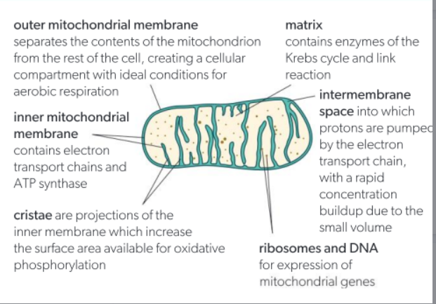
Inner membrane = folded into cristae
Between them is the narrow intermembrane space
2. Cristae (Folds of the Inner Membrane)
Cristae greatly increase surface area.
They provide space to embed proteins for:
- Electron Transport Chain (ETC)
- ATP Synthase
📌 More cristae = more surface area = more ATP.
3. Matrix (Inner Compartment)
Gel-like fluid inside the inner membrane containing:
- Enzymes for the Krebs cycle
- Substrates like pyruvate, NAD⁺, FAD
- Mitochondrial DNA and ribosomes
📌 This environment supports efficient Krebs cycle reactions.
4. Compartmentalization
Each part of respiration occurs in a specific mitochondrial compartment:
- Krebs cycle → Matrix
- ETC → Inner membrane
- Proton gradient → Intermembrane space
This separation keeps processes efficient and maintains different optimal conditions like pH across compartments.
🧠 Quick Recap Table
Feature | Role in Respiration |
---|---|
Double membrane | Creates intermembrane space for proton buildup |
Cristae | High surface area for ETC and ATP synthase |
Matrix | Contains Krebs cycle enzymes and substrates |
Compartmentalization | Separates steps and optimizes conditions |
B2.2.5 – Adaptations of the Chloroplast for Photosynthesis
🔬 What is the role of the chloroplast?
Chloroplasts are the site of photosynthesis in plant cells where light energy is converted into chemical energy (glucose). Their structure is highly adapted to carry out this complex process efficiently.
⚙️ Key Structural Adaptations
1. Thylakoid Membranes with Photosystems
- Thylakoids are flattened membrane sacs containing photosystems (with chlorophyll and accessory pigments) embedded in their membranes.
- More membrane = more space for photosystems, ATP synthase, and electron transport chains → boosts the rate of light reactions.
- 📌 Function: Absorb sunlight efficiently and capture light energy for the light-dependent reactions.
2. Grana = Stacked Thylakoids
- Thylakoids are often stacked into grana (like coins). This increases surface area even further.
- 📌 Function: Maximizes light absorption for efficient energy capture.
3. Small Volume Inside Thylakoids
- The lumen (inside of thylakoid) has a very small volume.
- 📌 Function: Builds up a proton gradient faster → powers ATP synthase during the light-dependent reactions.
4. Stroma = Fluid Around Thylakoids
- The stroma is the gel-like fluid surrounding thylakoids, containing:
- Enzymes for the Calvin cycle
- RuBP, ATP, and NADPH
- 📌 Function: Site of the Calvin cycle (light-independent stage) with optimal conditions for enzyme activity.
5. Compartmentalization
- Chloroplasts have a double membrane separating the internal thylakoid space from the stroma.
- 📌 Function: Keeps incompatible processes separate and maintains ideal pH for each stage of photosynthesis.
🧠 Summary Table
Feature | Adaptation Benefit |
---|---|
Thylakoid membranes | High surface area for photosystems and ETC |
Grana | Stacking maximizes light absorption |
Small thylakoid lumen | Faster proton buildup → efficient ATP synthesis |
Stroma | Contains Calvin cycle enzymes and substrates |
Compartmentalization | Organizes and separates different reactions |
B2.2.6 – Functional Benefits of the Double Membrane of the Nucleus
Structure Recap
The nucleus is enclosed by a double membrane called the nuclear envelope.
- Outer membrane is continuous with the rough ER
- Nuclear envelope has nuclear pores for selective exchange
🔎 Key Functions of the Double Membrane
1. Protects Genetic Material
- The envelope keeps DNA safely enclosed within the nucleus, shielding it from enzymes, chemicals, or signals in the cytoplasm.
- Ensures stable gene expression and prevents DNA damage or mutations.
2. Allows Selective Exchange Through Nuclear Pores
Nuclear pores regulate traffic in and out of the nucleus:
- Out: mRNA, tRNA, ribosomal subunits
- In: Enzymes, transcription factors, nucleotides
📌 This maintains control over nuclear-cytoplasmic transport.
3. Maintains a Specialized Environment
The nucleus has its own internal conditions optimized for:
- DNA replication
- Transcription of mRNA
- Chromatin organization and repair
📌 These conditions differ from the cytoplasm and support efficient nuclear function.
4. Disassembly During Cell Division
- In mitosis and meiosis, the nuclear envelope disassembles into vesicles.
📌 This allows chromosome segregation and spindle fiber access. After division, vesicles reassemble the envelope.
🧠 Summary Table
Feature | Functional Benefit |
---|---|
Double membrane | Protects DNA from harmful cytoplasmic components |
Nuclear pores | Enable controlled movement of molecules in/out |
Internal nuclear environment | Optimized for transcription and replication |
Disassembly in mitosis | Allows chromosome separation during division |
B2.2.7 – Structure and Function of Free Ribosomes vs Rough Endoplasmic Reticulum (RER)
🧬 What are Ribosomes?
Ribosomes are the sites of protein synthesis in all cells. They read mRNA and build polypeptides. In eukaryotic cells, they exist in two forms:
- Free in the cytoplasm
- Attached to the rough endoplasmic reticulum (RER)
⚙️ Free Ribosomes 🧫
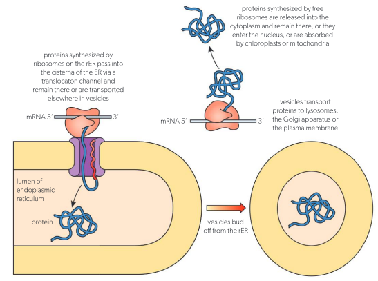
Structure: Small particles made of rRNA and protein, not attached to any membrane.
Function: Produce proteins used inside the cell, such as:
- Metabolic enzymes
- Proteins for nucleus or mitochondria
- Structural proteins (e.g., cytoskeleton)
🌊 Rough Endoplasmic Reticulum (RER)
Structure: Flattened membrane sacs called cisternae with ribosomes on the cytoplasmic surface, giving a rough appearance.
Function: Produces proteins that are:
- Secreted from the cell (e.g., hormones, enzymes)
- Inserted into the cell membrane
- Delivered to lysosomes or other compartments
Process: Proteins enter the RER lumen, are folded and modified, then packed into vesicles and sent to the Golgi apparatus.
🔍 Comparison Table: Free Ribosomes vs RER Ribosomes
Feature | Free Ribosomes | Rough ER (Bound Ribosomes) |
---|---|---|
Location | Free in cytoplasm | Attached to RER membrane |
Main function | Make proteins used inside the cell | Make proteins for secretion or membrane |
Protein destination | Cytoplasm, nucleus, mitochondria | Outside cell, plasma membrane, organelles |
Example products | Enzymes for glycolysis | Digestive enzymes, hormones, antibodies |
B2.2.8 – Structure and Function of the Golgi Apparatus
🏗️ Structure
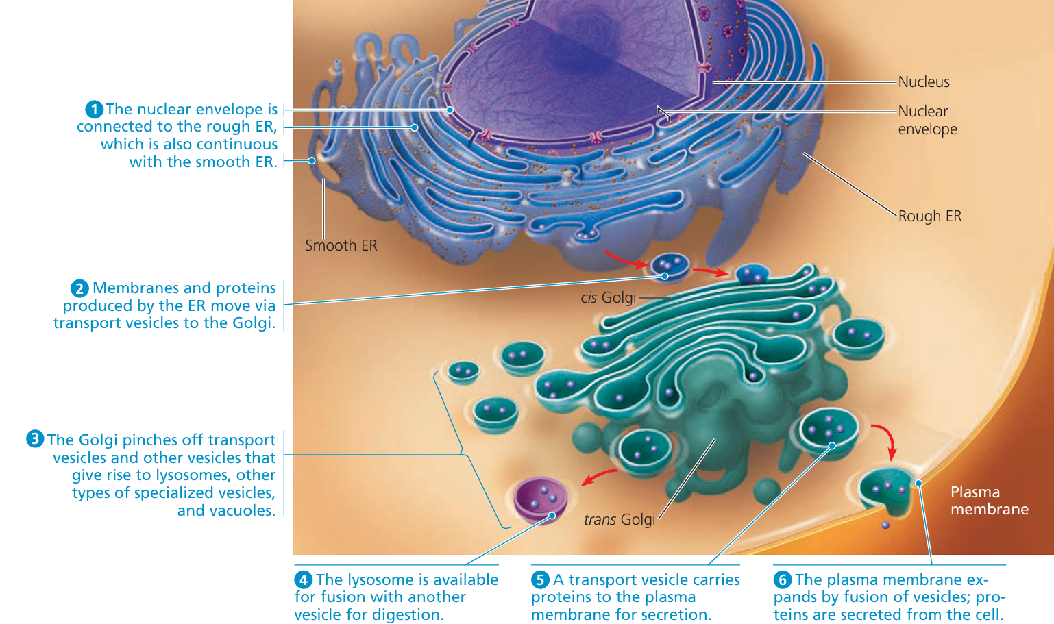
- The Golgi apparatus is made up of a stack of flattened membrane sacs called cisternae.
- It has a cis face (receives materials from rER) and a trans face (exports processed materials).
- No ribosomes on its surface (unlike the rER).
⚙️ Main Functions
The Golgi apparatus is like the post office of the cell – it modifies, sorts, and packages proteins made by the rough ER.
1. Protein Processing
Receives proteins from the rER in transport vesicles.
Modifies them:
- Adds carbohydrates → makes glycoproteins
- May also add lipids or phosphate groups
2. Packaging and Sorting
Proteins are sorted based on their destination.
Packaged into vesicles and sent to:
- Plasma membrane (for secretion)
- Lysosomes
- Other organelles
3. Quaternary Structure Assembly
If a protein needs multiple subunits (e.g. antibodies), the Golgi can help assemble the final product.
🧪 Examples of Golgi Function
Modifies insulin before it’s secreted.
Makes digestive enzymes active before sending them to lysosomes.
Packages neurotransmitters for secretion by neurons.
🔍 Note on Golgi Transport Models
Model | Description |
---|---|
Vesicle Transport Model | Proteins move between static cisternae in vesicles |
Cisternal Maturation Model | Whole cisternae move and mature from cis → trans |
Most evidence currently supports the cisternal maturation model.
🧠 Key Summary
Feature | Function |
---|---|
Cisternae | Flattened sacs for processing |
Protein Mod | Glycosylation, phosphorylation, etc. |
Sorting | Directs proteins to correct destinations |
Secretion Role | Prepares proteins for export from the cell |
B2.2.9 – Structure and Function of Vesicles in Cells
🔹 What are Vesicles?
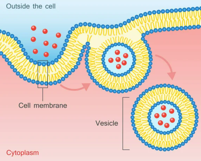
- Small, round sacs surrounded by a single membrane.
- They carry and transport materials inside the cell.
- Contain different substances like proteins, lipids, or waste.
🔄 How Are Vesicles Formed?
Formed by pinching off a part of a bigger membrane (e.g., plasma membrane or Golgi membrane).
This creates a small bubble that buds off with cargo inside.
🧩 Role of Clathrin in Vesicle Formation
- Clathrin is a protein that assists vesicle formation during endocytosis.
- It assembles into a basket-like structure on the inner membrane surface.
- This helps the membrane curve and pinch off to form vesicles.
- Without clathrin, vesicle formation would be inefficient.
⚙️ Functions of Vesicles
- Transport: Move proteins and molecules between organelles (e.g., Golgi to plasma membrane)
- Secretion: Carry substances out of the cell (exocytosis)
- Storage: Hold materials temporarily
- Endocytosis: Bring substances into the cell via membrane budding
🧠 Summary
Feature | Role |
---|---|
Vesicle Structure | Small membrane-bound sacs |
Clathrin Protein | Helps bend membrane to form vesicles |
Function | Transport, secretion, storage, endocytosis |