Home / Cell specialization Study Notes

IB DP Biology Cell specialization Study Notes | New Syllabus

IB DP Biology Cell specialization Study Notes

IB DP Biology Cell specialization Study Notes

IB DP Biology Cell specialization Study Notes Study Notes at  IITian Academy  focus on  specific topic and type of questions asked in actual exam. Study Notes focus on IB Biology syllabus with guiding questions of

  • What are the roles of stem cells in multicellular organisms?
  • How are differentiated cells adapted to their specialized functions?

Standard level and higher level: 4 hours

Additional higher level: 1 hour

IBDP Biology 2025 -Study Notes -All Topics

B2.3.1 – Production of Unspecialized Cells After Fertilization and Differentiation

What Happens After Fertilization?

The fertilized egg (zygote) divides to make many unspecialized cells (called stem cells or embryonic cells). These cells are all the same at first and have the potential to become any cell type.

Differentiation: How Cells Become Specialized

All cells have the same DNA and genes, but not all genes are active in every cell.
Differentiation means cells turn on some genes and turn off others to make proteins needed for their specific role.

  • White blood cells → fight infections
  • Heart muscle cells → pump blood
  • Skeletal muscle cells → move the body
  • Neurons → send nerve signals

Gene Expression and Protein Production

A gene is a DNA segment that gives instructions to make a specific protein.
Different cell types express only the genes they need.

For example:

  • Stomach cells produce proteins for digestion
  • Eye cells produce proteins for vision

Cells don’t waste energy making proteins they don’t need.

Role of Gradients in Early Embryo

  • Early in development, chemical gradients (varying concentrations of molecules) influence which genes turn on/off.
  • These gradients cause cells in different parts of the embryo to receive different signals.
  • This is a key factor in guiding cells to specialize in the right place.
ConceptExplanation
Unspecialized CellsAll cells start identical after fertilization
DifferentiationCells become specialized by expressing specific genes
Gene ExpressionOnly necessary genes are turned on per cell type
Gradients in EmbryoChemical signals guide early gene expression and specialization


B2.3.2 – Properties of Stem Cells

What Makes Stem Cells Special?

Stem cells are unique because they have two key properties:

  • Unlimited division: They can keep dividing and making more cells without stopping.
  • Differentiation: They can develop into different types of specialized cells depending on their type.

Types of Stem Cells Based on Potential

Stem Cell TypeCan Become…Where Found
TotipotentAny cell type (including whole organism)Zygote and early embryo (morula)
PluripotentAlmost any cell typeInner cell mass of embryo
MultipotentSeveral related cell typesSome adult tissues (e.g., bone marrow)
UnipotentOnly one cell typeSome adult tissues

Where Do Stem Cells Come From?

  • Embryonic stem cells: From leftover embryos during IVF (in vitro fertilization). Cells can be taken for genetic testing and research. Require legal and ethical permission.
  • Umbilical cord blood: Rich source of stem cells collected at birth.
  • Adult stem cells: Found in certain tissues to help repair and maintain.
Stem cells can divide endlessly and turn into different cell types. Their ability depends on their potency (totipotent → unipotent). Sources include embryos, umbilical cord blood, and some adult tissues.

B2.3.3 – Stem Cell Niches in Adult Humans

What is a Stem Cell Niche?

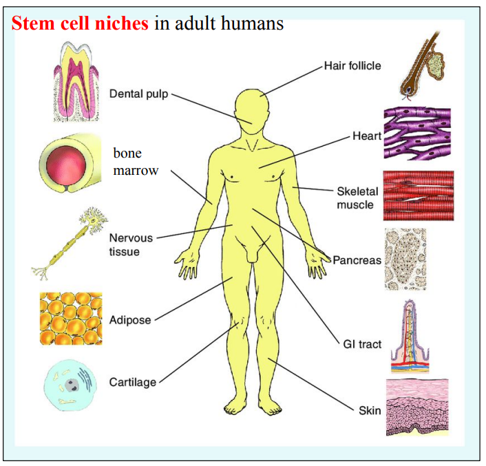
A stem cell niche is a specialized microenvironment within tissues where adult stem cells live and are controlled. It helps regulate whether the stem cells:

  • Stay inactive (quiescent)
  • Self-renew (make more stem cells)
  • Differentiate (become specialized cells for tissue repair or function)

Functions of a Stem Cell Niche

A stem cell niche sends signals that:

  • Support self-renewal
  • Trigger differentiation
  • Maintain quiescence (a resting, inactive state)
  • Protect stem cells from stress or damage

Two Key Niches in Adult Humans

LocationFunction of the NicheExample Role
Bone MarrowMaintains a balance between stem cell storage and the production of blood cells.Continuous renewal of RBCs, WBCs, platelets
Hair FollicleStores stem cells that control cycles of hair growth, rest, and regrowth.Hair regeneration and repair after injury

Why Are Stem Cell Niches Important?

  • Keep stem cells safe and protected from damage
  • Support tissue repair and regeneration after injury
  • Control stem cell behavior to prevent overgrowth (tumors)
  • Let stem cells respond to body signals, like during injury or stress

Real-Life Examples

  • Skin Injury: When you cut your skin, stem cells in the skin niche become active, divide, and make new cells to heal the wound.
  • Hair Growth: Hair grows in cycles because stem cells in hair follicles are activated and go dormant in a regulated pattern.
🧠 Key Takeaways:
Stem cell niches are the homes of adult stem cells.
They regulate when stem cells divide, rest, or differentiate.
Bone marrow and hair follicles are classic examples.
Niches are essential to ensure safe, controlled regeneration in tissues.

B2.3.4 – Differences Between Totipotent, Pluripotent and Multipotent Stem Cells

What is “Potency”?

Potency means the ability of a stem cell to develop into different types of specialized cells. The more cell types it can form, the higher its potency.

1. Totipotent Stem Cells – “Total Power”

  • Meaning: Can make every cell type in the body plus extra structures like the placenta.
  • Found in: Very early embryo (zygote and first few cell divisions)
  • Can become: Any body cell + extraembryonic tissue (like placenta)
  • Example: A fertilized egg (zygote)
🔑 Totipotent = Total potential (All body cells + placenta)

2. Pluripotent Stem Cells – “Plenty of Possibilities”

  • Meaning: Can make almost all cell types, just not placenta.
  • Found in: Early-stage embryo (blastocyst)
  • Can become: Any body cell (nerve, muscle, skin, etc.)
  • Limitation: Cannot form placenta or supporting tissues
  • Example: Embryonic stem cells
🔑 Pluripotent = All body cells, but not placenta

3. Multipotent Stem Cells – “Multiple but Limited”

  • Meaning: Can only make cells within a particular group or tissue type.
  • Found in: Adult tissues (e.g., bone marrow)
  • Can become: Related cells within one system only
  • Example: Blood stem cells (can make RBCs, WBCs, platelets – but not brain or liver cells)
🔑 Multipotent = Limited potential (within one tissue type)

Comparison Table – Types of Stem Cell Potency

TypeWhere FoundCan BecomeExample
TotipotentZygote, early embryoAll body cells + placentaFertilized egg
PluripotentBlastocyst (early embryo)All body cells (but not placenta)Embryonic stem cell
MultipotentAdult tissues (e.g., bone marrow)Limited related cells within a tissue systemBlood stem cells (RBCs etc.)

Key Recap

  • Totipotent ➜ All cells + placenta (most powerful)
  • Pluripotent ➜ All body cells (not placenta)
  • Multipotent ➜ A few related cells only (e.g., blood types)
As the embryo develops, stem cells lose potency:
Totipotent → Pluripotent → Multipotent

B2.3.5 – Cell Size as an Aspect of Specialization

Why Does Cell Size Matter?

Cell size and shape are not random. Each type of cell is specially designed (specialized) to perform its role efficiently. The structure supports the function.

Gametes: Sperm vs Egg
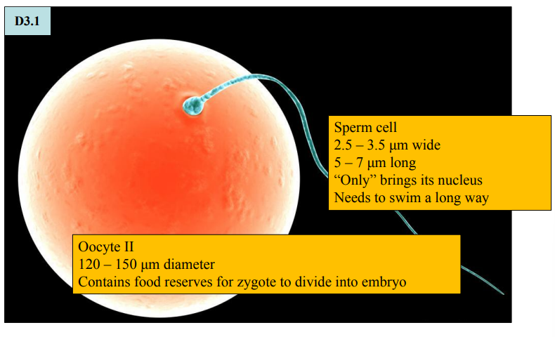
Cell TypeRelative SizeSpecial Features
Sperm CellVery small, long and thinTail (flagellum) for movement; minimal cytoplasm for speed
Egg Cell (Oocyte)One of the largest human cellsLarge, round; contains nutrients for early development
Sperm: Built for mobility
Egg: Built for nutrient storage and early development

Red Blood Cells vs White Blood Cells
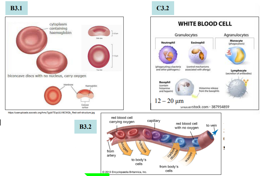
FeatureRed Blood Cell (RBC)White Blood Cell (WBC)
Diameter~6–8 μm~12–17 μm (varies by type)
ShapeBiconcave discIrregular, can change shape
FunctionTransport oxygenFight infections
Special AdaptationNo nucleus = more space for hemoglobinCan enlarge when active for protein synthesis
Only RBCs can squeeze through tiny capillaries (lumen = 5–10 μm)

Neurons: Communication Cells
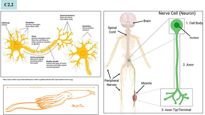
Neuron TypeSize RangeSpecial Role
Granule Cells (Cerebellum)Very small (~4–5 μm)Allows packing of millions of cells
Motor NeuronsAxon up to 1 meter longTransmit signals to muscles over long distances
Long axons = fast communication
Tiny bodies = dense networking in the brain

Striated Muscle Cells (Skeletal Muscle)
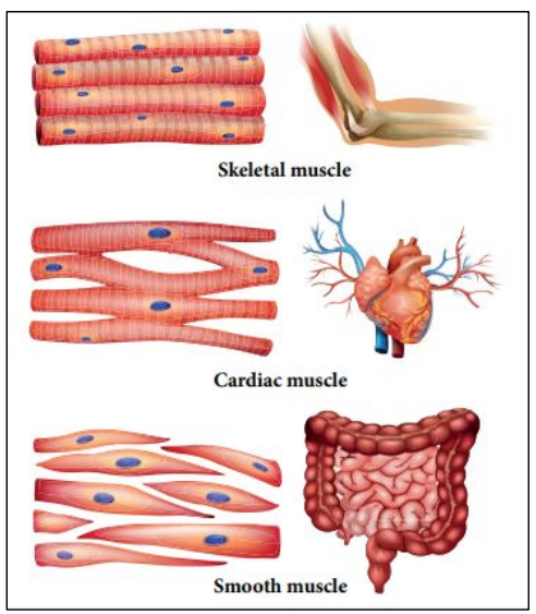
FeatureDetails
LengthUp to 40 mm
Diameter10–50 μm
Special TraitsLong, multinucleated fibers
FunctionGenerate powerful contractions to move bones
Large size = more force
Multiple nuclei = greater protein synthesis capacity

Summary – Size Supports Function

Cell TypePurposeSize Adaptation
Sperm CellReach eggSmall, fast, tail for movement
Egg CellSupport early embryoLarge, nutrient-rich
Red Blood CellCarry oxygenSmall, flexible, no nucleus
White Blood CellFight infectionIrregular, size changes with activity
NeuronSend electrical messagesVery long axon or small dense bodies
Muscle CellContract and move body partsLong, thick, multinucleated for strength
📌 Key Takeaway:
Form follows function – Every cell’s size and shape is specialized to help it do its job efficiently. This diversity is essential for the proper working of tissues and organs in the human body.

B2.3.6 – Surface Area-to-Volume Ratios & Cell Size Constraints

Why Does Surface Area-to-Volume Ratio Matter?

As cells grow, their volume increases faster than surface area. This affects how well materials like oxygen, nutrients, and waste move in and out of the cell.

  • Surface Area = where exchange happens
  • Volume = where materials are used or produced

If a cell is too big, it may not exchange substances fast enough to stay alive.

Key Concept: Surface Area Grows Slower Than Volume

Let’s consider cubes as models for cells:

Cube Side LengthSurface Area (6 × side²)Volume (side³)SA:V Ratio
1 unit616 : 1
2 units2483 : 1
3 units54272 : 1

As size increases:

  • Volume increases much faster than surface area
  • SA:V ratio decreases significantly
  • Less surface area per unit of volume

Consequences of a Low Surface Area-to-Volume Ratio

  • Nutrient Uptake Drops – Not enough space for nutrients to enter fast enough
  • Waste Builds Up – Can’t remove waste quickly, risking toxicity
  • Overheating – Heat produced inside isn’t lost quickly enough

This is why most cells stay small or adopt shapes (like being flat or long) to maintain a high surface area-to-volume ratio.

Modeling with Cubes – Simplifying Complexity

  • Real cells have irregular shapes and internal complexity
  • But cubes are used as models to simplify and visualize the SA:V relationship
  • Even though cubes aren’t realistic, the scale effect is the same – bigger size means lower SA:V
NOS Insight: Models simplify real systems so we can understand the core principles better, even if they aren’t perfect.
🧠 Key Takeaways:
A high surface area-to-volume ratio is essential for efficient exchange of substances in and out of cells.
As cells grow, this ratio drops, limiting their function and setting a constraint on cell size.
Mathematical models using simple shapes like cubes help students understand these limitations, even though real cells are more complex.

B2.3.7 – Adaptations to Increase Surface Area-to-Volume Ratio in Cells

📐 Why Increase Surface Area-to-Volume Ratio?

Cells involved in rapid exchange of materials like gases, nutrients, and waste need a large surface area compared to their volume. A higher surface area-to-volume (SA:V) ratio improves the efficiency of absorption, secretion, and diffusion.

🧬 Common Adaptations to Increase SA:V Ratio

AdaptationHow It HelpsWhere It’s Found
FlatteningReduces thickness, spreading the cell to expose more surfaceEpithelial cells lining blood capillaries, alveoli
MicrovilliFinger-like projections that multiply the cell’s surfaceProximal tubule cells in the nephron
Infoldings (Invaginations)Folds on the cell membrane that expand surface inwardBasal side of kidney tubule cells
Biconcave shapeIncreases external surface area while keeping volume lowRed blood cells (erythrocytes)

🔬 Example 1: Red Blood Cells (Erythrocytes)

  • Adaptation: Biconcave (disc-shaped) structure
  • Function: Maximizes surface area for rapid oxygen exchange
  • Effect: More efficient loading/unloading of O2 and CO2

The biconcave shape gives red blood cells a high SA:V ratio, crucial for their role in gas transport.

🔬 Example 2: Proximal Convoluted Tubule Cells (Kidney Nephron)

These cells are highly active in reabsorption of useful substances like glucose, ions, and water from the filtrate.

Key Adaptations:

  • Microvilli (on apical surface):
    Drastically increase surface area facing the filtrate
    Allow efficient reabsorption into the cell
  • Basal Infoldings (on underside):
    Increase area for exchange with blood capillaries
    Provide space for mitochondria to fuel active transport

These features ensure maximum efficiency in reabsorbing valuable substances back into the bloodstream.

🧠 Key Takeaways:
Cells adapt their structure to maximize surface area, improving efficiency of exchange processes.
Flattening, microvilli, infoldings, and biconcave shapes are common adaptations.
Red blood cells and proximal tubule cells are excellent examples of structural specialization for high SA:V ratio.

B2.3.8 – Adaptations of Type I and Type II Pneumocytes in Alveoli

🌬️ What Are Pneumocytes?

Pneumocytes are the specialized epithelial cells lining the alveoli (air sacs) of the lungs. They form the alveolar epithelium, a tissue made of two distinct cell types that together optimize gas exchange.

🔬 Type I Pneumocytes – Designed for Diffusion

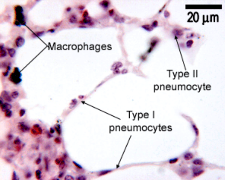
  • Structure: Extremely thin and flat
  • Function: Minimize the diffusion distance for oxygen and carbon dioxide
  • Coverage: Make up ~95% of the alveolar surface
  • Cell Division: Do not divide – replaced by Type II cells if damaged

Their thinness helps maximize gas exchange efficiency by reducing the barrier between air and blood.

🧪 Type II Pneumocytes – Surfactant Secretion

  • Structure: Cuboidal cells with many secretory vesicles (called lamellar bodies)
  • Function: Secrete pulmonary surfactant, a lipid-protein mixture

Role of Surfactant:

  • Reduces surface tension inside alveoli
  • Prevents alveolar collapse during exhalation
  • Keeps alveoli open and functional

Repair Function: Type II cells can divide and replace damaged Type I cells.

Type II cells are essential for stabilizing alveoli and ensuring proper lung function.

🧬 A Tissue with Two Specialized Cell Types

The alveolar epithelium is a great example of a tissue where:

  • Different cell types (Type I and II pneumocytes) work together
  • Each cell has unique adaptations serving the overall function: gas exchange
🧠 Key Takeaways:
Type I pneumocytes are extremely thin to reduce diffusion distance for gases.
Type II pneumocytes contain lamellar bodies that release surfactant to keep alveoli open.
Both types are crucial for efficient oxygen and carbon dioxide exchange in the lungs.
The alveolar epithelium demonstrates how multiple cell types within a tissue can cooperate for optimal function.

B2.3.9 – Adaptations of Cardiac Muscle Cells and Striated Muscle Fibres

🧬 What Are Striated Muscles?

Striated muscles include:

  • Skeletal muscle – moves the body
  • Cardiac muscle – powers the heartbeat

They are called “striated” because they have visible bands (stripes) under the microscope, caused by the regular arrangement of contractile units called myofibrils.

🔬 Shared Feature: Myofibrils in Both Types

  • Both skeletal and cardiac muscle cells contain myofibrils made of actin and myosin
  • These proteins form sarcomeres, the repeating contractile units responsible for muscle contraction
  • The presence of myofibrils gives both types their striped (striated) appearance

🔑 Myofibrils = Essential for contraction in all striated muscles

🧠 Skeletal Muscle Fibres – Powerful & Multinucleated

FeatureSkeletal Muscle
ShapeLong, cylindrical
NucleiMultiple nuclei per cell (located at edges)
BranchingUnbranched
LengthCan be very long (several cm)
FunctionVoluntary movement of body parts

These muscle fibres are formed by fusion of many cells, which explains their multinucleated structure. Their large size allows them to generate powerful, rapid contractions.

❤️ Cardiac Muscle Cells – Coordinated & Branched

FeatureCardiac Muscle
ShapeShorter, often branched
NucleiUsually 1–2 nuclei per cell (central)
BranchingBranched
Special JunctionsIntercalated discs connect cells
FunctionInvoluntary, rhythmic heartbeat

Intercalated discs contain gap junctions and desmosomes, allowing electrical signals to pass rapidly between cells — enabling the synchronized contraction of the heart.

Cardiac cells work together as a unit, even though each cell is individually separate.

Is a Skeletal Muscle Fibre a Cell?

Yes, but it’s not a typical cell.
A skeletal muscle fibre is technically a single cell, but:

  • It is multinucleated
  • It is very long, formed by the fusion of multiple embryonic cells (myoblasts)

Because of its unusual structure, some textbooks describe it as a “fibre” instead of a “cell” — but biologically, it is one giant cell.

📊 Summary Table – Skeletal vs Cardiac Muscle

FeatureSkeletal MuscleCardiac Muscle
AppearanceStriatedStriated
MyofibrilsPresentPresent
NucleiMany, peripherally located1–2, centrally located
BranchingNoYes
Intercalated DiscsNoYes – for synchronized contraction
Voluntary?Yes (under conscious control)No (involuntary heartbeat)
Cell or Fibre?Single multinucleated cellSingle cell (mononucleated or binucleated)
🧠 Key Takeaways:
Both muscle types have contractile myofibrils for force generation.
Skeletal fibres are long, unbranched, and multinucleated for powerful, voluntary movement.
Cardiac cells are branched, shorter, and connected by intercalated discs for coordinated, rhythmic contractions.
Skeletal muscle fibres, though unusual, are still considered single cells due to their common membrane.

B2.3.10 – Adaptations of Sperm and Egg Cells (Human Gametes)

🧠Remember:
– Sperm cells are fast and motile, built for speed and delivery.
– Egg cells are large, nutrient-rich, and protected to support early development.
– Both are haploid, contributing half the genetic material to form a diploid zygote.

👨‍🔬 What Are Gametes?

Gametes are specialized reproductive cells that combine during fertilization to form a zygote. In humans:

  • Male gamete = Sperm
  • Female gamete = Egg (Ovum)

Each gamete is uniquely adapted for its role in reproduction.

🔹 Sperm Cell – Built for Mobility and Delivery

AdaptationFunction
Streamlined ShapeReduces water resistance, aids fast swimming
Flagellum (tail)Propels the sperm using whip-like motion
Mitochondria (midpiece)Provides ATP for tail movement
AcrosomeContains enzymes to penetrate egg’s outer layers
Small SizeEnables fast movement over long distances
Haploid NucleusCarries 23 chromosomes

🔸 Egg Cell – Designed for Protection and Nourishment

AdaptationFunction
Large SizeContains nutrients for embryo development
Zona PellucidaProtects egg and blocks multiple sperm entry
Cortical GranulesHarden zona after fertilization to prevent polyspermy
Cytoplasm Rich in OrganellesSupports early embryo growth
Haploid NucleusCarries 23 chromosomes

🔍 Comparison of Human Gametes

FeatureSperm CellEgg Cell
SizeVery smallVery large
MobilityMotile (flagellum)Immobile
Energy SupplyMitochondria in midpieceStored in cytoplasm
NucleusHaploid (23 chromosomes)Haploid (23 chromosomes)
Protective LayersAcrosome (enzyme layer)Zona pellucida + cortical granules
Number ProducedMillions per day~1 per menstrual cycle
FunctionDeliver DNAProvide DNA + nourishment + protection
Scroll to Top