Additional Higher Level
B3.3.1 – Adaptations for Movement in Living Organisms

🌍 Movement: A Universal Feature of Life
Movement is a fundamental characteristic of living organisms.
Occurs at multiple biological levels:
| Type of Movement | Example | Description |
|---|---|---|
| Intracellular | Cytoplasmic streaming | Movement within cells (e.g. of organelles) |
| Internal (organ level) | Heart contractions | Movement of internal organs or fluids |
| Whole-body movement | Locomotion | Walking, flying, swimming using muscles |
🧬 Three Basic Locomotion Mechanisms in Animals
1. Amoeboid Movement

- Seen in: Amoeba, white blood cells
- Involves: pseudopodia (temporary extensions of cytoplasm)
- Function: Crawling-like movement on surfaces
2. Movement Using Cilia or Flagella

- Cilia = Short, hair-like projections
- Flagella = Long, whip-like tails
- Seen in: Paramecium (cilia), sperm cells (flagella)
3. Muscular Locomotion

- Common in: Vertebrates and most invertebrates
- Involves: Muscles contracting, attached to skeletal structures → Enables walking, swimming, flying, running, etc.
🌱 Motile vs Sessile Organisms
| Type | Definition | Example |
|---|---|---|
| Motile | Can move from place to place | Animals, Euglena, sperm |
| Sessile | Fixed in one place, but may move parts | Plants, corals |
Some sessile organisms can move parts of their bodies or respond to stimuli (e.g. sunflowers turning to sunlight – phototropism).
❓ Why Do Organisms Move?
| Reason for Movement | Explanation |
|---|---|
Foraging for food
| To find nutrients or capture prey |
Escaping danger
| From predators, toxic environments |
Finding a mate
| Essential for sexual reproduction |
🧭 Migration
| Seasonal movement to favorable climates |
Movement occurs at all biological levels: inside cells, within organs, or whole-body.
3 locomotion mechanisms: Amoeboid, Cilia/Flagella, Muscular.
Motile organisms move entirely; sessile organisms don’t relocate but can move parts.
Movement serves key survival functions like food, safety, mating, and migration.
B3.3.2 – Sliding Filament Model of Muscle Contraction
🔍 What is the Sliding Filament Model?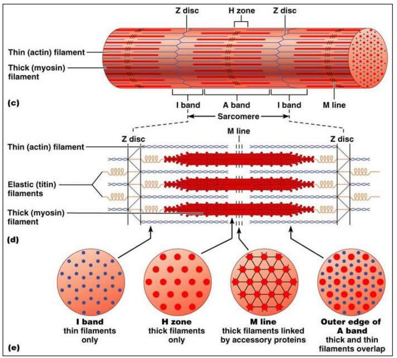
Explains how muscles contract at the microscopic level.
Focuses on the interaction between two protein filaments:
- Actin (thin filaments)
- Myosin (thick filaments)
📐 Structure of a Sarcomere
Sarcomere = basic unit of contraction in a myofibril
Found between two Z-lines
Contains:
- A-band (overlap of actin and myosin)
- I-band (actin only)
- H-zone (myosin only)
⚙️ How Contraction Happens (Step-by-Step)
| Step | Process |
|---|---|
| 1. Stimulation | Motor neuron releases Ca²⁺ ions into the muscle |
| 2. Cross-Bridge Formation | Myosin heads bind to actin forming cross-bridges |
| 3. Power Stroke | Myosin heads pull actin filaments inward, shortening the sarcomere |
| 4. Detachment | ATP binds to myosin, causing it to release actin |
| 5. Resetting | Myosin uses ATP to re-cock for the next stroke |
Note: The filaments themselves do not shorten – they slide past each other, which shortens the sarcomere!
🔁 What Happens During Contraction?
| Before Contraction | After Contraction |
|---|---|
| Z-lines farther apart | Z-lines closer together |
| H-zone visible | H-zone reduced or disappears |
| I-band wide | I-band narrows |
| A-band stays same | A-band stays same |
⚡ Role of Calcium & ATP
- Calcium ions: Uncover actin’s binding sites by removing tropomyosin
- ATP is required for:
- Cross-bridge detachment
- Resetting of myosin heads
- Active transport of Ca²⁺ back into sarcoplasmic reticulum
Muscles contract by sliding actin and myosin past each other – not by shortening filaments.
ATP and calcium are essential for contraction and relaxation.
Sarcomeres shorten → muscle shortens → movement occurs.
B3.3.3 – Role of the Protein Titin and Antagonistic Muscles in Muscle Relaxation
🧵 Titin: he Giant Protein of the Sarcomere
Titin is the largest known protein in the human body.
It spans half a sarcomere, from Z-line to M-line.
Anchors myosin (thick filaments) in place.
🔧 Functions of Titin
| Function | Explanation |
|---|---|
| Elastic recoil | Helps sarcomere return to original length after contraction |
| Prevents overstretching | Acts like a spring to resist excessive stretch |
| Structural support | Maintains alignment of myosin within the sarcomere |
Without titin, muscles would overstretch and fail to regain shape, leading to damage and weakness.
🔁 Antagonistic Muscles: The Push-Pull System
Muscles only contract – they can’t actively extend themselves.
Antagonistic muscles are pairs of muscles that work opposite each other.
💡 Examples:
| Muscle Pair | Action |
|---|---|
| Biceps | Contract to flex the arm |
| Triceps | Contract to extend the arm |
⚖️ How They Work Together
- One muscle contracts → produces movement
- The opposing muscle relaxes or gets stretched
- To return or reverse the motion, roles are switched
Example in Legs: Quadriceps contract to extend the knee; Hamstrings contract to flex the knee.
This coordinated system allows for smooth, controlled motion.
📦 Summary Box:
Titin acts like a spring inside sarcomeres:
– Restores length after stretching
– Prevents damage from overstretching
Antagonistic muscles:
– Enable movement in opposite directions
– Are essential because muscles can’t push, only pull
B3.3.4 – Structure and Function of Motor Units in Skeletal Muscle
⚙️ What is a Motor Unit?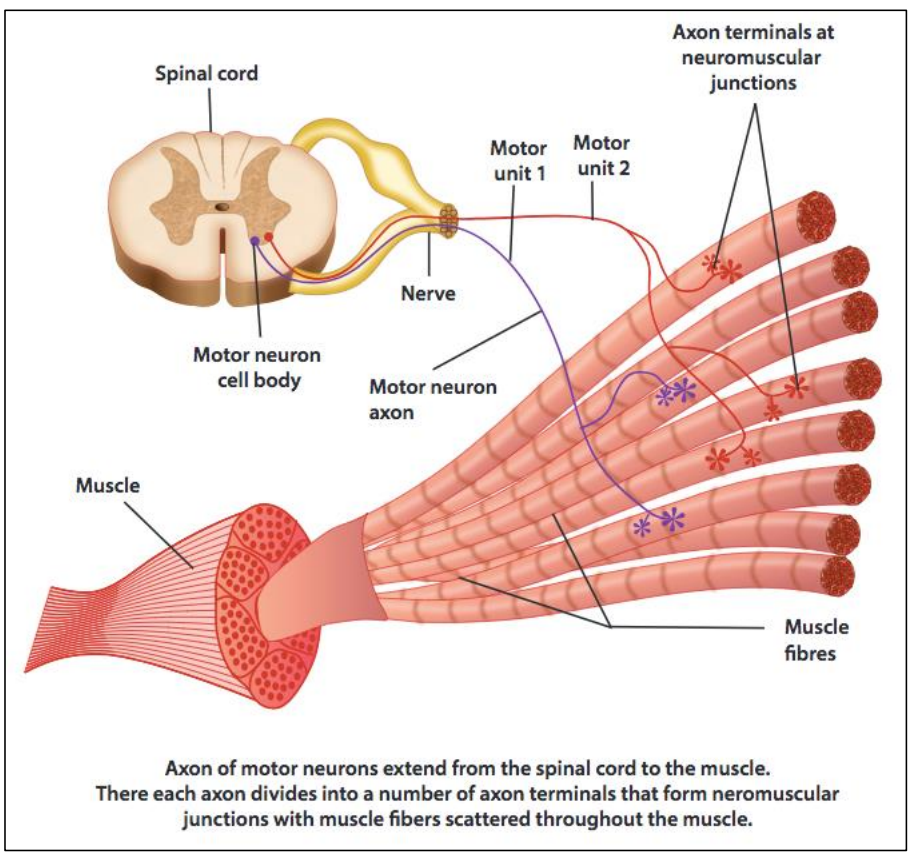
A motor unit is the basic functional unit of skeletal muscle control.
It consists of:
- One motor neuron
- All the muscle fibres it stimulates
- Neuromuscular junctions (NMJs) – the synapses between them
🧩 Structure of a Motor Unit
| Component | Description |
|---|---|
| Motor Neuron | A nerve cell that carries electrical signals from the spinal cord to the muscle |
| Muscle Fibres | Long, cylindrical cells that contract when stimulated |
| Neuromuscular Junction | Specialized synapse where a motor neuron communicates with a muscle fibre |
Each muscle fibre is stimulated by only one motor neuron, but one motor neuron can stimulate many muscle fibres.
⚡ How Motor Units Work
- Electrical impulse (action potential) travels down the motor neuron
- Acetylcholine (ACh) is released at the neuromuscular junction
- ACh binds to receptors on the muscle fibre membrane
- This causes the muscle fibre to contract
🔍 Small vs. Large Motor Units
| Type | Number of fibres per neuron | Function | Example |
|---|---|---|---|
| Small | Few fibres | Fine, precise control | Eye muscles 👁️ |
| Large | Hundreds of fibres | Powerful, gross movements | Leg muscles 🦵 |
🧠 Function of Motor Units
Control strength of contraction by:
- Recruiting more motor units for stronger contractions
- Using fewer motor units for gentle or fine movements
This is called motor unit recruitment.
A motor unit = motor neuron + its muscle fibres + neuromuscular junctions
One neuron can control many fibres, but each fibre is controlled by only one neuron
Motor units allow:
– Graded strength control
– Efficient, coordinated muscle contractions
– Fine movements use small units, while powerful movements use large units
B3.3.6 – Movement at a Synovial Joint
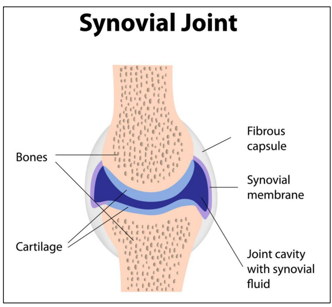
🦿 What is a Synovial Joint?
A synovial joint is a freely movable joint found in limbs and the vertebral column.
Example: Hip joint (between femur and pelvis).
These joints are designed to provide smooth, controlled movement while reducing friction and shock.
⚙️ Key Structures in a Synovial Joint and Their Functions
| Structure | Function |
|---|---|
| Bones | Act as levers for movement. In hip joint: femur and pelvis. |
| Cartilage | Covers bone ends, prevents friction, acts as a shock absorber. |
| Synovial fluid | Lubricates the joint; nourishes cartilage. |
| Synovial membrane | Secretes synovial fluid. |
| Ligaments | Connect bone to bone, maintain joint stability. |
| Tendons | Connect muscle to bone, allow muscles to move bones. |
| Muscles | Contract to pull on tendons, creating movement at the joint. |
🧍♂️ Human Hip Joint as an Example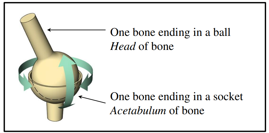
- Type: Ball-and-socket joint
- Structure: Femur head fits into the acetabulum of the pelvis
- Movement: Flexion, extension, rotation in multiple directions
🔁 How Movement Happens
- Muscles contract → pull tendons
- Tendons pull on bones → joint moves
- Synovial fluid and cartilage ensure movement is smooth and frictionless
- Ligaments keep bones aligned during motion
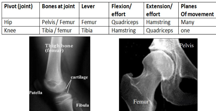
Synovial joints like the hip allow free movement using a combination of bones, cartilage, fluid, ligaments, tendons, and muscles.
The femur and pelvis work together in the hip joint to enable flexible and strong movement.
B3.3.7 – Range of Motion of a Joint
🤸♀️ What is “Range of Motion”?
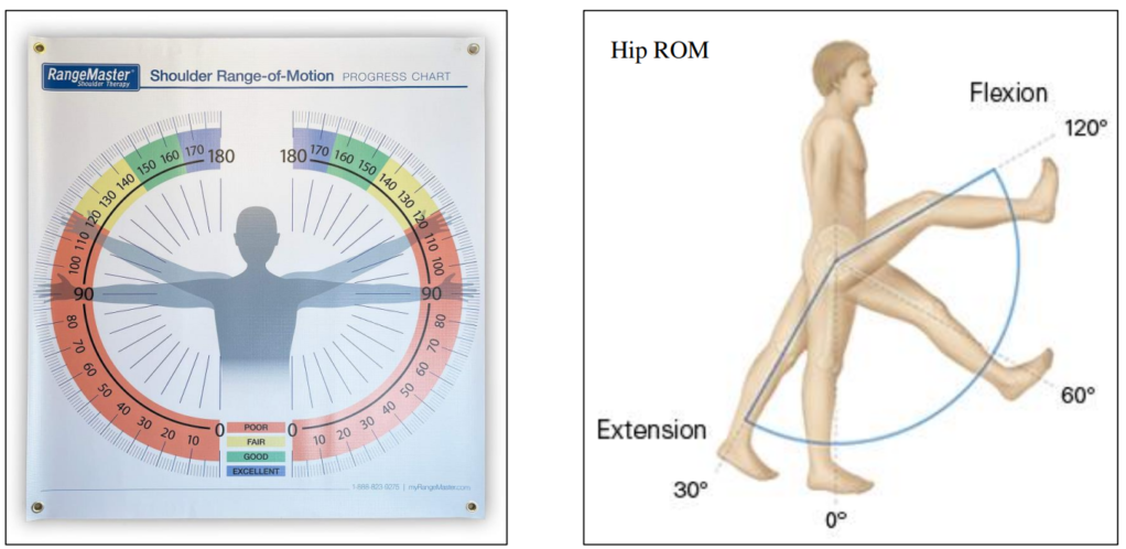
Range of motion (ROM) = how far a joint can move in different directions.
It depends on the joint type, muscle flexibility, and ligament tension.
For example: The shoulder has a wider ROM than the knee.
📊 Measuring Joint Movement
- Goniometer – like a protractor for joints!
- Computer image analysis – tracks angles during motion using video or images
📏 Types of Joint Movement Dimensions
| Type of Movement | Description | Example Joint |
|---|---|---|
| Flexion / Extension | Decreasing / increasing the angle of the joint | Elbow, knee |
| Abduction / Adduction | Moving away from / toward the body’s midline | Shoulder, hip |
| Rotation | Bone moves around its own axis | Neck, shoulder |
| Circumduction | Circular movement (a combo of flexion, etc.) | Hip, shoulder |
🧪 Application Skill: Measuring with a Goniometer
- Align center of goniometer at joint
- One arm stays still, the other follows limb movement
- Measure the angle of movement (e.g. elbow flexes ~145°)
🔄 Comparing Joints
| Joint | Main Movements | Range of Motion |
|---|---|---|
| Shoulder | Flexion, rotation | Very wide (multi-axial) |
| Hip | Flexion, abduction | Wide, but less than shoulder |
| Knee | Mainly flexion/extension | Narrow (hinge joint) |
| Elbow | Flexion/extension | Moderate range |
The range of motion tells us how much a joint can move in different directions. It varies by joint type and can be measured using a goniometer or digital tools. Comparing joints helps us understand functional differences in movement.
B3.3.8 – Intercostal Muscles as an Example of Antagonistic Muscle Action
🫁 What are Intercostal Muscles?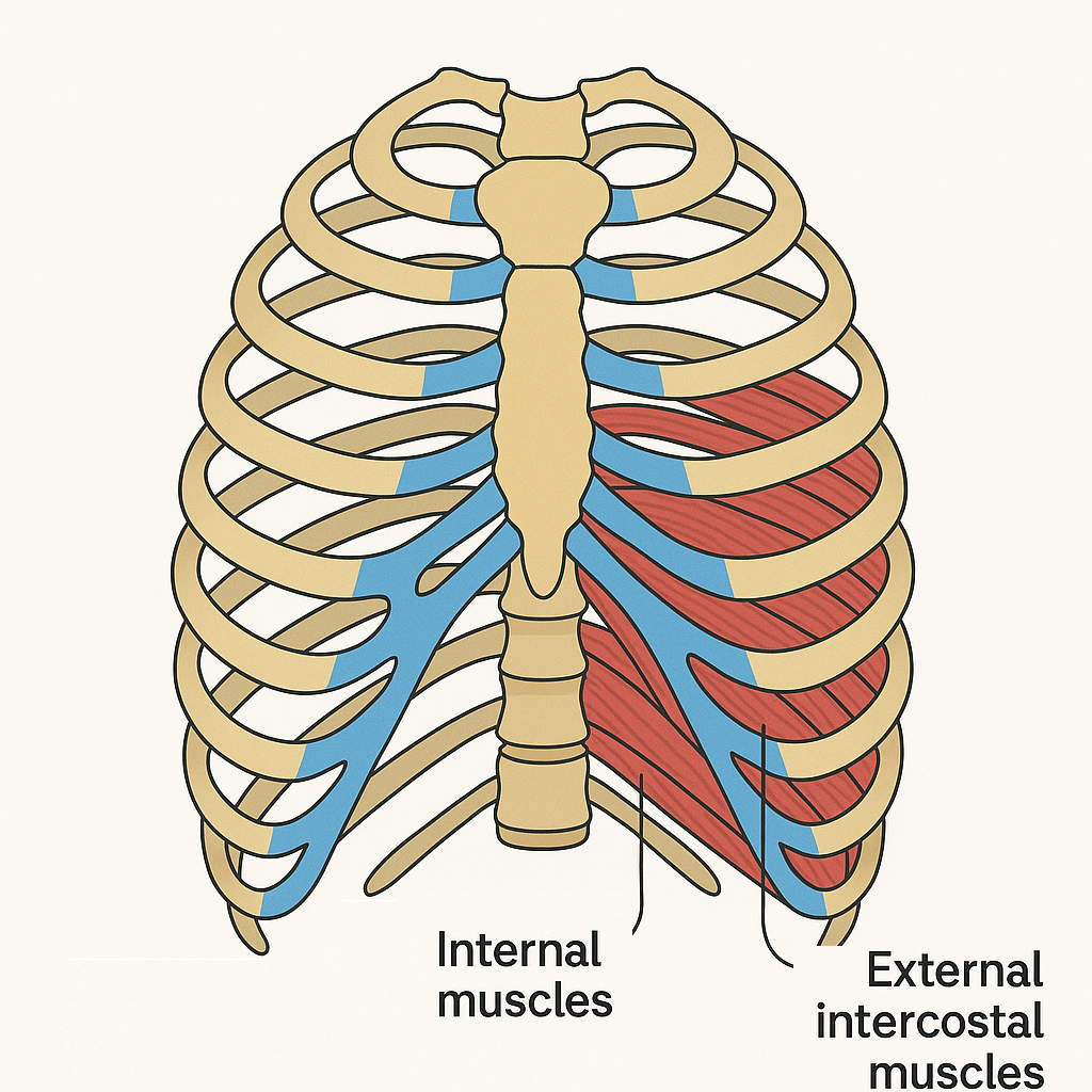
Intercostal muscles are found between the ribs.
They are arranged in two layers:
- External intercostal muscles
- Internal intercostal muscles
These muscles help move the ribcage to allow breathing (ventilation).
🔄 Antagonistic Muscle Action
Antagonistic muscles work in pairs:
- When one contracts, the other relaxes.
- This opposing action allows controlled movement and efficient return.
📏 Fiber Orientation & Opposing Movements
| Muscle Type | Fiber Direction | Function |
|---|---|---|
| External intercostals | Downward & forward (↘️) | Lift ribs up & out → inhalation |
| Internal intercostals | Upward & backward (↖️) | Pull ribs down & in → forced exhalation |
This cross-orientation allows them to work against each other like a pulley system.
⚙️ Titin and Stored Potential Energy
- When one muscle contracts, it stretches the opposing muscle.
- This stretches titin in the sarcomeres of the relaxed muscle.
- Titin acts like a spring, storing potential energy.
- This stored energy helps with smooth relaxation and muscle recoil.
🧠 Real-Life Analogy
Think of external and internal intercostals like biceps and triceps for the ribs one lifts while the other pulls down!
Intercostal muscles show how antagonistic pairs allow precise ribcage movement. The fiber direction ensures they move the ribs in opposite directions, while titin stores energy during stretching, making breathing efficient and controlled.
B3.3.9 – Reasons for Locomotion
🚶♀️ What is Locomotion?
Locomotion is the movement of an organism from one place to another.
It’s a key feature of motile organisms and supports survival and reproduction.
🔍 Why Do Organisms Move?
Organisms move for several essential reasons:
1. Foraging for Food
Locomotion helps organisms find food sources in different locations.
Example:
- Honeybees fly from flower to flower to collect nectar.
- Lions roam savannahs to hunt prey.
2. Escaping from Danger
Movement allows organisms to flee predators or harmful environments.
Example:
- Gazelles run swiftly to escape lions.
- Octopuses use jet propulsion to evade threats.
3. Searching for a Mate
Many species move to locate and attract mates, increasing reproductive success.
Example:
- Peacocks roam and display plumage during mating season.
- Frogs migrate to ponds to find partners for reproduction.
4. Migration
Some species travel long distances seasonally for better living conditions.
Example:
- Monarch butterflies migrate thousands of kilometers between Canada and Mexico.
- Salmon swim upstream to spawn in freshwater.
📊 Quick Summary Table
| Reason | Purpose | Example Organism |
|---|---|---|
| Foraging for food | To find nourishment | Honeybee, Lion |
| Escaping danger | Avoid predators/environmental threats | Gazelle, Octopus |
| Searching for a mate | Reproduction | Peacock, Frog |
| Migration | Seasonal movement for survival | Monarch butterfly, Salmon |
Locomotion helps organisms survive and reproduce. It plays critical roles in finding food, avoiding predators, finding mates, and migrating to better environments.