Additional Higher Level
C2.1.1 – Receptors as Proteins with Binding Sites for Specific Signalling Chemicals
🔑 What Are Receptors?
Receptors are proteins found on:
- The cell surface membrane
- The cytoplasm (in some cases)
They have a specific shape that allows them to bind only to one type of signalling molecule.
💬 What Are Ligands?
A ligand is a signalling chemical that binds to a receptor.
This binding is usually highly specific – like a lock and key.
🧠 Examples of ligands:
- Hormones (e.g. insulin)
- Neurotransmitters (e.g. dopamine)
- Cytokines or growth factors
🎯 How It Works
- A ligand travels through blood or extracellular fluid.
- It binds to a receptor on/in a target cell.
- This causes a change inside the cell, called a signal transduction.
- It might activate an enzyme, open a channel, or switch on a gene.
🔬 Real-World Example
| Ligand | Receptor Location | Response |
|---|---|---|
| Insulin | Cell membrane of liver cells | Increases glucose uptake |
| Adrenaline | Muscle cell membrane | Triggers fight-or-flight response |
| Dopamine | Neurons | Affects mood and movement |
– Receptors are proteins that bind specific ligands.
– Ligands are the signalling molecules (like hormones).
– Binding triggers a cellular response – like turning on a gene or changing enzyme activity.
– The interaction is specific, like a lock and key.
C2.1.2 – Cell Signalling by Bacteria in Quorum Sensing
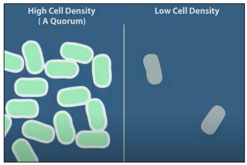
🧬 What Is Quorum Sensing?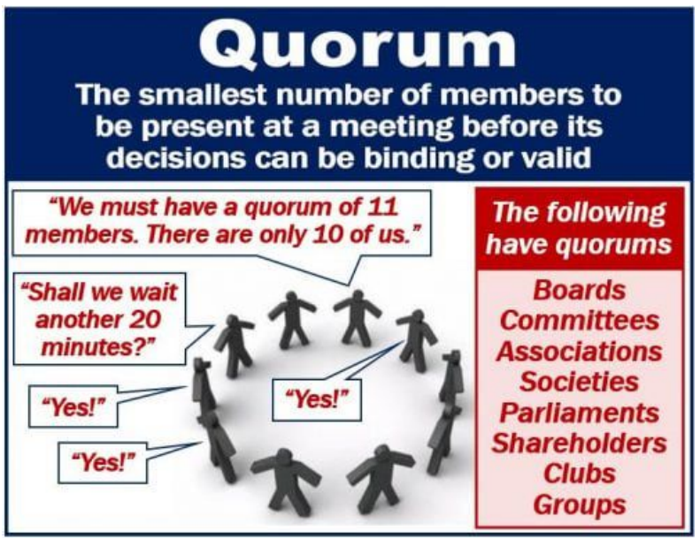
Quorum sensing is how bacteria communicate with each other using chemical signals.
It helps bacteria detect their population density.
When enough bacteria are present, they change behavior as a group – like a coordinated team!
💬 How It Works:
Each bacterium releases a signalling molecule called an autoinducer.
As the bacterial population grows, the concentration of autoinducers increases.
When the concentration crosses a threshold:
- The autoinducers bind to receptors on/in the bacteria.
- This triggers a change in gene expression.
📌 Result? All the bacteria start doing the same thing at the same time!
🌟 Real Example: Bioluminescence in Vibrio fischeri
| Feature | Description |
|---|---|
| Bacterium | Vibrio fischeri |
| Environment | Lives in the light organ of certain marine animals like the Hawaiian bobtail squid |
| Signal | Autoinducer molecule |
| Response | Turns on genes that produce light (bioluminescence) |
| Purpose | Helps the squid hide its shadow at night – camouflage! |
📊 Why Quorum Sensing Matters
- Coordinates virulence (infection-causing ability) in pathogens
- Controls biofilm formation (slimy protective layers)
- Regulates antibiotic resistance
- Useful in synthetic biology and biotechnology
– Quorum sensing = bacterial communication based on population density
– Uses autoinducers to send and detect signals
– Triggers a group response when threshold levels are reached
– Example: Vibrio fischeri produces light only when enough bacteria are present
C2.1.3 – Functional Categories of Signalling Chemicals in Animals
🧪 What Are Signalling Chemicals?
Signalling chemicals carry messages from one cell to another.
They bind to receptors on target cells to trigger a response.
The type, speed, and target of the response depends on the category of the chemical.
The 4 Key Categories:
| Category | Function | Speed | Target Range | Example |
|---|---|---|---|---|
| Hormones | Long-distance messengers released into the bloodstream | Slower (minutes to hours) | Whole body | Insulin, adrenaline |
| Neurotransmitters | Chemical signals between neurons or to muscles | Very fast (milliseconds) | Specific, local synapses | Acetylcholine, dopamine |
| Cytokines | Immune system messengers that regulate inflammation and cell communication | Moderate speed | Local or whole-body | Interleukins, interferons |
| Calcium ions (Ca²⁺) | Second messengers inside cells for quick signalling | Very fast | Intracellular (within cells) | Triggers muscle contraction, enzyme activation |
🧠 Comparison: Who Does What?
| Feature | Hormones | Neurotransmitters | Cytokines | Calcium Ions |
|---|---|---|---|---|
| Released by | Endocrine glands | Neurons | Immune cells | Stored in organelles (e.g., ER) |
| Transport method | Bloodstream | Synaptic cleft | Blood or local tissue | Cytoplasm |
| Target distance | Long | Very short | Short or long | Within same cell |
| Response speed | Slow | Very fast | Moderate | Instant |
| Example function | Regulate blood sugar | Move a muscle | Trigger fever | Trigger muscle contraction |
– Hormones: Long-distance, slow-acting signals in the bloodstream
– Neurotransmitters: Fast, local messengers at nerve synapses
– Cytokines: Immune signalling molecules for inflammation and immunity
– Calcium ions: Act inside cells as second messengers for quick responses
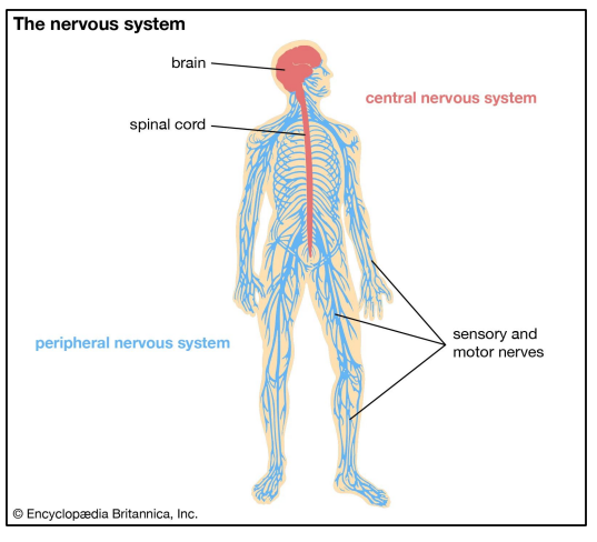
C2.1.8 – Transmembrane Receptors for Neurotransmitters and Changes to Membrane Potential
🧬 Neurotransmitters and Transmembrane Receptors
Neurotransmitters are chemical messengers released by neurons to communicate with other cells.
They bind to specific receptors on the postsynaptic cell membrane, triggering a response.
Some receptors are transmembrane proteins that also act as ion channels.
🎯 Key Example: The Acetylcholine Receptor
Acetylcholine (ACh) is a common neurotransmitter found at neuromuscular junctions and in the brain.
The acetylcholine receptor is a ligand-gated ion channel:
- It spans the membrane (transmembrane receptor).
- When ACh binds, the channel opens.
⚙️ How the Process Works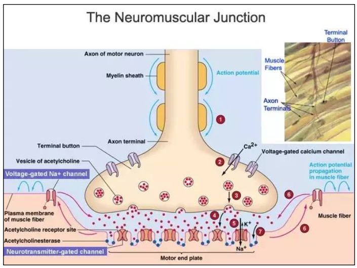
- Neurotransmitter Release: A neuron releases acetylcholine into the synaptic cleft.
- Binding to Receptor: ACh binds to its receptor on the postsynaptic membrane.
- Ion Channel Opens: The receptor changes shape and opens a channel for ions to flow through.
- Ions Diffuse In: Na⁺ (sodium ions) rush into the cell. Sometimes K⁺ may flow out too, but sodium influx dominates.
- Change in Membrane Potential: The inside of the cell becomes less negative (this is called depolarization).
- May trigger an action potential (if threshold is reached)
- Activate other proteins or channels
- Cause a muscle contraction (if it’s a muscle cell)
🧪 Acetylcholine Receptor at a Glance
| Feature | Description |
|---|---|
| Type of receptor | Ligand-gated ion channel |
| Neurotransmitter | Acetylcholine (ACh) |
| Ions involved | Mainly Na⁺, sometimes K⁺ |
| Result of activation | Depolarization (change in membrane potential) |
| Possible outcome | Action potential, muscle contraction, signaling |
💡 Did You Know?
The nicotinic acetylcholine receptor is also activated by nicotine, which mimics ACh.
Blocking this receptor (e.g., with curare) can cause paralysis!
– Acetylcholine binds to a transmembrane receptor that acts as an ion channel.
– This allows positively charged ions (mainly Na⁺) to enter the cell.
– The result is a change in membrane potential, which may trigger further cellular responses.
C2.1.9 – Transmembrane Receptors That Activate G Proteins
🧠 What Are G Protein-Coupled Receptors (GPCRs)?
GPCRs are a large family of transmembrane proteins.
Found across all tissues in the human body.
Their job: Detect external signals (like hormones or neurotransmitters) and start a response inside the cell.
🔗 Structure of GPCRs
- GPCRs span the plasma membrane 7 times (they are also called 7-transmembrane receptors).
- Outside: Binding site for the signaling molecule (ligand).
- Inside: Interacts with a G protein (made of α, β, and γ subunits).
🔄 How GPCRs Work — Step-by-Step
- Signal Arrival: A ligand (e.g. adrenaline, dopamine) binds to the extracellular part of the GPCR.
- Receptor Activation: The GPCR changes shape and activates the G protein on the inner membrane surface.
- G Protein Activation: The α-subunit exchanges GDP for GTP, becoming active. It separates from the βγ subunits.
- Message Relay: The active α-subunit or βγ complex interacts with target proteins like:
- Enzymes (e.g. adenylate cyclase → makes cAMP)
- Ion channels
- Second messengers
- Signal Termination: GTP is hydrolyzed back to GDP, and the subunits rejoin, resetting the system.
🔍 Why Are GPCRs Important?
- Involved in vision, smell, taste, mood, heart rate, and more.
- ~800 GPCRs in humans!
- Targeted by many medicines (e.g., antihistamines, beta blockers).
🧪 GPCR Signaling Pathway Overview
| Step | Event | Location |
|---|---|---|
| 1 | Ligand binds to GPCR | Outside cell |
| 2 | GPCR activates G protein | Inner membrane |
| 3 | G protein subunits split | Cytoplasm |
| 4 | Target protein is activated | Cytoplasm or membrane |
| 5 | Cellular response triggered | Inside cell |
💡 Real Example: Adrenaline and Heart Rate
Adrenaline binds to β-adrenergic receptors (a type of GPCR).
Activates G protein → adenylate cyclase → cAMP → faster heart rate.
– GPCRs detect external signals and activate G proteins inside the cell.
– The G protein then triggers internal responses, often through second messengers.
– GPCRs are involved in many body systems and are major drug targets.
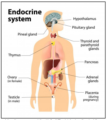
C2.1.10 – Mechanism of Action of Epinephrine (Adrenaline) Receptors
🧬 What Is Epinephrine / Adrenaline?
A hormone and neurotransmitter.
Released by the adrenal glands (above the kidneys).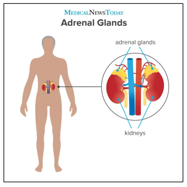
Prepares the body for “fight or flight” — increases heart rate, dilates pupils, boosts glucose.
📌 Naming Note (NOS):
- Adrenaline = Latin: ad (at) + ren (kidney)
- Epinephrine = Greek: epi (above) + nephros (kidney)
- Both mean the same thing – naming reflects international cooperation in science!
- “Adrenaline” is common in the UK.
- “Epinephrine” is more common in the US.
How Epinephrine Triggers a Response in Cells
This is an example of G protein-coupled receptor (GPCR) signaling.
🔄 Step-by-Step Mechanism:
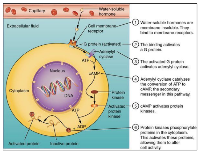
- Epinephrine Binds to Receptor: The hormone binds to β-adrenergic receptor (a GPCR) on the plasma membrane.
- Activation of G Protein: Receptor changes shape → activates a G protein inside the cell.
GDP is replaced with GTP on the G protein’s α-subunit. - Activation of Adenylate Cyclase: The GTP-bound α-subunit activates adenylate cyclase (an enzyme in the membrane).
- Production of Second Messenger: cAMP: Adenylate cyclase converts ATP → cyclic AMP (cAMP).
cAMP is a second messenger — it carries the signal deeper into the cell. - Activation of Protein Kinase A (PKA): cAMP activates PKA, which then phosphorylates (adds phosphate groups to) target proteins.
- Cellular Responses:
- Increased heart rate (cardiac muscle)
- Breakdown of glycogen → glucose (liver)
- Relaxation of airways (smooth muscle in lungs)
📊 Signal Amplification
One epinephrine molecule can activate many G proteins, leading to thousands of cAMP molecules.
This causes a huge cellular response from a tiny hormone signal!
🧪 Epinephrine Signaling Pathway Overview
| Step | Event | Molecules Involved |
|---|---|---|
| 1 | Epinephrine binds to GPCR | Epinephrine, β-adrenergic receptor |
| 2 | G protein is activated | GTP replaces GDP |
| 3 | Adenylate cyclase is activated | Enzyme at membrane |
| 4 | cAMP is produced | Second messenger from ATP |
| 5 | Protein kinase A is activated | Phosphorylation of target proteins |
| 6 | Cell responds | e.g., energy release, heart rate |
– Epinephrine binds to a GPCR, starting a signal transduction cascade.
– G proteins activate adenylate cyclase, which creates cAMP from ATP.
– cAMP activates protein kinases, causing changes like increased heart rate or glucose release.
– This is a great example of signal amplification and international naming conventions in biology.
C2.1.11 – Transmembrane Receptors with Tyrosine Kinase Activity
🧪 What Is a Tyrosine Kinase Receptor?

- A type of transmembrane receptor with built-in enzyme activity.
- Specifically: it phosphorylates tyrosine residues on proteins inside the cell.
- These receptors help transmit signals from protein hormones like insulin.
Insulin: A Classic Example
- Insulin is a protein hormone secreted by the pancreas when blood glucose levels are high.
- Its main job is to promote glucose uptake into cells, especially muscle and fat cells.
🔗 How Insulin Triggers a Cellular Response
🧬 Step-by-Step Mechanism:
- Insulin Binds to Receptor: Insulin binds to the insulin receptor on the plasma membrane.

This receptor is a tyrosine kinase receptor. - Receptor Dimerization & Autophosphorylation: Binding causes the receptor to dimerize (two parts join).
The receptor autophosphorylates (adds phosphate groups to its own tyrosine residues inside the cell). - Signal Cascade Begins: These phosphorylated tyrosines act as docking sites for intracellular proteins.
A signal transduction cascade begins — a sequence of reactions inside the cell. - Vesicle Movement: The cascade causes vesicles containing GLUT4 (glucose transporters) to move to the cell membrane.
- Glucose Uptake: GLUT4 transporters are inserted into the membrane.
Glucose enters the cell via facilitated diffusion → blood sugar levels drop.

📦 Summary Table: Insulin Signaling Pathway
| Step | Event | Key Molecules |
|---|---|---|
| 1 | Insulin binds receptor | Insulin, insulin receptor |
| 2 | Receptor phosphorylates tyrosines | ATP, tyrosine residues |
| 3 | Signal transduction cascade starts | Intracellular proteins |
| 4 | Vesicles with GLUT4 move to membrane | GLUT4-containing vesicles |
| 5 | Glucose transporters allow glucose uptake | Glucose enters cell |
– The insulin receptor is a tyrosine kinase receptor found in the plasma membrane.
– Binding of insulin triggers phosphorylation of tyrosine residues inside the cell.
– This activates a cascade that leads to GLUT4 transporters moving to the membrane.
– Result: Glucose enters cells, helping to regulate blood sugar levels.
C2.1.12 – Intracellular Receptors That Affect Gene Expression
🧪 What Are Intracellular Receptors?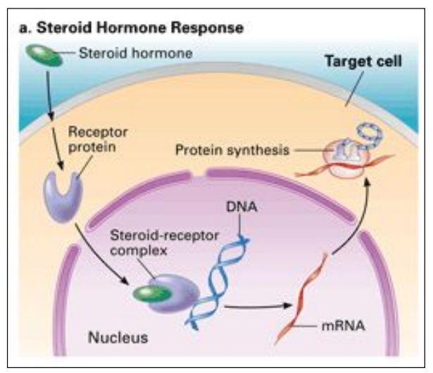
These are receptor proteins inside the cell, usually in the cytoplasm or nucleus.
They’re activated by lipid-soluble signalling chemicals (like steroid hormones) that can pass through the cell membrane.
💡 How Do They Work?
- Steroid hormone (e.g., oestradiol, progesterone, testosterone) diffuses through the cell membrane.
- It binds to its specific intracellular receptor inside the cell.
- This binding activates the receptor.
- The hormone-receptor complex moves into the nucleus (if not already there).
- It binds to specific DNA sequences (called hormone response elements).
- This activates gene transcription – turning genes on or off!
🧬 Examples of Steroid Hormones
| Hormone | Function |
|---|---|
| Oestradiol | Regulates development of female secondary sex characteristics and menstrual cycle. |
| Progesterone | Prepares the uterus for pregnancy and maintains early stages. |
| Testosterone | Regulates development of male secondary sex characteristics and sperm production. |
🧠 Key Idea: Hormones Can Control Gene Expression
These steroid hormones act as gene switches, telling the cell to make specific proteins.
This is slower than neurotransmitter signalling but causes long-lasting changes in cell behavior.
– Intracellular receptors are found in the cytoplasm or nucleus.
– They bind to steroid hormones like oestradiol, progesterone, and testosterone.
– Once activated, they bind DNA and affect gene expression.
– This leads to long-term changes in cell function by changing which proteins are made.
C2.1.13 – Effects of the Hormones Oestradiol and Progesterone on Target Cells
🧬 What Are Target Cells?

Target cells are cells that have specific receptors for a hormone.
When the hormone binds, it triggers a response unique to that cell type.
🌸 Oestradiol – Effects on Hypothalamic Cells
- Target: Cells in the hypothalamus that secrete gonadotropin-releasing hormone (GnRH).
- Effect:
- Oestradiol stimulates these cells to increase secretion of GnRH.
- GnRH then acts on the pituitary to trigger FSH and LH release – both key to the menstrual cycle and ovulation.
In short: Oestradiol → Hypothalamus → ↑ GnRH → ↑ FSH & LH → Ovulation
🌙 Progesterone – Effects on Endometrial Cells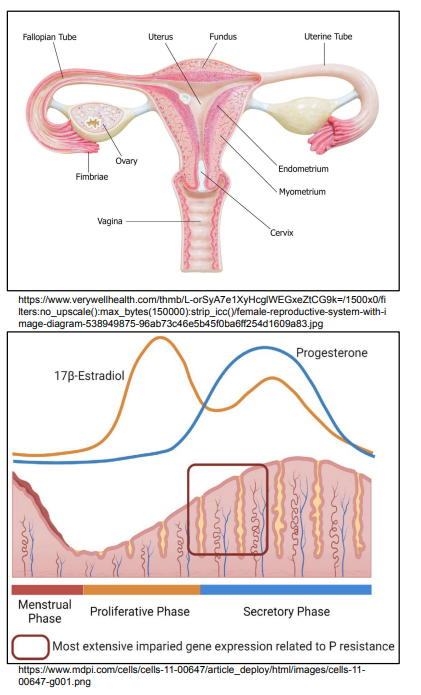
- Target: Cells in the endometrium (lining of the uterus).
- Effect:
Stimulates endometrial cells to prepare for implantation by:- Thickening the uterine lining
- Increasing blood vessel growth
- Promoting secretion of nutrients
- Helps maintain the lining during early pregnancy.
In short: Progesterone → Endometrium → Thickens & nourishes uterine lining
🔁 Hormonal Coordination Summary
| Hormone | Target Cell | Main Effect |
|---|---|---|
| Oestradiol | Hypothalamic cells (GnRH-secreting) | Increases GnRH → Stimulates ovulation |
| Progesterone | Endometrial cells | Prepares uterus for pregnancy by thickening the lining |
– Oestradiol acts on the hypothalamus, boosting GnRH and influencing the menstrual cycle.
– Progesterone acts on the endometrium, preparing it for possible implantation and pregnancy.
– Both hormones are essential for proper reproductive function in females.
C2.1.14 – Regulation of Cell Signalling Pathways by Positive and Negative Feedback
⚙️ What Is Feedback in Cell Signalling?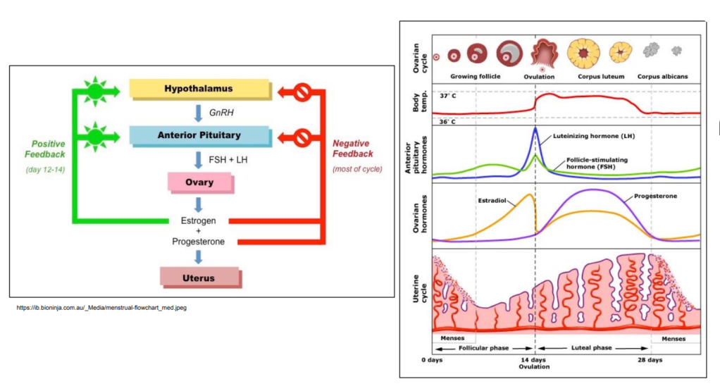
Feedback refers to how the output of a signalling pathway affects its own activity.
It helps cells regulate responses, keeping them balanced or amplifying them when needed.
➕ Positive Feedback
- Definition: A process where the response enhances or amplifies the original signal.
- Effect: Makes the signal stronger and usually leads to a rapid change or self-perpetuating loop.
- Example: Oestrogen and LH Surge
- Oestradiol increases LH secretion via positive feedback on the hypothalamus.
- Result: LH surge triggers ovulation.
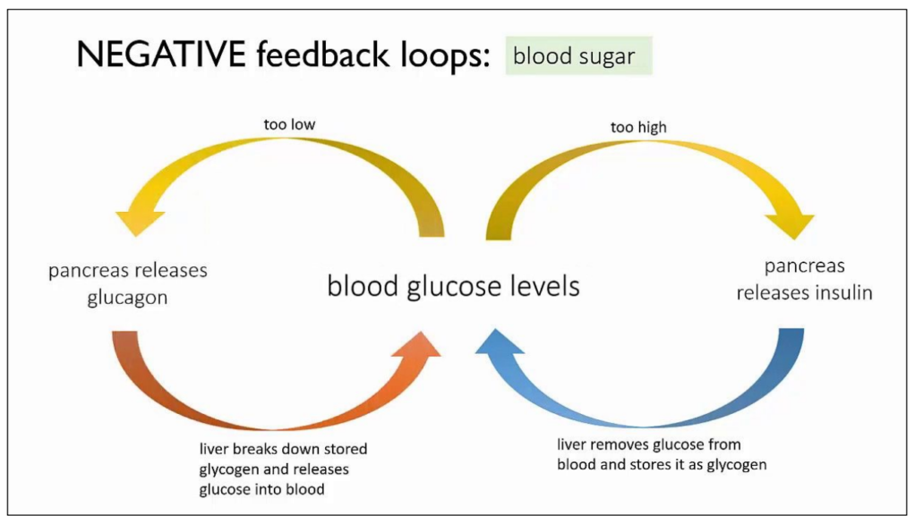
➖ Negative Feedback
- Definition: A process where the response reduces or suppresses the original signal.
- Effect: Maintains homeostasis by preventing overreaction or overstimulation.
- Example: Insulin Regulation
- High blood glucose → insulin released → glucose uptake by cells → blood glucose drops
- Drop in glucose reduces insulin secretion (negative feedback).
🔁 Feedback Regulation Summary Table
| Feedback Type | Definition | Effect | Example |
|---|---|---|---|
| Positive | Enhances the original signal | Amplifies the response | Oestradiol → LH surge |
| Negative | Suppresses the original signal | Maintains balance | Insulin → lowers blood glucose |
– Positive feedback amplifies signalling and promotes a rapid or irreversible response.
– Negative feedback stabilizes the system by limiting further signalling.
– These mechanisms are vital for precise control of cellular processes like reproduction and metabolism.