D3.3.1 – Homeostasis: Maintenance of the Internal Environment
🧬 What is Homeostasis?
- Homeostasis is the process by which an organism keeps its internal environment stable.
- It involves keeping key variables within narrow, preset limits, even when the external environment changes.
- This stability is vital for cells and organs to function properly.
📌 Key Homeostatic Variables in Humans
| Variable | Normal Range / Set Point | Importance |
|---|---|---|
| Body temperature | Around 37°C | Enzymes work best at this temperature; too high or low damages cells |
| Blood pH | Around 7.4 (slightly alkaline) | pH affects enzyme activity and oxygen transport |
| Blood glucose concentration | ~4-6 mmol/L (fasting) | Provides steady energy supply; too high or low is harmful |
| Blood osmotic concentration | ~300 mOsm/L | Maintains fluid balance and cell size |
🌿 How Does Homeostasis Work?
Body uses feedback systems to monitor and adjust these variables.
Typically involves three parts:
- Receptor: Detects change (e.g., temperature sensors in skin).
- Coordinator (Control center): Usually brain or endocrine glands; processes info.
- Effector: Produces response to restore balance (e.g., sweat glands, muscles).
🔍 Examples of Homeostasis in Action
| Variable | Change | Response | Effect |
|---|---|---|---|
| Body temperature | Rises (hot environment) | Sweat production, vasodilation of skin blood vessels | Heat loss, temperature returns to normal |
| Blood glucose | Increases after meal | Insulin released by pancreas | Cells take up glucose, blood level lowers |
| Blood pH | Drops (acidosis) | Increased breathing rate to expel CO₂ | Raises blood pH back to normal |
| Blood osmotic concentration | Increases (dehydration) | Thirst stimulated, ADH hormone released | Water intake and retention restore balance |
🧠 Why Is Homeostasis Important?
- Ensures enzymes and cells function optimally.
- Protects against harmful extremes (too hot/cold, acidic/basic).
- Maintains fluid and electrolyte balance.
- Supports overall health and survival.
D3.3.2 – Negative Feedback Loops in Homeostasis
🧬 What is Negative Feedback?
- Negative feedback is a control mechanism that reverses any change in a homeostatic variable to bring it back to its normal level (set point).
- It acts like a thermostat in your home: if the temperature goes too high or too low, it triggers actions to restore balance.
- Negative feedback maintains stability by correcting deviations both above and below the set point.
🌿 Why Negative Feedback?
- It prevents extreme fluctuations in the body.
- Keeps variables within narrow, safe limits.
- Ensures a stable internal environment, which is essential for cell function.
- Positive feedback, in contrast, amplifies changes, which is usually unstable and rare in homeostasis (except in special cases like blood clotting or childbirth).
🔍 How Negative Feedback Works: Basic Steps
| Step | Description | Example |
|---|---|---|
| 1. Stimulus | Change occurs, e.g., body temperature rises | Heat from environment |
| 2. Receptor | Detects change, e.g., temperature sensors in skin | Thermoreceptors |
| 3. Control center | Processes info, e.g., hypothalamus in brain | Receives signal |
| 4. Effector | Produces response to reverse change | Sweat glands activate to cool skin |
| 5. Response | Variable returns toward set point | Body temperature lowers to normal |
📌 Example: Blood Glucose Regulation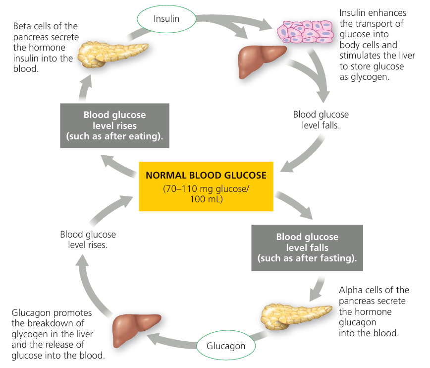
If blood glucose rises after eating:
Pancreas releases insulin.
Insulin promotes glucose uptake by cells.
Blood glucose falls back to normal.
If blood glucose drops:
Pancreas releases glucagon.
Glucagon triggers glucose release from liver.
Blood glucose rises to normal.
📊 Negative Feedback vs Positive Feedback
| Feature | Negative Feedback | Positive Feedback |
|---|---|---|
| Effect on change | Reverses change | Amplifies change |
| Role in homeostasis | Maintains stability | Rare, triggers rapid events |
| Examples | Temperature, blood glucose | Blood clotting, childbirth contractions |
| Outcome | Returns variable to set point | Drives process to completion |
D3.3.4 – Physiological Changes in Type 1 and Type 2 Diabetes
🧠 What is Diabetes?
- Diabetes is a chronic condition where the body cannot regulate blood glucose properly.
- Results in high blood glucose (hyperglycemia).
- Two main types: Type 1 and Type 2 diabetes.
🌿 Type 1 Diabetes
| Aspect | Details |
|---|---|
| Cause | Autoimmune destruction of beta cells in the pancreas |
| Effect | Little or no insulin production |
| Result | Blood glucose remains high because glucose can’t enter cells |
| Risk Factors | Genetic predisposition, autoimmune triggers (unknown exact cause) |
| Common Age of Onset | Usually childhood or adolescence |
| Treatment | Requires insulin injections to regulate blood glucose |
| Prevention | Currently no known prevention |
🌿 Type 2 Diabetes
| Aspect | Details |
|---|---|
| Cause | Insulin resistance: body cells respond poorly to insulin Eventually, pancreas may produce less insulin |
| Effect | High blood glucose due to ineffective glucose uptake |
| Risk Factors | Obesity, sedentary lifestyle, poor diet, age, genetics |
| Common Age of Onset | Usually adults, but increasing in younger people due to lifestyle |
| Treatment | Lifestyle changes (diet, exercise), oral medications, sometimes insulin |
| Prevention | Healthy diet, regular physical activity, weight management |
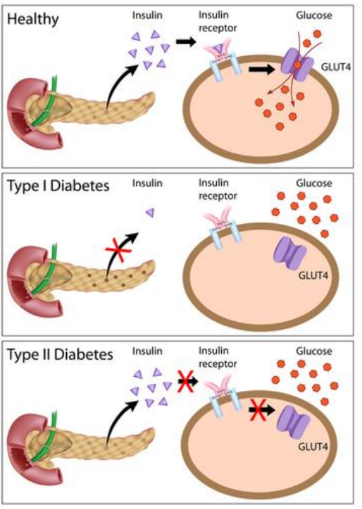
🔬 Physiological Changes in Both Types
| Change | Type 1 Diabetes | Type 2 Diabetes |
|---|---|---|
| Insulin levels | Low or absent | Normal, high initially, then may decrease |
| Blood glucose | High | High |
| Glucose uptake by cells | Severely reduced | Reduced due to resistance |
| Effects on metabolism | Cells starve for glucose, use fats → weight loss, ketoacidosis risk | Cells starve for glucose but less severe initially |
🔍 Why Diabetes is Dangerous
- High blood glucose damages blood vessels and nerves.
- Leads to complications: heart disease, kidney failure, blindness, poor wound healing.
- Managing blood glucose is crucial to prevent these.
📌 Summary Table: Differences Between Type 1 & Type 2 Diabetes
| Feature | Type 1 Diabetes | Type 2 Diabetes |
|---|---|---|
| Cause | Autoimmune beta cell destruction | Insulin resistance + eventual insulin deficiency |
| Insulin Production | None or very low | Initially normal/high, then decreases |
| Onset Age | Usually young | Usually adult (but younger cases rising) |
| Risk Factors | Genetics, autoimmunity | Obesity, lifestyle, genetics |
| Treatment | Insulin injections | Lifestyle changes, meds, insulin if needed |
| Prevention | No known prevention | Healthy lifestyle can prevent/delay |
Type 1 diabetes is caused by loss of insulin production due to immune attack.
Type 2 diabetes is caused mainly by cells becoming resistant to insulin.
Both cause high blood glucose but differ in causes, risk factors, and treatment.
Prevention focuses on lifestyle changes for type 2; type 1 currently has no prevention.
Managing blood glucose is vital to avoid serious health complications.
D3.3.5 – Thermoregulation as an Example of Negative Feedback Control
🧬 What is Thermoregulation?
Thermoregulation is the process by which the body maintains a stable internal temperature (~37°C), despite changes in the external environment.
It is a classic example of negative feedback control.
🌿 Key Components of Thermoregulation
| Component | Role |
|---|---|
| Peripheral thermoreceptors | Detect temperature changes in the skin and send signals to the brain |
| Hypothalamus | Acts as the control center; compares signals with the set point and coordinates responses |
| Pituitary gland | Releases hormones like thyroid-stimulating hormone (TSH) to regulate metabolism |
| Thyroxin | Hormone from the thyroid gland that increases metabolic rate, producing heat |
🔬 How the Body Responds to Temperature Changes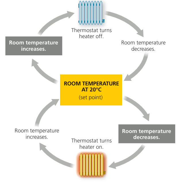
When Body Temperature Rises (Too Hot):
- Peripheral thermoreceptors detect increased skin temperature.
- Signals sent to hypothalamus trigger cooling mechanisms:
- Sweat glands produce sweat → evaporative cooling.
- Vasodilation of skin blood vessels → more heat lost through skin.
- Metabolic rate may decrease to reduce heat production.
When Body Temperature Falls (Too Cold):
- Peripheral thermoreceptors detect low temperature.
- Hypothalamus activates heat-producing responses:
- Muscle shivering: rapid muscle contractions generate heat.
- Vasoconstriction: skin blood vessels constrict to reduce heat loss.
- Thyroxin secretion increases, raising metabolic rate for more heat.
- Brown adipose tissue (brown fat) generates heat by burning fats (non-shivering thermogenesis).
📍 Role of Effectors: Muscle and Adipose Tissue
| Effector | Function |
|---|---|
| Skeletal muscles | Shivering produces heat by rapid contractions |
| Brown adipose tissue | Specialized fat that generates heat without muscle movement (especially in infants) |
🔍 Summary of Negative Feedback Loop in Thermoregulation
| Step | Description |
|---|---|
| 1. Stimulus | Body temperature deviates from set point |
| 2. Receptors | Peripheral thermoreceptors detect change |
| 3. Control center | Hypothalamus processes info and coordinates response |
| 4. Effectors | Sweat glands, muscles, blood vessels, thyroid gland respond |
| 5. Response | Body temperature returns to normal |
🧠 Why Is Thermoregulation Important?
Keeps enzymes working optimally.
Prevents damage from extreme temperatures.
Maintains overall homeostasis and survival.
Thermoregulation is controlled by a negative feedback system involving sensors (thermoreceptors), control center (hypothalamus), and effectors.
Sweating, vasodilation, shivering, vasoconstriction, and hormone regulation work together to maintain temperature.
Thyroxin increases metabolic heat production.
Muscle activity and brown fat play important roles in generating heat when cold.
D3.3.8 – Role of Glomerulus, Bowman’s Capsule, and Proximal Convoluted Tubule in Excretion
🧬 Key Structures in Kidney Excretion
| Structure | Location | Function |
|---|---|---|
| Glomerulus | Network of capillaries inside Bowman’s capsule | Filters blood plasma under high pressure (ultrafiltration) |
| Bowman’s capsule | Cup-shaped structure surrounding the glomerulus | Collects the filtrate (filtered fluid) from blood |
| Proximal convoluted tubule (PCT) | Twisted tubule after Bowman’s capsule | Reabsorbs useful substances back into the blood |
🌿 Ultrafiltration at the Glomerulus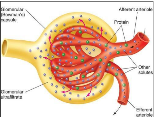
- Blood enters the glomerulus at high pressure.
- Small molecules (water, glucose, ions, urea) pass through capillary walls into Bowman’s capsule.
- Large molecules (proteins, blood cells) stay in the blood.
- This process is called ultrafiltration—it filters plasma based on size.
🔍 Filtrate in Bowman’s Capsule
The filtrate contains:
Water
Glucose
Ions (Na⁺, K⁺, Cl⁻)
Urea and other wastes
This fluid moves from Bowman’s capsule into the proximal convoluted tubule.
🧪 Reabsorption in the Proximal Convoluted Tubule
Most useful substances are actively and passively reabsorbed into blood:
- Glucose (all of it, normally)
- Ions (sodium, chloride, potassium)
- Water (by osmosis)
- Waste products like urea and toxins remain in the filtrate.
- Reabsorption keeps body fluids balanced and prevents loss of valuable molecules.
📌 Summary of Filtration and Reabsorption
| Process | What Happens | Purpose |
|---|---|---|
| Ultrafiltration | Blood plasma filtered through glomerulus into Bowman’s capsule | Removes plasma fluid and small solutes from blood |
| Reabsorption | Useful molecules reabsorbed in proximal tubule | Retains nutrients and balances ions and water |
| Excretion | Waste and excess substances remain in filtrate to be excreted | Removes toxins from body |
🧠 Why These Steps Are Important
Prevents loss of valuable substances like glucose and ions.
Ensures removal of metabolic wastes and toxins.
Maintains fluid and chemical balance in the body.
Glomerulus and Bowman’s capsule perform ultrafiltration to remove plasma fluid and small solutes.
Proximal convoluted tubule reabsorbs useful substances back into blood.
This process separates waste from useful molecules for excretion in urine.
Essential for maintaining body’s chemical balance and health.
D3.3.10 – Osmoregulation by Water Reabsorption in the Collecting Ducts
🧬 Key Concept: Osmoregulation via Water Reabsorption
The collecting ducts in the kidney control water reabsorption to maintain blood osmotic concentration.
This process is regulated by the hormone antidiuretic hormone (ADH).
🌿 Role of Osmoreceptors in the Hypothalamus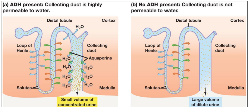
- Osmoreceptors are specialized nerve cells in the hypothalamus.
- They detect changes in blood osmotic concentration (how concentrated the blood is).
- If blood becomes too concentrated (high osmolarity), osmoreceptors send signals to increase ADH secretion.
- If blood is too dilute (low osmolarity), ADH secretion is reduced.
🔬 ADH Secretion by the Pituitary Gland
- ADH is released from the posterior pituitary gland into the bloodstream.
- The amount of ADH secreted depends on signals from osmoreceptors.
- More ADH → more water reabsorbed; less ADH → less water reabsorbed.
🧪 Aquaporins and Water Permeability
- Cells in the collecting ducts have aquaporin proteins that form water channels.
- Without ADH, aquaporins are mostly stored inside cells in intracellular vesicles → low water permeability → dilute urine.
- With ADH, aquaporins move to the cell membrane, increasing water permeability → more water reabsorbed → concentrated urine.
📍 How This Process Works
| Condition | Osmoreceptor Response | ADH Level | Aquaporins | Urine | Blood Osmolarity |
|---|---|---|---|---|---|
| High blood osmolarity (dehydrated) | Stimulated | Increased | Inserted into membrane | Concentrated (less water) | Decreases toward normal |
| Low blood osmolarity (overhydrated) | Inhibited | Decreased | Removed from membrane | Dilute (more water) | Increases toward normal |
🧠 Why This Mechanism Is Vital
- Prevents dehydration by conserving water.
- Prevents overhydration by excreting excess water.
- Maintains stable osmotic concentration of blood for normal cell function.
Osmoreceptors in the hypothalamus detect blood osmolarity changes and control ADH release.
ADH regulates water permeability of collecting duct cells by moving aquaporins to/from the membrane.
This system adjusts urine concentration to maintain water balance and osmotic homeostasis.