IB DP Biology Cell and nuclear division Study Notes
IB DP Biology Cell and nuclear division Study Notes at IITian Academy focus on specific topic and type of questions asked in actual exam. Study Notes focus on IB Biology syllabus with guiding questions of
- How can large numbers of genetically identical cells be produced?
- How do eukaryotes produce genetically varied cells that can develop into gametes?
Standard level and higher level: 3 hours
Additional higher level: 1 hour
D2.1.1 – Generation of New Cells in Living Organisms by Cell Division 🔬🧬
🧬 What Is Cell Division?
Cell division is the process by which one parent (mother) cell divides into two daughter cells.
Essential for growth, tissue repair, and reproduction in all living organisms.
Ensures continuity of life by passing genetic information to new cells.
🌱 Why Is Cell Division Important?
Allows organisms to grow larger by increasing cell numbers.
Helps replace damaged or dead cells.
Enables asexual reproduction in some organisms.
📌 The Cell Cycle: The Life Cycle of a Cell
The cell cycle is a series of ordered events leading to cell division.
Divided into two main phases:
Phase | Description |
---|---|
Interphase | Cell grows, performs normal functions, and prepares for division. Includes DNA replication. |
Mitotic (M) Phase | The cell physically divides into two daughter cells. |
🌿 Interphase (Preparation Phase)
The longest phase of the cell cycle.
Consists of three sub-phases:
- G₁ phase: Cell grows and performs normal activities.
- S phase: DNA replication—each chromosome duplicates to form sister chromatids.
- G₂ phase: Cell prepares for mitosis, making proteins and organelles needed for division.
🔬 Mitotic (M) Phase: Actual Cell Division
The cell divides in a multi-step process called mitosis.
Mitosis stages:
Stage | Key Events |
---|---|
Prophase | Chromosomes condense; nuclear envelope starts breaking down; spindle fibers form. |
Metaphase | Chromosomes line up along the cell’s equator (metaphase plate). |
Anaphase | Sister chromatids are pulled apart to opposite poles of the cell. |
Telophase | Nuclear envelopes re-form; chromosomes decondense; spindle breaks down. |
Followed by cytokinesis: the cytoplasm divides, forming two separate daughter cells.
Cell division produces two genetically identical daughter cells.
The cell cycle ensures cells grow, replicate DNA, and divide properly.
Proper cell division is vital for growth, repair, and reproduction.
D2.1.2 – Cytokinesis: Splitting of Cytoplasm Between Daughter Cells
🧬 What Is Cytokinesis?
- Cytokinesis is the final step in cell division where the cytoplasm of the parent cell is split into two daughter cells.
- Happens after nuclear division (mitosis or meiosis).
- Ensures each daughter cell gets enough cytoplasm and organelles to survive and function.
🌿 Cytokinesis in Animal Cells
- Animal cells don’t have a rigid cell wall, so the cell membrane can be pinched.
- A ring of contractile proteins (mainly actin and myosin) forms just beneath the cell membrane around the cell’s middle.
- These proteins contract like a muscle, creating a cleavage furrow that pinches the membrane inward.
- This process continues until the cell membrane splits completely, forming two separate daughter cells.
🌱 Cytokinesis in Plant Cells
- Plant cells have a rigid cell wall, so they cannot pinch in like animal cells.
- Instead, vesicles from the Golgi apparatus carry cell wall materials and assemble at the center of the cell.
- These vesicles fuse to form a cell plate between the two nuclei.
- The cell plate grows outward until it fuses with the existing cell wall, dividing the parent cell into two.
- This forms two daughter cells each with their own cell wall.
📌 Key Differences Between Animal and Plant Cytokinesis
Feature | Animal Cell Cytokinesis | Plant Cell Cytokinesis |
---|---|---|
Cell membrane | Pinched inward by contractile ring (actin & myosin) | Cell plate formed by fused vesicles |
Cell wall | No rigid cell wall, so membrane can be pinched | Rigid cell wall prevents pinching |
Result | Cleavage furrow forms, splits cytoplasm | Cell plate develops into new cell wall |
Cytokinesis is the division of the cytoplasm to complete cell division.
In animals, a contractile ring pinches the membrane.
In plants, vesicles form a new cell wall (cell plate) to separate cells.
Both processes ensure two fully functional daughter cells.
D2.1.3 – Equal and Unequal Cytokinesis
🧬 What Is Cytokinesis?
- Cytokinesis is the division of the cytoplasm between two daughter cells after nuclear division.
- Usually, the cytoplasm divides equally, giving each daughter cell roughly the same amount.
- However, unequal cytokinesis also occurs in some cases.
⚖️ Equal Cytokinesis
Most cell divisions produce two daughter cells with equal amounts of cytoplasm and organelles.
Both cells must receive essential organelles, especially those that cannot be made from scratch, like:
- Mitochondria
- Chloroplasts (in plant cells)
Ensures both daughter cells are viable and can function independently.
⚖️ Unequal Cytokinesis
- Sometimes, cytoplasm is divided unequally, producing daughter cells with different sizes and contents.
- One daughter cell gets most of the cytoplasm; the other is smaller and may have a different fate.
🌱 Examples of Unequal Cytokinesis
Example | Description |
---|---|
Oogenesis (Humans) | The process of egg formation. Produces one large egg cell with most cytoplasm and small polar bodies that usually degenerate. The large egg cell has all the nutrients and organelles needed for early development. |
Budding (Yeast) | A small bud forms on the parent cell. The bud grows and eventually detaches, receiving some cytoplasm and organelles, but less than the parent. |
📌 Why Does Unequal Cytokinesis Occur?
In oogenesis, unequal division helps concentrate resources in the egg to support early embryo growth.
In yeast budding, it allows a new organism to form while the parent remains largely intact.
Cytokinesis usually divides cytoplasm equally but can be unequal in special cases.
Both daughter cells must receive key organelles like mitochondria.
Oogenesis and yeast budding are classic examples of unequal cytokinesis.
Unequal division supports specialized functions or reproduction.
D2.1.4 – Roles of Mitosis and Meiosis in Eukaryotes
🧠 Why Nuclear Division Happens Before Cell Division
- Nuclear division (mitosis or meiosis) must occur before the cell divides.
- This prevents formation of anucleate cells (cells without a nucleus), which are non-functional.
- Ensures each daughter cell gets the right set of chromosomes.
🌿 Mitosis
Aspect | Details |
---|---|
Purpose | Growth, repair, and asexual reproduction |
Number of divisions | One |
Daughter cells | Two |
Chromosome number | Diploid (2n) – same as parent cell |
Genetic makeup | Genetically identical to the parent cell |
Maintains chromosome number and genome stability.
Produces clones for growth and tissue repair.
Essential for organisms growing and replacing damaged cells.
🌱 Meiosis
Aspect | Details |
---|---|
Purpose | Production of gametes for sexual reproduction |
Number of divisions | Two |
Daughter cells | Four |
Chromosome number | Haploid (n) – half the parent’s chromosome number |
Genetic makeup | Genetically different from parent cell |
Halves chromosome number to maintain species’ chromosome count after fertilization.
Generates genetic diversity through:
- Crossing over (exchange of chromosome segments)
- Independent assortment of chromosomes
Enables evolution and adaptation through sexual reproduction.
Mitosis: One division, two identical diploid cells for growth and repair.
Meiosis: Two divisions, four genetically diverse haploid cells for reproduction.
Nuclear division ensures each daughter cell gets the correct genetic material before cytoplasm divides.
D2.1.5 – DNA Replication as a Prerequisite for Both Mitosis and Meiosis
🧠 Why DNA Replication is Essential
- DNA replication must occur before mitosis and meiosis.
- Ensures each daughter cell gets a complete and identical set of genetic information.
- Without replication, daughter cells would receive incomplete chromosomes.
🌿 What Happens During DNA Replication
- Each chromosome duplicates its DNA, forming two identical sister chromatids.
- The sister chromatids are joined together at a specific region called the centromere.
- Chromatids remain attached until they are separated later in cell division.
🌱 Role in Mitosis and Meiosis
Process | Outcome of DNA Replication | Purpose |
---|---|---|
Mitosis | Two sister chromatids per chromosome held by centromere | Ensures daughter cells are genetically identical with full chromosome sets |
Meiosis | Same duplication, but chromatids separate over two divisions | Halves chromosome number and generates diversity |
Sister chromatids are held together by cohesin proteins until anaphase.
In mitosis, chromatids separate to produce two identical diploid cells.
In meiosis, chromatids separate over two divisions, resulting in four haploid cells.
DNA replication creates sister chromatids essential for correct chromosome segregation.
The number of chromosomes is species-specific and maintained through replication.
This process underpins genetic continuity and variation in eukaryotes.
D2.1.6 – Condensation and Movement of Chromosomes in Mitosis and Meiosis
🧠 Why Condensation and Movement Are Important
During both mitosis and meiosis, chromosomes must be:
- Condensed to fit inside the nucleus.
- Moved to opposite poles of the cell.
- This ensures each daughter cell receives a complete, accurate set of chromosomes.
🧬 Chromosome Condensation
DNA packaging:
- DNA is a very long, thin molecule and needs to be tightly packed.
- DNA wraps around histone proteins forming nucleosomes.
- Nucleosomes coil further to form chromatin fibers, which supercoil into compact chromosomes.
Purpose:
- Compact chromosomes can be efficiently moved.
- Protects DNA from damage during division.
🔄 Chromosome Movement
Spindle formation:
- Microtubules made of tubulin proteins assemble into the spindle apparatus.
- The spindle extends from the two poles of the cell.
Attachment:
- Microtubules attach to chromosomes at a special protein structure on the centromere called the kinetochore.
Separation:
- Microtubules shorten and pull sister chromatids (in mitosis) or homologous chromosomes (in meiosis I) toward opposite poles.
- Motor proteins help move chromosomes along microtubules.
Histones facilitate DNA supercoiling to form compact chromosomes.
Microtubules and motor proteins coordinate the movement of chromosomes.
This process is crucial for accurate chromosome segregation in both mitosis and meiosis.
D2.1.7 – Phases of Mitosis
🔍 What is Mitosis?
- Mitosis is the nuclear division process that produces two genetically identical daughter cells from one parent cell.
- Essential for growth, repair, and asexual reproduction.
- Maintains the diploid chromosome number.
🕰️ Phases of Mitosis
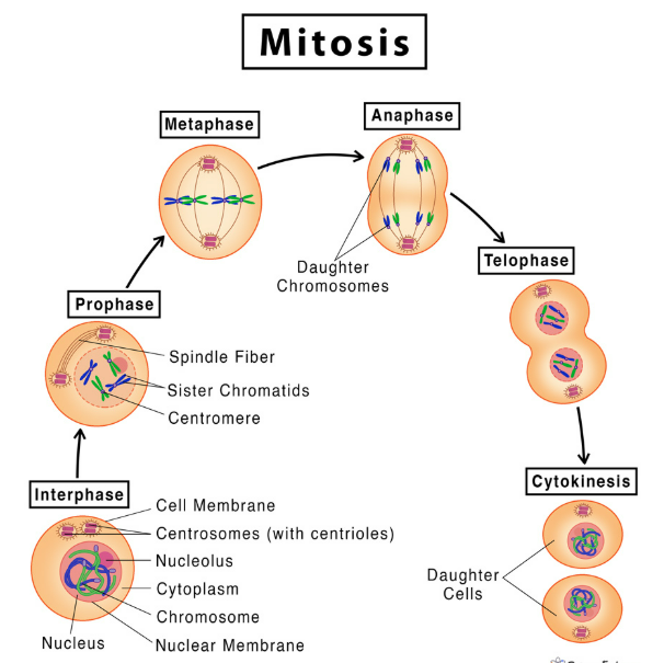
- Prophase
Chromosomes condense and become visible as sister chromatids.
The nuclear envelope breaks down.
The spindle apparatus begins to form from microtubules.
Centrioles (in animal cells) move to opposite poles. - Metaphase
Chromosomes line up along the metaphase plate (cell equator).
Spindle fibers attach to the kinetochores on centromeres. - Anaphase
Sister chromatids separate as spindle fibers shorten.
Chromatids (now individual chromosomes) are pulled to opposite poles. - Telophase
Chromosomes arrive at poles and begin to decondense.
Nuclear envelopes re-form around each set of chromosomes.
Spindle apparatus disassembles.
🧩 Result of Mitosis
- The nucleus has divided.
- Two genetically identical daughter nuclei are produced.
- Followed by cytokinesis, splitting the cytoplasm to form two separate daughter cells.
D2.1.8 – Identification of Phases of Mitosis
🧬 Skill Focus
Students should be able to identify the phases of mitosis by observing:
Diagrams of mitotic stages
Microscope slides of cells undergoing mitosis
Micrographs (photographs taken through a microscope)
🔎 Key Features to Identify Each Phase
Phase | What to Look For | Visual Clues in Cells / Micrographs |
---|---|---|
Prophase | – Chromosomes condense, become visible as thick strands – Nuclear envelope begins to disappear – Spindle starts forming | Dark, thick chromosomes scattered in the nucleus; fuzzy or no visible nuclear envelope |
Metaphase | – Chromosomes align at the cell center (equator) – Spindle fibers attached to centromeres | Chromosomes lined up clearly in a straight line across the middle of the cell |
Anaphase | – Sister chromatids pulled apart to opposite poles – Chromatids look like V-shaped moving structures | Chromatids moving away from the center, toward opposite poles of the cell |
Telophase | – Chromosomes reach poles – Nuclear envelope reforms – Chromosomes begin to uncoil | Two groups of chromosomes at opposite ends; faint nuclear envelopes reappearing |
🔧 Tips for Identification
Look for chromosome shape and position.
Notice changes in the nuclear envelope.
Identify spindle fibers if visible.
Use staining techniques to make chromosomes more visible under microscope.
D2.1.9 – Meiosis as a Reduction Division
🧠 Key Terms
Diploid (2n): A cell with two sets of chromosomes – one from each parent.
Example: Human body cells have 46 chromosomes (23 pairs).
Haploid (n): A cell with one set of chromosomes.
Example: Human gametes (sperm and egg) have 23 chromosomes.
🌿 What is Meiosis?
- Meiosis is a special type of cell division that reduces chromosome number by half.
- It produces four haploid nuclei (gametes) from one diploid nucleus.
- This is why it’s called reduction division.
🔬 Why Is Meiosis Important in Sexual Life Cycles?
- Sexual reproduction involves fusion of two haploid gametes (fertilization).
- Meiosis ensures gametes have half the chromosome number, so after fertilization, the diploid number is restored.
- Without meiosis, chromosome number would double each generation, causing genetic chaos.
🧪 Two Divisions in Meiosis: Overview
Division | Name | What Happens | Result |
---|---|---|---|
1st division | Meiosis I | Homologous chromosomes separate | Two haploid cells, each chromosome still has two chromatids |
2nd division | Meiosis II | Sister chromatids separate (like mitosis) | Four haploid cells with single chromatids |
🧬 Outline of the Two Rounds of Segregation
Meiosis I: Homologous Chromosome Separation
Homologous pairs (one from each parent) line up in the middle of the cell.
They are then pulled apart to opposite poles.
This reduces chromosome number from diploid to haploid.
Meiosis II: Sister Chromatid Separation
Similar to mitosis.
Sister chromatids separate and move to opposite poles.
Results in 4 genetically distinct haploid cells.
📌 Summary Table: Diploid vs Haploid and Meiosis Divisions
Term/Process | Definition/Description | Chromosome Number | Number of Cells Produced |
---|---|---|---|
Diploid (2n) | Two sets of chromosomes | 2n (e.g., 46 in humans) | 1 cell (starting) |
Haploid (n) | One set of chromosomes | n (e.g., 23 in humans) | 4 cells (end of meiosis) |
Meiosis I | Separation of homologous chromosomes | Reduction from 2n to n | 2 haploid cells |
Meiosis II | Separation of sister chromatids | Remains haploid | 4 haploid cells |
🔍 Real-Life Example
In humans, meiosis occurs in the ovaries (egg formation) and testes (sperm formation).
This keeps chromosome number constant across generations and introduces genetic variation (through crossing over and independent assortment).
Meiosis is a reduction division: 1 diploid cell → 4 haploid cells.
Two rounds of division:
Meiosis I separates homologous chromosomes.
Meiosis II separates sister chromatids.
Produces gametes necessary for sexual reproduction and genetic diversity.
D2.1.10 – Down Syndrome and Non-Disjunction
🧠 What is Non-Disjunction?
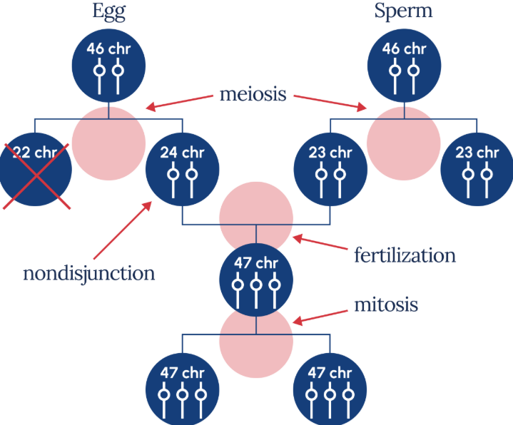
- Non-disjunction is an error during meiosis when homologous chromosomes or sister chromatids fail to separate properly.
- This results in gametes with an abnormal number of chromosomes – either too many or too few.
- When such a gamete fertilizes or is fertilized, it leads to chromosomal abnormalities in the offspring.
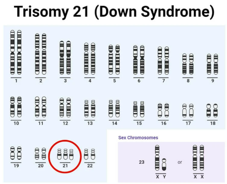
🌿 Down Syndrome: An Example of Non-Disjunction
- Down syndrome occurs when there is an extra copy of chromosome 21 – called trisomy 21.
- Instead of two copies, the individual has three copies of chromosome 21.
- This extra chromosome disrupts norm al development.
🔬 How Non-Disjunction Causes Down Syndrome
Stage | What Should Happen | What Happens in Non-Disjunction |
---|---|---|
Meiosis I | Homologous chromosomes separate | Both homologous chromosome 21 go to same cell |
Meiosis II | Sister chromatids separate | Both sister chromatids go to same cell |
Result | Gametes with one copy of chromosome 21 | Gametes with two copies or zero copies of chromosome 21 |
When a gamete with two copies of chromosome 21 fertilizes a normal gamete (one copy), the zygote has 3 copies = trisomy 21.
📌 Features of Down Syndrome
Physical traits: distinct facial features, short stature, and poor muscle tone.
Intellectual disability of varying degrees.
Higher risk of heart defects and other health issues.
🧪 Why Does Non-Disjunction Occur?
The exact cause isn’t always clear.
Risk increases with maternal age (older mothers have higher chance).
Meiosis errors during egg formation are more common than in sperm.
🔍 Other Examples of Non-Disjunction
Turner syndrome: missing one X chromosome (XO).
Klinefelter syndrome: extra X chromosome in males (XXY).
Non-disjunction = failure of chromosomes to separate during meiosis.
Leads to abnormal chromosome numbers in gametes.
Down syndrome is caused by trisomy 21, an extra chromosome 21.
Results in physical and intellectual challenges.
Risk factors include maternal age and random meiotic errors.
D2.1.11 – Meiosis as a Source of Variation
🧠 Why Is Variation Important?
- Genetic variation is essential for evolution and adaptation.
- Meiosis creates new combinations of genes in gametes.
- This increases the chance that some offspring will survive environmental changes.
🌿 How Does Meiosis Create Variation?
1. Random Orientation of Bivalents (Independent Assortment)
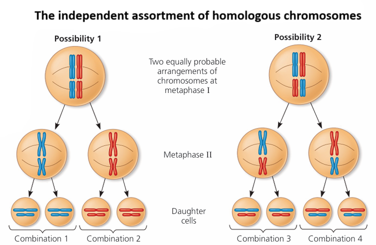
- During metaphase I, homologous chromosome pairs (bivalents) line up randomly along the cell equator.
- This means maternal and paternal chromosomes mix differently in each gamete.
- Each pair’s orientation is independent of others, creating millions of possible chromosome combinations.
2. Crossing Over (Genetic Recombination)
- During prophase I, homologous chromosomes pair tightly.
- Sections of chromatids swap segments at points called chiasmata.
- This process exchanges alleles between maternal and paternal chromosomes.
- Result: chromosomes with new allele combinations different from parents.
🔬 Combined Effect
Independent assortment and crossing over together ensure that every gamete is genetically unique.
This leads to vast genetic diversity in sexually reproducing populations.
📊 Summary Table: Sources of Genetic Variation in Meiosis
Mechanism | When it Occurs | How it Creates Variation |
---|---|---|
Random orientation (independent assortment) | Metaphase I | Different combinations of maternal & paternal chromosomes in gametes |
Crossing over | Prophase I | Exchange of DNA segments between homologous chromosomes, creating new allele combinations |
🔍 Real-Life Example
Humans produce over 8 million possible chromosome combinations from independent assortment alone (2^23).
Crossing over increases this number even more.
This is why siblings (except identical twins) are genetically different.
Meiosis increases genetic diversity through:
Random orientation of chromosomes (independent assortment).
Crossing over (exchange of DNA between homologues).
Genetic variation is vital for natural selection and survival of species.
Additional Higher Level
D2.1.12 – Cell Proliferation for Growth, Cell Replacement, and Tissue Repair
🧬 What is Cell Proliferation?
Cell proliferation means cells dividing to produce new cells.
It is essential for:
- Growth of organisms
- Replacing damaged or old cells
- Repairing tissues after injury
🌿 Cell Proliferation in Growth
🌱 Plants: Meristems
Meristems are regions of actively dividing cells in plants.
Located at tips of roots and shoots.
Provide new cells for:
- Plant growth in length (primary growth)
- Formation of new tissues and organs
🐣 Animals: Early Embryos
Early-stage embryos undergo rapid cell division.
Cells multiply quickly to form the tissues and organs of the developing organism.
🧪 Cell Replacement in Animals
Example: Skin
Skin cells continuously divide to replace dead or damaged cells.
This keeps the skin healthy and functioning as a protective barrier.
This process is called routine cell replacement.
🔬 Tissue Repair (Wound Healing)
When skin is wounded:
- Cells around the injury proliferate rapidly to fill the gap.
- New cells replace damaged tissue.
- This restores the skin’s protective function.
📊 Summary Table: Cell Proliferation Examples
Purpose | Example | Where? | Why? |
---|---|---|---|
Growth | Plant meristems | Root & shoot tips | Lengthening & new tissues |
Growth | Early animal embryos | Developing embryo | Form tissues & organs |
Routine cell replacement | Skin | Outer layer of skin | Replace dead skin cells |
Tissue repair | Skin wound healing | At site of injury | Repair damaged tissue |
Cell proliferation is vital for growth, maintenance, and repair.
Plants use meristems for continuous growth.
Animals rely on rapid division in early embryos and in tissues like the skin.
During wound healing, cell proliferation restores damaged tissue to keep the body protected.
D2.1.13 – Phases of the Cell Cycle
🧬 What is the Cell Cycle?
The cell cycle is a series of events that cells go through to grow and divide.
It explains how cell proliferation happens in a controlled, organized way.
🌿 Main Stages of the Cell Cycle
Stage | Description | Purpose |
---|---|---|
Interphase | The cell prepares for division; longest phase | Cell grows and copies DNA |
Mitosis (M phase) | The nucleus divides | Ensures each new cell gets a full set of chromosomes |
Cytokinesis | The cytoplasm divides, forming two separate cells | Completes cell division |
🧪 Details of Interphase
Sub-phase | What Happens |
---|---|
G1 (Gap 1) | Cell grows larger and makes new proteins and organelles |
S (Synthesis) | DNA is replicated — each chromosome duplicates |
G2 (Gap 2) | Cell checks DNA for errors and makes final preparations for division |
🔬 Mitosis & Cytokinesis
Mitosis: The duplicated chromosomes are equally separated into two nuclei.
Cytokinesis: The cytoplasm splits, forming two distinct daughter cells.
📊 Summary Table: Cell Cycle Phases Overview
Phase | Main Activity | Result |
---|---|---|
G1 | Cell growth, protein/organelle synthesis | Larger cell ready for DNA copy |
S | DNA replication | Two copies of each chromosome |
G2 | Final prep, error check | Ready for mitosis |
Mitosis (M) | Nuclear division | Two nuclei, identical chromosomes |
Cytokinesis | Cytoplasm division | Two separate daughter cells |
The cell cycle controls cell growth and division.
Interphase (G1, S, G2) is when the cell grows and duplicates DNA.
Mitosis divides the nucleus; cytokinesis splits the cell.
This cycle allows organisms to grow, repair tissues, and replace cells.
D2.1.14 – Cell Growth During Interphase
🧬 What Happens During Interphase?
Interphase is a metabolically active period where the cell is busy preparing for division.
It’s not a resting phase – lots of important growth and activity happen here.
🌿 Key Processes in Cell Growth During Interphase
Biosynthesis of cell components occurs, including:
- Proteins (enzymes, structural proteins)
- Lipids (for membranes)
- DNA replication during the S phase (synthesis phase)
The cell increases in size as it produces new organelles and molecules.
🧪 Organelle Growth and Division
Mitochondria and chloroplasts grow and divide independently during interphase.
This increases their numbers to meet the energy and metabolic needs of daughter cells after division.
🔍 Why Is Organelle Division Important?
Both mitochondria and chloroplasts have their own DNA and can replicate on their own.
Having enough organelles is essential for:
- Energy production (mitochondria)
- Photosynthesis (chloroplasts in plants)
- Ensures each daughter cell gets enough to function properly.
📊 Summary Table: Growth Activities in Interphase
Activity | Purpose |
---|---|
Protein biosynthesis | Prepare enzymes and cell structures |
DNA replication | Ensure identical genetic info for daughter cells |
Organelle growth & division | Meet energy and metabolic demands of new cells |
Cell size increase | Accommodate duplicated DNA and organelles |
Interphase is a busy growth phase, not rest.
The cell makes proteins, lipids, and duplicates DNA.
Mitochondria and chloroplasts grow and divide to supply energy and functions.
These processes prepare the cell for successful division.
D2.1.15 – Control of the Cell Cycle Using Cyclins
🧠 What Are Cyclins?
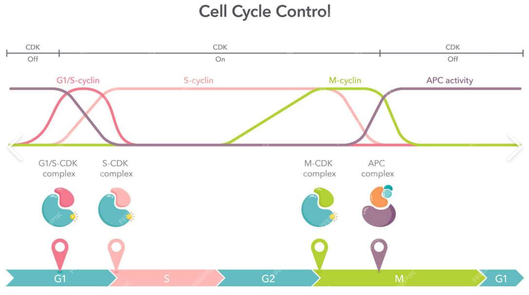
Cyclins are proteins that regulate the progression of the cell cycle.
Their concentrations rise and fall at specific points during the cycle.
🌿 How Cyclins Control the Cell Cycle
- Different cyclins increase in amount during certain phases of the cycle.
- When a cyclin reaches a threshold level, it triggers the cell to pass a checkpoint and move to the next phase.
- After the checkpoint is passed, cyclin levels fall.
🔬 Checkpoints Controlled by Cyclins
Checkpoint | Cyclin Level Role |
---|---|
G1 to S phase | Cyclin concentration rises to allow DNA replication to start |
G2 to Mitosis (M) | Cyclin reaches threshold to trigger mitosis |
During Mitosis | Cyclin levels control progression through mitosis |
📌 Why Are Cyclins Important?
- They ensure the cell only moves to the next stage when it is ready.
- Prevent errors like DNA damage or incomplete replication.
- Help coordinate the complex sequence of events in the cycle.
Cyclins are regulatory proteins whose levels change during the cell cycle.
A specific cyclin must reach a threshold concentration to pass checkpoints.
This system ensures controlled, orderly cell division.
D2.1.16 – Consequences of Mutations in Genes That Control the Cell Cycle
🧠 What Are Proto-Oncogenes and Tumour Suppressor Genes?
- Proto-oncogenes are normal genes that help cells grow and divide properly.
- Tumour suppressor genes are genes that slow down cell division, repair DNA mistakes, or tell cells when to die.
🌿 Mutations and Their Effects
Gene Type | Normal Role | Effect of Mutation |
---|---|---|
Proto-oncogenes | Promote cell division | Mutation converts them to oncogenes → cause cells to divide uncontrollably |
Tumour suppressor genes | Inhibit cell division or repair DNA | Mutation inactivates them → loss of control over cell division |
🔬 Result: Uncontrolled Cell Division
- Mutations disrupt the normal checks and balances in the cell cycle.
- This leads to excessive cell division and can form a tumour.
- If tumours invade other tissues, this causes cancer.
📌 How These Mutations Lead to Cancer
Oncogenes act like a “gas pedal stuck down” – constantly telling the cell to divide.
Damaged tumour suppressor genes are like a “broken brake” – unable to stop uncontrolled division.
Together, these mutations upset the careful control of the cell cycle.
Mutations in proto-oncogenes turn them into oncogenes that cause too much division.
Mutations in tumour suppressor genes remove inhibition, allowing unchecked growth.
Both lead to uncontrolled cell division, increasing cancer risk.
D2.1.17 – Differences Between Tumours: Cell Division, Growth, and Metastasis
🧠 Key Terms
Tumour: A mass of abnormal cells formed by uncontrolled cell division.
Benign tumour:
Slow-growing, cells stay localized (do not invade nearby tissues).
Usually not cancerous.
Often have a clear boundary and may be removed surgically.
Malignant tumour:
Fast-growing and can invade nearby tissues.
Capable of metastasis (spreading to other parts of the body).
Causes cancer.
🌿 Primary and Secondary Tumours
Term | Meaning |
---|---|
Primary tumour | Original tumour formed at the site where cancer started |
Secondary tumour | Tumour formed when cancer cells spread to a new site (metastasis) |
🔬 Differences in Tumour Characteristics
Feature | Benign Tumour | Malignant Tumour |
---|---|---|
Rate of cell division | Slower | Faster |
Growth | Limited, localized | Rapid, invasive |
Ability to invade tissue | No | Yes |
Capacity for metastasis | No | Yes |
Cancer causing? | Usually no | Yes |
📊 Mitotic Index: Measuring Cell Division Rate
Mitotic index = (Number of cells in mitosis) ÷ (Total number of cells) × 100
Higher mitotic index means more cells are dividing, indicating a more aggressive tumour.
Used by scientists and doctors to assess tumour growth rate.
🔍 Why Is This Important?
- Understanding tumour type helps in diagnosis and treatment decisions.
- Benign tumours may only need removal.
- Malignant tumours often require more aggressive treatments (chemotherapy, radiotherapy).
Benign tumours grow slowly and don’t spread; malignant tumours grow fast, invade tissues, and metastasize.
Primary tumours start at the original site; secondary tumours form after spreading.
Mitotic index measures how fast tumour cells divide and helps determine aggressiveness.