▶️ Answer/Explanation
(a)
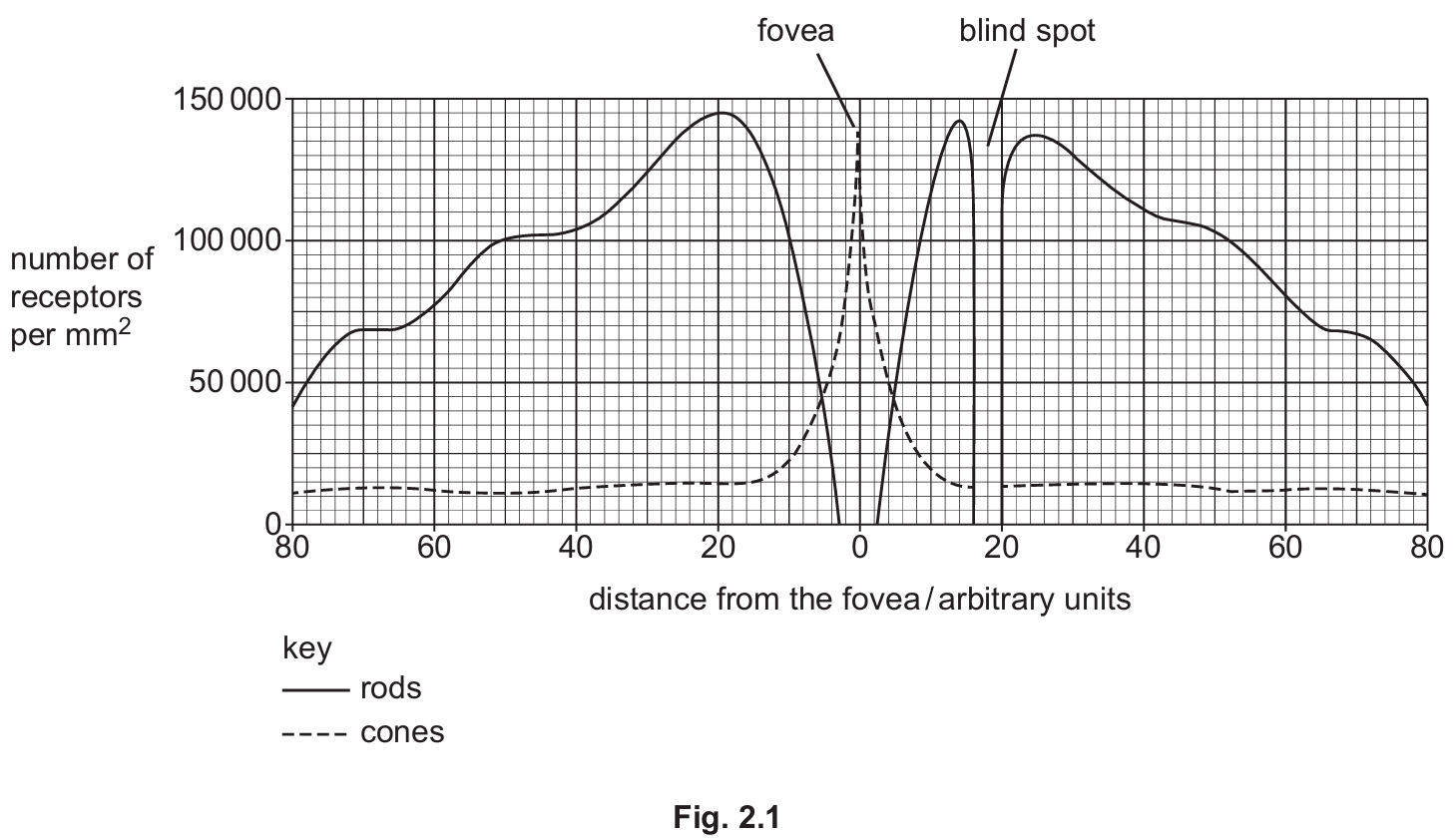
Based on Fig. 2.1, the differences are:
- Overall, there are significantly more rods than cones across the retina.
- The number of cones peaks at the fovea (at \(0\) arbitrary units on the x-axis).
- There are no rods present at the fovea.
- The number of rods peaks on either side of the fovea (around \(20\) arbitrary units) and generally increases as you move towards the fovea before dropping to zero.
- Cones are present in low, relatively constant numbers outside the fovea.
(b)
A nocturnal animal would have a greater number (or proportion) of rod cells compared to a diurnal animal.
Explanation: Rod cells are highly sensitive to low light intensities, making them essential for night vision. Cones, which require brighter light to function and provide colour vision, would be less important; thus, nocturnal animals may have fewer cones.
(c)
(i) Pupil reflex.
(ii) Circular muscle (of the iris).
(iii) Antagonistic action.
Explanation: In bright light, the pupil constricts to limit light entry. This is achieved by the contraction of the circular muscles and the relaxation of the radial muscles. Since these two muscle sets work in opposition, their action is described as antagonistic.
(d)
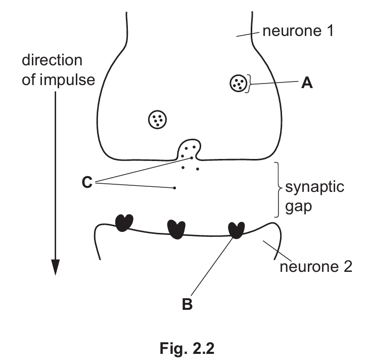
(i)
A: Vesicle
B: Receptor (protein)
C: Neurotransmitters
(ii) Part C (neurotransmitters) moves by diffusion. This occurs due to the random movement of particles from an area of higher concentration (released from the vesicle) to an area of lower concentration (across the synaptic gap towards the postsynaptic membrane).