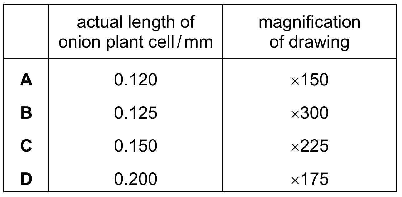
▶️ Answer/Explanation
To find the length of the drawing, we use the magnification formula: $\text{Image size} = \text{Actual size} \times \text{Magnification}$. Calculating for each student:
- (A) $0.120 \text{ mm} \times 150 = 18 \text{ mm}$
- (B) $0.125 \text{ mm} \times 300 = 37.5 \text{ mm}$
- (C) $0.150 \text{ mm} \times 225 = 33.75 \text{ mm}$
- (D) $0.200 \text{ mm} \times 175 = 35 \text{ mm}$
Comparing these values, Student (B) produced the longest drawing at $37.5 \text{ mm}$.
✅ Answer: (B)
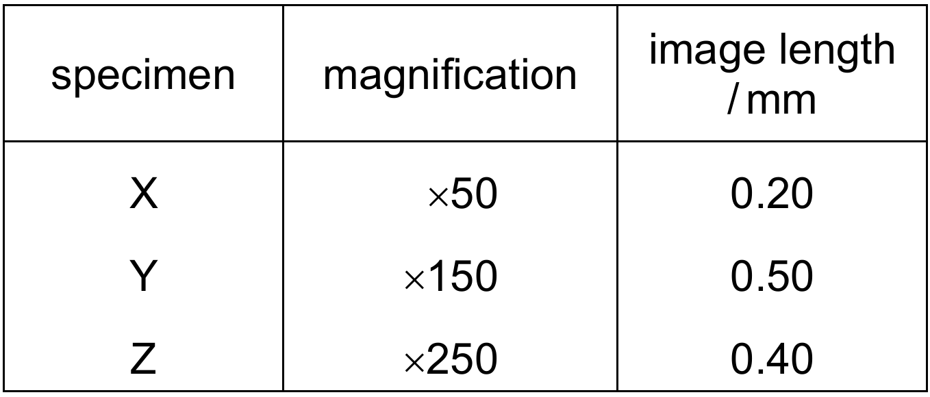
▶️ Answer/Explanation
To find the actual length, use the formula: $\text{Actual Length} = \frac{\text{Image Length}}{\text{Magnification}}$.
* For specimen $X$: $\frac{0.20}{50} = 0.0040$ $mm$.
* For specimen $Y$: $\frac{0.50}{150} \approx 0.0033$ $mm$.
* For specimen $Z$: $\frac{0.40}{250} = 0.0016$ $mm$.
Comparing these values, the smallest is $Z$ ($0.0016$ $mm$), followed by $Y$ ($0.0033$ $mm$), and the largest is $X$ ($0.0040$ $mm$).
Therefore, the order of increasing actual length is $Z \rightarrow Y \rightarrow X$.
✅ Answer: (D)