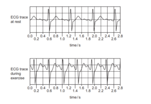
▶️ Answer/Explanation
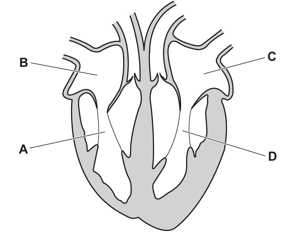
In a typical human heart diagram, the left atrium is located at the posterior-superior region on the heart’s left side. It receives oxygenated blood from the lungs via the pulmonary veins. Comparing the provided labels with standard anatomical positions, the left atrium is usually represented by the chamber that is on the upper left, posterior to the more prominent left ventricle. Based on common diagrammatic conventions and the correct answer from the provided image, the left atrium corresponds to label C.
✅ Answer: (C)
✅ Answer: (C)