Home / iGCSE Biology (0610)-9.2 Heart – iGCSE Style Questions Paper 2

iGCSE Biology (0610)-9.2 Heart – iGCSE Style Questions Paper 2

Question

The diagram shows a heart defect that some babies are born with. The arrows indicate direction of blood flow.

Which statement explains why this heart defect might cause the baby to have poor growth?

(A) Blood flows to the lungs at high pressure increasing rate of gas exchange.
(B) Blood flows to the lungs at low pressure decreasing rate of gas exchange.
(C) Mixing of blood results in less oxygen available for respiration.
(D) Mixing of blood results in more glucose available for respiration.
▶️ Answer/Explanation
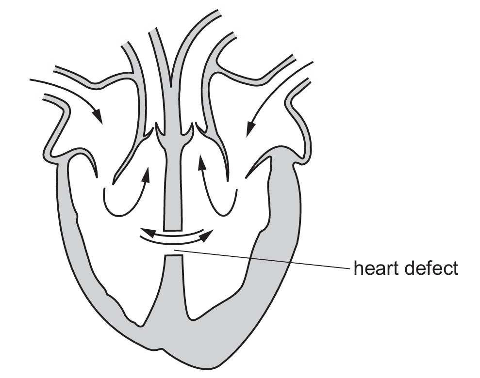
The diagram illustrates a ventricular septal defect (VSD), where a hole in the septum allows oxygenated blood from the left ventricle to mix with deoxygenated blood in the right ventricle. This mixing means that the blood pumped out to the body via the aorta contains a lower concentration of oxygen than normal. Since oxygen is a vital reactant for aerobic respiration, a reduced supply limits the amount of energy released in the baby’s cells. Without sufficient energy, cellular processes such as protein synthesis and cell division are hindered, leading to stunted or poor growth. Option (D) is incorrect because the defect affects gas transport, not glucose levels.
Answer: (C)

Question

Through which pathway does blood leave the heart?

(A) through arteries from the atria
(B) through arteries from the ventricles
(C) through veins from the atria
(D) through veins from the ventricles
▶️ Answer/Explanation

To answer this, we must look at the direction of blood flow and the type of vessels involved. Arteries are defined as blood vessels that carry blood away from the heart, while veins bring blood back. In the cardiac cycle, the atria act as receiving chambers that collect blood, whereas the ventricles are the powerful pumping chambers. When the heart contracts, the right ventricle pumps deoxygenated blood to the lungs via the pulmonary artery, and the left ventricle pumps oxygenated blood to the rest of the body via the aorta. Therefore, blood leaves the heart through arteries starting from the ventricles.

Answer: (B)

Question

The diagram shows a section through a human heart.

Which structure is an atrioventricular valve in the right side of the heart?

▶️ Answer/Explanation
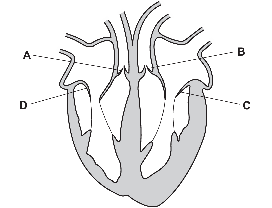
To identify the correct structure, we must first orient the heart: the left side of the diagram represents the right side of the heart. Atrioventricular (AV) valves are located between the atria and the ventricles. Structure D represents the tricuspid valve, which is the AV valve on the right side. In contrast, C is the bicuspid (mitral) valve on the left side, while A and B represent the semilunar valves (pulmonary and aortic respectively) that lead out of the ventricles into the major arteries.
Answer: (D)
Scroll to Top