▶️ Answer/Explanation
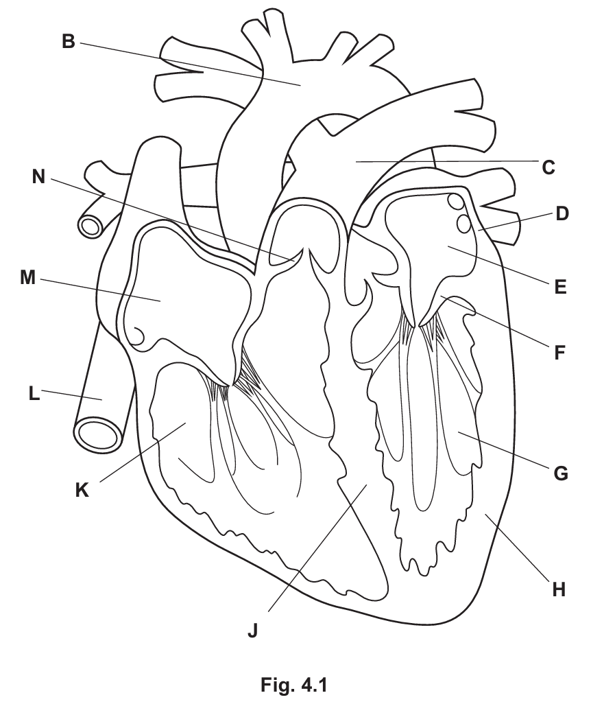
(a) (i)
Structure separating oxygenated and deoxygenated blood: J (Septum)
An atrioventricular valve: F (Bicuspid/Mitral valve)
Explanation: The septum (J) divides the left and right sides of the heart to prevent the mixing of oxygenated and deoxygenated blood. The structure F is the valve situated between the left atrium and the left ventricle.
(a) (ii)
H (the left ventricle) has a much thicker muscular wall than D (the left atrium). This is because the ventricle needs to generate higher pressure to pump blood a longer distance (to the entire body/systemic circulation). The atrium only pumps blood a short distance into the ventricle.
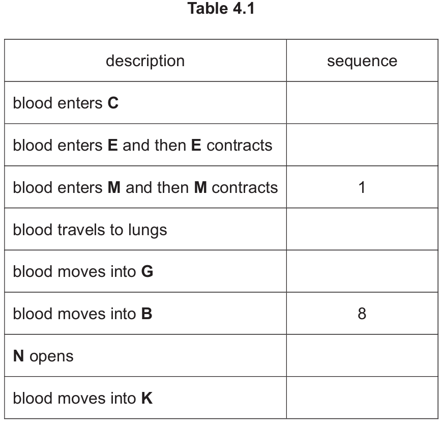
(a) (iii)
The correct sequence is as follows:
| description | sequence |
|---|---|
| blood enters C (Pulmonary Artery) | 4 |
| blood enters E and then E contracts (Left Atrium) | 6 |
| blood enters M and then M contracts (Right Atrium) | 1 |
| blood travels to lungs | 5 |
| blood moves into G (Left Ventricle) | 7 |
| blood moves into B (Aorta) | 8 |
| N opens (Pulmonary Valve) | 3 |
| blood moves into K (Right Ventricle) | 2 |
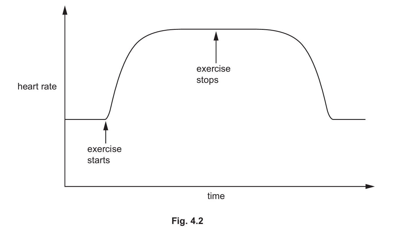
(b) (i)
Exercise increases the heart rate. This occurs because:
• Muscles require more oxygen and glucose for increased aerobic respiration to release energy for contraction.
• Increased blood flow is needed to remove carbon dioxide (and lactic acid) produced by the muscles.
• Adrenaline may be released, stimulating the heart rate to rise.
(b) (ii)
Any three from:
• Diet (high in saturated fats, salt, or cholesterol)
• Smoking (tobacco)
• Stress (uncontrolled)
• Genetic predisposition (family history)
• Age (risk increases with age)
• Sex (males are generally at higher risk)