▶️ Answer/Explanation
(a)(i) Table 2.1 Completion:
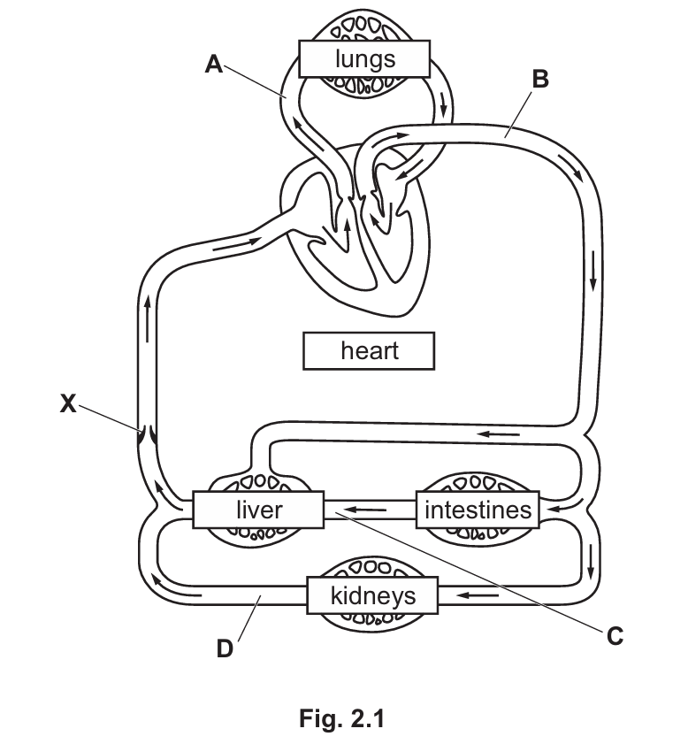
| Letter | Name of Blood Vessel | Carries Oxygenated Blood? |
| A | Pulmonary artery | No |
| B | Aorta | Yes |
| C | Hepatic portal vein | No |
| D | Renal vein | No |
Explanation:
• A (Pulmonary artery): Carries blood from the right ventricle of the heart to the lungs to be oxygenated. It is the only artery that carries deoxygenated blood.
• B (Aorta): The main artery carrying oxygenated blood from the left ventricle to the rest of the body.
• C (Hepatic portal vein): Connects the intestines to the liver. It carries nutrient-rich but deoxygenated blood.
• D (Renal vein): Carries deoxygenated blood away from the kidneys back toward the vena cava.
(a)(ii)
Name: Valve
Function: Prevents the back flow of blood.
Explanation: Veins (where X is located) operate under low pressure. Valves are essential to ensure blood flows in only one direction (towards the heart) and does not pool due to gravity.
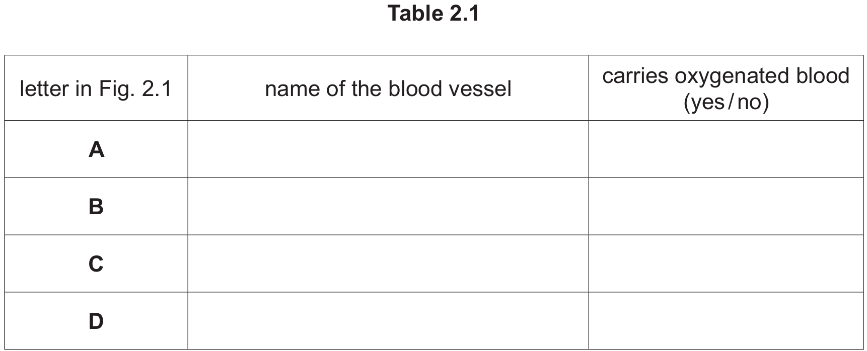
(b)(i) Differences in Heart Structure:
• The fish heart has only two chambers (one atrium and one ventricle), whereas the human heart has four.
• The fish heart lacks a septum separating left and right sides (since it is a single loop), while the human heart has a septum.
• The fish heart has fewer valves and fewer blood vessels attached compared to the human heart.
(b)(ii) Advantages of Human Circulatory System (Double Circulation):
• Separation of blood: Oxygenated and deoxygenated blood are kept separate by the septum, maintaining a steep concentration gradient for efficient gas exchange.
• High Pressure to Body: Blood loses pressure in the capillaries of the lungs. By returning to the heart (left side), it can be pumped out to the body at a much higher pressure, ensuring efficient delivery of oxygen and nutrients to tissues to support a high metabolic rate.
• Low Pressure to Lungs: The pulmonary circuit remains at lower pressure, preventing damage to the delicate capillaries in the lungs.
(c)(i)
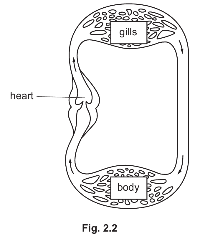
Structure P: Red blood cell (Erythrocyte).
Note: These are recognizable by their biconcave disc shape and location inside the vessel lumen.
(c)(ii)
Formula: $$\text{Actual size} = \frac{\text{Image size}}{\text{Magnification}}$$
(c)(iii)
Answer: $31.5 \, \mu\text{m}$
Calculation: To convert millimeters (mm) to micrometers ($\mu\text{m}$), multiply by 1000.
$$0.0315 \, \text{mm} \times 1000 = 31.5 \, \mu\text{m}$$